Comparative Study
. 2007 Feb 7:8:45.
doi: 10.1186/1471-2164-8-45.

Patterns of gene recombination shape var gene repertoires in Plasmodium falciparum: comparisons of geographically diverse isolates

Affiliations
Comparative Study

Patterns of gene recombination shape var gene repertoires in Plasmodium falciparum: comparisons of geographically diverse isolates

Susan M Kraemer et al. BMC Genomics. .

Abstract

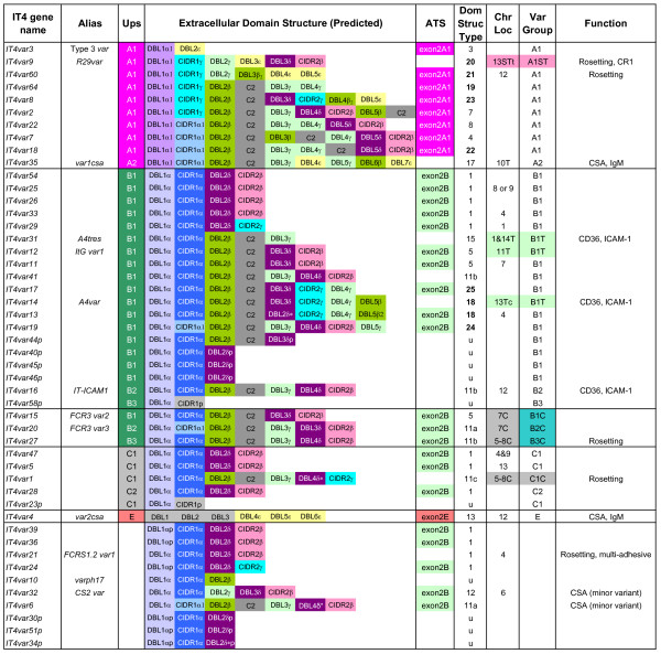
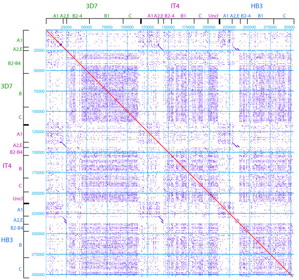
Background: Var genes encode a family of virulence factors known as PfEMP1 (Plasmodium falciparum erythrocyte membrane protein 1) which are responsible for both antigenic variation and cytoadherence of infected erythrocytes. Although these molecules play a central role in malaria pathogenesis, the mechanisms generating variant antigen diversification are poorly understood. To investigate var gene evolution, we compared the variant antigen repertoires from three geographically diverse parasite isolates: the 3D7 genome reference isolate; the recently sequenced HB3 isolate; and the IT4/25/5 (IT4) parasite isolate which retains the capacity to cytoadhere in vitro and in vivo.

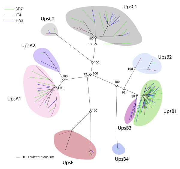
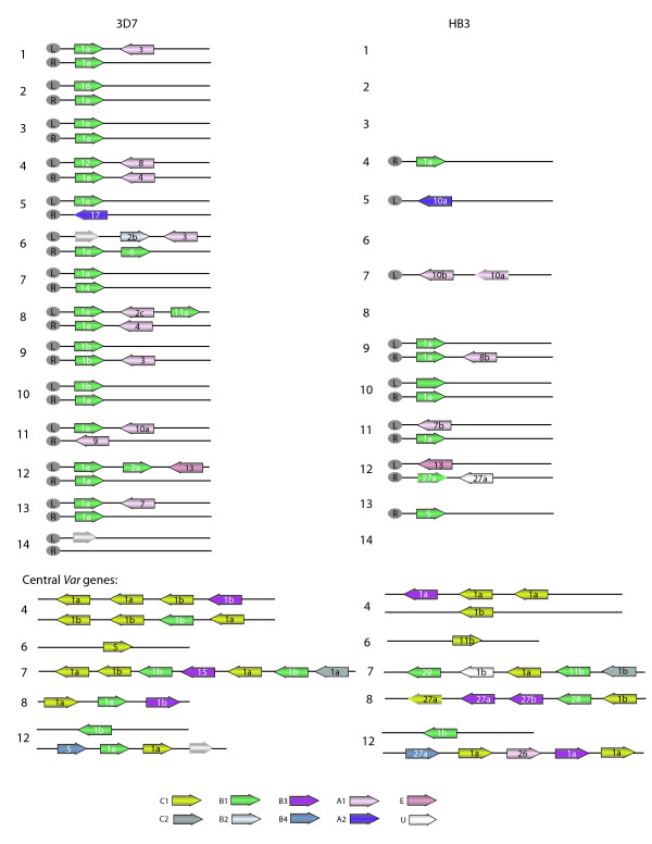
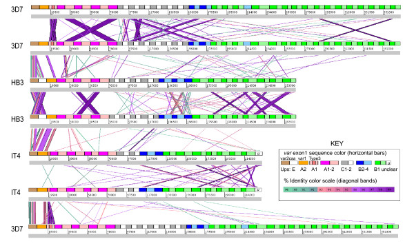
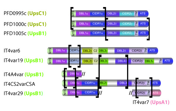
Results: These comparisons revealed that only two var genes (var1csa and var2csa) are conserved in all three isolates and one var gene (Type 3 var) has homologs in IT4 and 3D7. While the remaining 50 plus genes in each isolate are highly divergent most can be classified into the three previously defined major groups (A, B, and C) on the basis of 5' flanking sequence and chromosome location. Repertoire-wide sequence comparisons suggest that the conserved homologs are evolving separately from other var genes and that genes in group A have diverged from other groups.

Conclusion: These findings support the existence of a var gene recombination hierarchy that restricts recombination possibilities and has a central role in the functional and immunological adaptation of var genes.


Figures

Figure 1
Figure 1
Schematic representation of the IT4 var gene repertoire. Gene names, Ups sequence type, domain architecture, chromosomal location, transcription orientation, and binding functions are listed. IT4 var genes are primarily assigned to different groups on the basis of 5' flanking sequence (Ups type) and chromosomal location when known. PfEMP1 proteins are comprised of multiple domains termed N-termimal segment (NTS), Duffy binding-like (DBL), cysteine-rich interdomain region (CIDR), C2, transmembrane (TM), and acidic terminal segment (ATS or exon2) which have been classified by sequence criteria into different types. The PfEMP1 proteins in the 3D7 clone were arbitrarily classified into 17 different protein architectural types on the basis of domain composition [6]. Types 18–25 (bolded) are unique to IT4. Chromosome locations are indicated as T, ST, SST: first, second, and third var genes from the telomere respectively. C: internal var genes. t: transcribed towards telomere, c: transcribed towards centromere. The chromosomal location of var2csa was determined in [78]. Accession numbers for newly sequenced genes are EF158071-EF158105.
Figure 2
Figure 2
Schematic representation of HB3 var genes. Genes are organized as in figure 1 and grouped according to 5' flanking sequence (Ups type) and chromosomal location. Partial (p) and pseudogenes (Ψ) are labeled. Bolded domain structure types are unique to the HB3 parasite line. Binding properties have not been mapped to HB3 PfEMP1 proteins.
Figure 3
Figure 3
Phylogenetic comparison of var gene flanking regions from IT4, HB3, and 3D7 parasite isolates. A neighbor-joining tree was generated based upon 500 bp of 5' gene flanking sequence. Upstream groupings (Ups groups) with bootstrap support out of 1000 replicates are color shaded and labeled. Gene names have been removed from the figure for simplification.
Figure 4
Figure 4
Chromosomal distribution of var genes in the 3D7 and HB3 parasite isolates. Var genes are color shaded according to 5' gene flanking Ups type (U indicates unknown) and labeled according to protein architecture. The chromosomal locations were predicted for 36 of the 54 HB3 PfEMP1 proteins based upon gene flanking sequence and comparison to the 3D7 reference genome (see methods). Arrows without an outline indicate pseudogenes.
Figure 5
Figure 5
ACT nucleotide comparison of var gene repertoires. Concatamers of var gene exon1 sequences were arranged sequentially by Ups type: UpsE, A, C, B2-4, and B1, (colored as indicated) with one genome per horizontal line. The isolate-transcendent var genes var2csa, var1csa and Type 3 var are positioned at the left end of the concatemer. BLASTN was performed with word length set at 90 nucleotides (filter for low complexity removed). The comparisons were viewed in ACT with a window size of 120 nucleotides, at minimum 90% identity to show segments of similarity between var genes. Diagonal bands connecting individual var genes are colored according to percent identity, shown in the scale diagram (inset). Band width corresponds to region of sequence identity. Var names are listed, in order of appearance, in Additional file 4: Table S7.
Figure 6
Figure 6
Examples of chimeric genes in the 3D7 and IT4 parasite isolates. Identical or nearly identical regions are indicated by brackets.
Figure 7
Figure 7
Dotplot comparisons of PfEMP1 protein coding sequence. The extracellular binding region of PfEMP1 proteins are organized by parasite isolate and 5' Ups sequence type. Uncl (unclassified) refers to sequences in which the Ups sequences have not been determined. Dot plot parameters include a window length of 30 amino acids and percent identity of 80% or greater.

Similar articles

Cited by

References

    1. Snow RW, Guerra CA, Noor AM, Myint HY, Hay SI. The global distribution of clinical episodes of Plasmodium falciparum malaria. Nature. 2005;434:214–217. doi: 10.1038/nature03342. - DOI - PMC - PubMed
    1. Miller LH, Baruch DI, Marsh K, Doumbo OK. The pathogenic basis of malaria. Nature. 2002;415:673–9. doi: 10.1038/415673a. - DOI - PubMed
    1. Baruch DI, Pasloske BL, Singh HB, Bi X, Ma XC, Feldman M, Taraschi TF, Howard RJ. Cloning the P. falciparum gene encoding PfEMP1, a malarial variant antigen and adherence receptor on the surface of parasitized human erythrocytes. Cell. 1995;82:77–87. doi: 10.1016/0092-8674(95)90054-3. - DOI - PubMed
    1. Su XZ, Heatwole VM, Wertheimer SP, Guinet F, Herrfeldt JA, Peterson DS, Ravetch JA, Wellems TE. The large diverse gene family var encodes proteins involved in cytoadherence and antigenic variation of Plasmodium falciparum-infected erythrocytes. Cell. 1995;82:89–100. doi: 10.1016/0092-8674(95)90055-1. - DOI - PubMed
    1. Smith JD, Chitnis CE, Craig AG, Roberts DJ, Hudson-Taylor DE, Peterson DS, Pinches R, Newbold CI, Miller LH. Switches in expression of Plasmodium falciparum var genes correlate with changes in antigenic and cytoadherent phenotypes of infected erythrocytes. Cell. 1995;82:101–10. doi: 10.1016/0092-8674(95)90056-X. - DOI - PMC - PubMed

Publication types

Substances