. 2007 Feb 7:2:3.
doi: 10.1186/1750-1326-2-3.

APP-BP1 inhibits Abeta42 levels by interacting with Presenilin-1

Affiliations

APP-BP1 inhibits Abeta42 levels by interacting with Presenilin-1

Yuzhi Chen et al. Mol Neurodegener. .

Abstract

Background: The beta-amyloid precursor protein (APP) is sequentially cleaved by the beta- and then gamma-secretase to generate the amyloid beta-peptides Abeta40 and Abeta42. Increased Abeta42/Abeta40 ratios trigger amyloid plaque formations in Alzheimer's disease (AD). APP binds to APP-BP1, but the biological consequence is not well understood.

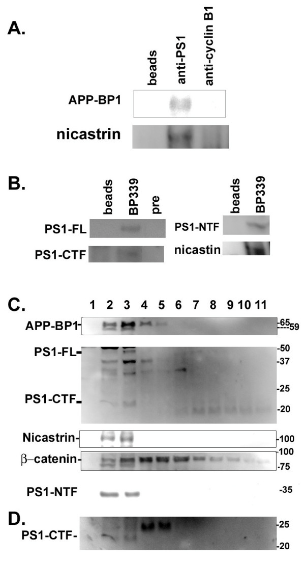
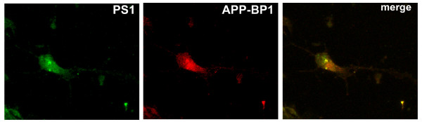
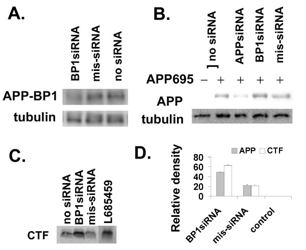
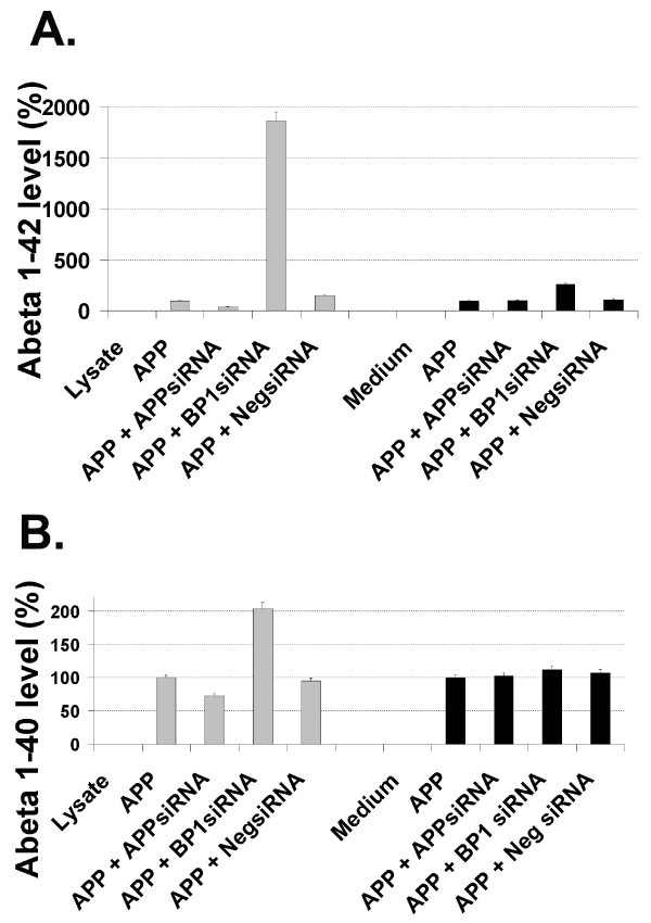
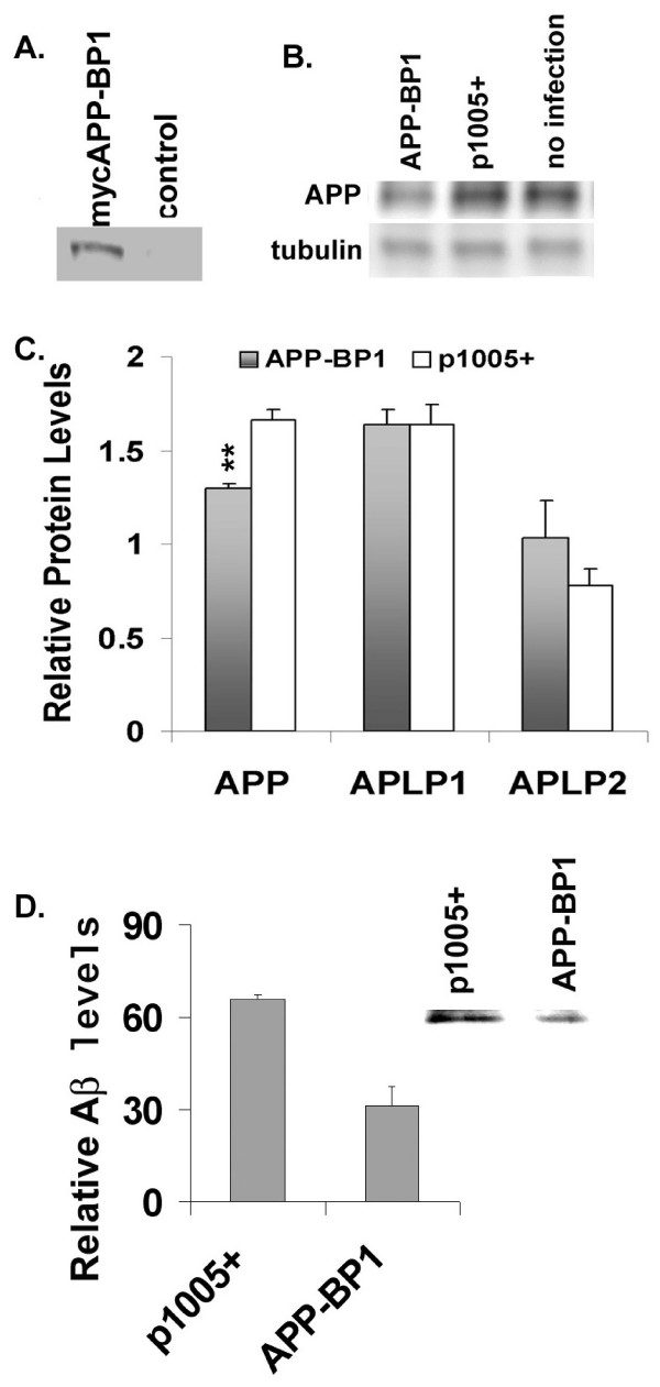
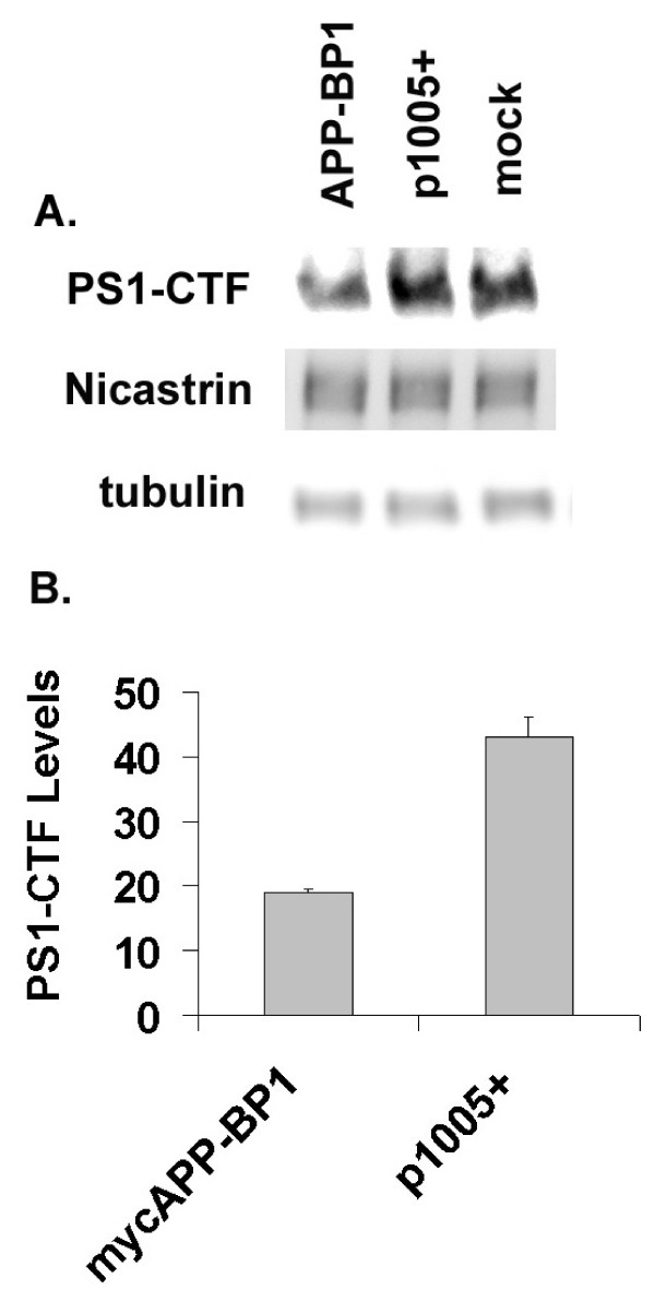
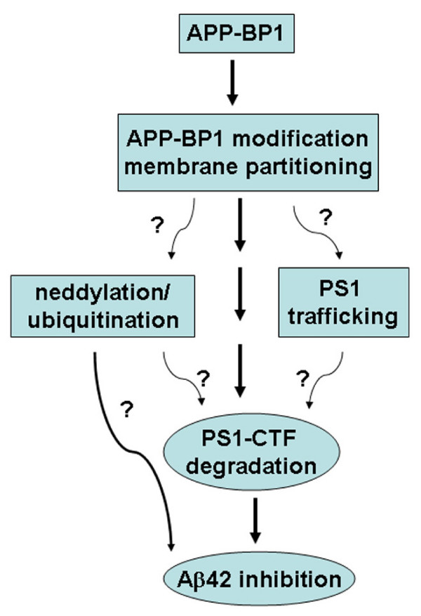
Results: We report that when the endogenous APP-BP1 was suppressed by small interfering RNAs (siRNAs), cell-associated Abeta42 was dramatically increased in APP695 expressing primary neurons. The accumulation of Abeta42 was accompanied by significant increases in APP and APP-CTF in APP-BP1 siRNA expressing neurons. In contrast, APP-BP1 overexpression in primary neurons significantly decreased the levels of Abeta and endogenous APP but not APLPs. We also investigated the potential mechanism of APP-BP1-mediated APP processing. APP-BP1 co-precipitated with Presenilin-1 (PS1) in native rat brain extracts, co-migrated with the gamma-secretase components in brain membrane extracts in glycerol gradient centrifugation, and colocalized in primary neurons. Further, the endogenous PS1-CTF was significantly downregulated by APP-BP1 expression.

Conclusion: Our data suggest that APP-BP1 may inhibit Abeta42 production by interacting with PS1 under physiological conditions.


Figures

Figure 1
Figure 1
APP-BP1, PS1 and nicastrin co-immunoprecipitated and co-migrated in brain protein extracts. A. APP-BP1 co-immunoprecipitated with PS1 in brain lysates. Adult rat brain lysates were immunoprecipitated with the rabbit anti-PS1 antibody (middle lane), or a non-related antibody (rabbit anti-cyclin B1). The blot was probed with the anti-APP-BP1 antibody, BP339 or with rabbit anti-nicastrin. Anti-PS1 specifically precipitated APP-BP1 and nicastrin which were absent in control lanes. This experiment has been repeated three times. B. PS1 co-precipitated with APP-BP1 in membrane fractions. Proteins extracted from membrane fractions were precipitated with the APP-BP1 antibody, BP339 or with the BP339 preimmune serum (pre) or beads alone. The blot was probed with the PS1 antibody (AB5308) for PS1-FL and PS1-CTF, or with antibodies against PS1-NTF or nicastrin. C. APP-BP1 co-migrated with PS1 in glycerol gradient of membrane protein extracts isolated from adult rat brains. Brain membrane proteins were subjected to glycerol gradient centrifugation. Proteins in equal volumes from each fraction were resolved by SDS-PAGE gels. The blots were incubated with specific antibodies followed by ChemiGlow detections. Fractions 2 to 4 contain the majority of each protein analyzed. APP-BP1 was found to co-migrate with PS1 (FL, NTF, and CTF), and nicastrin. The location of β-catenin was also shown in the glycerol gradient. BioRad Precision Plus protein standards are shown on the right. PS1-CTF identified by AB5308 antibody was verified by a PS1-C-specific antibody (D).
Figure 2
Figure 2
APP-BP1 and PS1 molecules were colocalized in primary neurons. Primary neurons were fixed, permeablized, blocked and stained with 9E10 for mycAPP-BP1 and the rabbit anti-PS1 (AB5308). Primary antibodies were coupled to Alexa Fluor 594 and Alexa Fluor 488 goat secondary antibodies. The images presented were obtained under a 40× objective with a Nikon confocal microscope using the EZ-C12.20 scanning program. Independent experiments using DAPI staining along with the above two antibodies did not show nuclear co-localization of APP-BP1 and PS1 (not shown).
Figure 3
Figure 3
Downregulation of APP-BP1 by siRNAs in primary neurons affected APP processing. A. APP-BP1 was suppressed by APP-BP1 siRNAs but not by missense siRNAs. Primary neurons were infected with 0.5 IU of siRNA virus per cell. Equal amount of proteins extracted by RIPA buffer was analyzed by western blotting for APP-BP1 expression. The same blot was reprobed with a mouse anti-γ-tubulin antibody. No obvious changes in γ-tubulin levels were detected. BP1siRNA, APP-BP1siRNA; mis-siRNA, misssense siRNA. B. Suppression of APP-BP1 by siRNAs caused increases in exogenous human APP. Primary neurons were infected with or without APP and siRNA constructs. APP was probed with the antibody 369. The same membrane was reprobed for γ-tubulin with a mouse anti-γ-tubulin for loading controls. In each sample, 10 μg of proteins was analyzed. The data is representative of three-independent experiments. C. APP-BP1 siRNA inhibited APP-CTF processing in primary neurons expressing human APP. APP-CTFs were resolved on 16% Tris-Tricine gels. The blot was probed with 369. A representative of 4 independent experiments was shown. The intervening lanes between mis-siRNA and L685459 were cut off. D. APP-BP1 siRNA expression significantly blocked APP processing. Quantitative western blot analyses of 3 (APP) or 4 (CTF) independent experiments were carried out using two-tail t-Test (APP: BP1siRNA vs mis-siRNA, p < 0.003; CTF: BP1siRNA vs mis-siRNA, p < 0.001).
Figure 4
Figure 4
Suppression of APP-BP1 protein expression by APP-BP1 siRNAs in primary neurons resulted in increases of intracellular Aβ42. Intracellular (from 50 μg of protein from lysates) and secreted (from 1/15 volume of conditioned medium) Aβ42 in primary neurons were determined by Aβ42 ELISA (A). Intracellular (from 50 μg of protein) and secreted (from 1/30 volume of medium of conditioned medium) Aβ40 in primary neurons was determined by Aβ40 ELISA (B). The amount of Aβ in samples that expressed APP without any siRNA interference was used as 100% of Aβ production. All the rest of the samples were normalized to this sample, and presented as percentage in respective experimental conditions. Data is representative of three independent experiments. BP1siRNA, APP-BP1 siRNA; NegsiRNA, Negative siRNA (Stratagene).
Figure 5
Figure 5
APP-BP1 expression increased APP processing in primary neurons. A. Expression of myc-tagged APP-BP1. Equal amount of total proteins from neurons infected with APP-BP1 virus or p1005+ virus was analyzed by western blotting using the mouse ant-myc antibody, 9E10. B. APP-BP1 overexpression downregulated rat endogenous APP. APP expression from 15 μg of proteins was analyzed by immunoblots using the anti-APP antibody 369. The amount of γ-tubulin from the same blot was reprobed with a mouse anti-γ-tubulin antibody. Representative blots from the same experiments are shown. C. Quantitative western blot analyses revealed that APP-BP1 significantly downregulated the endogenous APP but not APLPs in neurons overexpressing myc APP-BP1. mycAPP-BP1 expression significantly decreased APP levels compared to the sample infected with the vector (p1005+) (n = 4, p < 0.02, one-tail t-Test). APLP1 levels stayed the same, and APLP2 did not showed a significant change by t-Tests (n = 3, p = 0.1, one tail t-Test). D. RIPA buffer soluble protein extracts from primary neurons co-expressing APP-BP1 and APP695 or p1005+ and APP695 were precipitated with 6E10 followed by western blot analyses using 4G8. Mean levels of Aβ is presented in the graph (n = 4, p < 0.04). A representative of 4 independent blots was shown as an insert.
Figure 6
Figure 6
APP-BP1 expression decreases PS1-CTF stability in primary neurons. Primary neurons were infected with mycAPP-BP1 or p1005+ vector virus for 14 hrs before harvesting. Levels of the rat endogenous PS1 and PS1-CTF were examined by western blots. A representative PS1-CTF blot was shown in A. The same blot was reprobed with the rabbit anti-nicastrin or mouse anti-γ-tubulin antibodies after stripping. B, Quantitative western blot analyses show significant reduction of PS1-CTF in APP-BP1 expressing neurons (n = 3, p < 0.049, one tail t-Test).
Figure 7
Figure 7
Diagram of APP-BP1-mediated Aβ42 inhibition. Potential pathways through which APP-BP1 inhibits Aβ42 are presented with a question mark.

References

    1. Chow N, Korenberg JR, Chen XN, Neve RL. APP-BP1, a novel protein that binds to the carboxyl-terminal region of the amyloid precursor protein. J Biol Chem. 1996;271:11339–11346. doi: 10.1074/jbc.271.19.11339. - DOI - PubMed
    1. Chen Y, McPhie DL, Hirschberg J, Neve RL. The amyloid precursor protein-binding protein APP-BP1 drives the cell cycle through the S-M checkpoint and causes apoptosis in neurons. J Biol Chem. 2000;275:8929–8935. doi: 10.1074/jbc.275.12.8929. - DOI - PubMed
    1. Osaka F, Kawasaki H, Aida N, Saeki M, Chiba T, Kawashima S, Tanaka K, Kato S. A new NEDD8-ligating system for cullin-4A. Genes Dev. 1998;12:2263–2268. - PMC - PubMed
    1. Hori T, Osaka F, Chiba T, Miyamoto C, Okabayashi K, Shimbara N, Kato S, Tanaka K. Covalent modification of all members of human cullin family proteins by NEDD8. Oncogene. 1999;18:6829–6834. doi: 10.1038/sj.onc.1203093. - DOI - PubMed
    1. Wu JT, Lin HC, Hu YC, Chien CT. Neddylation and deneddylation regulate Cul1 and Cul3 protein accumulation. Nat Cell Biol. 2005. - PubMed