. 2007 Apr;81(8):4080-90.
doi: 10.1128/JVI.02545-06. Epub 2007 Feb 7.

PATJ, a tight junction-associated PDZ protein, is a novel degradation target of high-risk human papillomavirus E6 and the alternatively spliced isoform 18 E6

Affiliations

PATJ, a tight junction-associated PDZ protein, is a novel degradation target of high-risk human papillomavirus E6 and the alternatively spliced isoform 18 E6

Carina H Storrs et al. J Virol. 2007 Apr.

Abstract

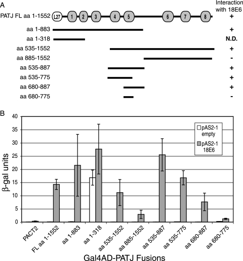
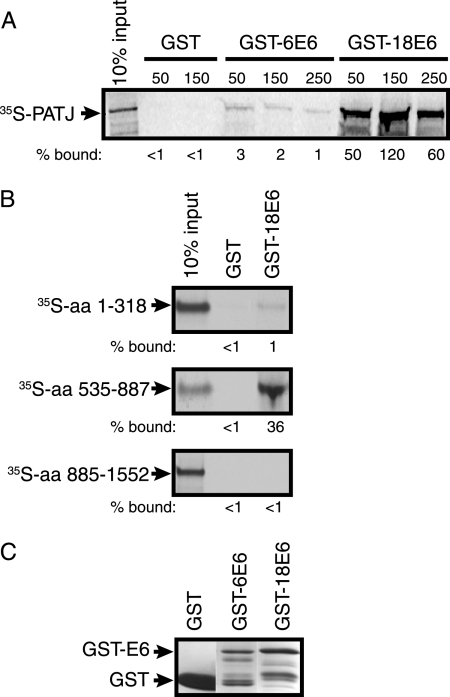
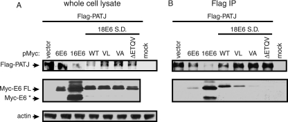
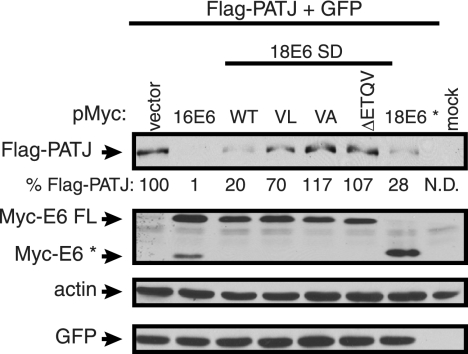
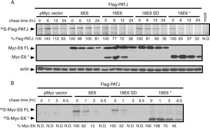
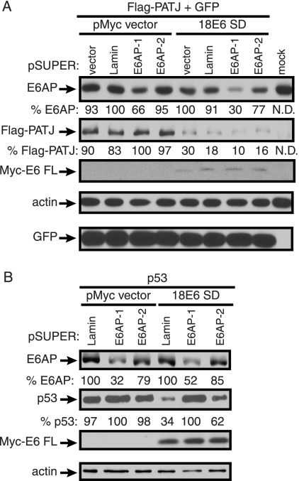
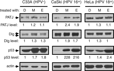
The E6 protein from high-risk human papillomavirus types interacts with and degrades several PDZ domain-containing proteins that localize to adherens junctions or tight junctions in polarized epithelial cells. We have identified the tight junction-associated multi-PDZ protein PATJ (PALS1-associated TJ protein) as a novel binding partner and degradation target of high-risk types 16 and 18 E6. PATJ functions in the assembly of the evolutionarily conserved CRB-PALS1-PATJ and Par6-aPKC-Par3 complexes and is critical for the formation of tight junctions in polarized cells. The ability of type 18 E6 full-length to bind to, and the subsequent degradation of, PATJ is dependent on its C-terminal PDZ binding motif. We demonstrate that the spliced 18 E6* protein, which lacks a C-terminal PDZ binding motif, associates with and degrades PATJ independently of full-length 18 E6. Thus, PATJ is the first binding partner that is degraded in response to both isoforms of 18 E6. The ability of E6 to utilize a non-E6AP ubiquitin ligase for the degradation of several PDZ binding partners has been suggested. We also demonstrate that 18 E6-mediated degradation of PATJ is not inhibited in cells where E6AP is silenced by shRNA. This suggests that the E6-E6AP complex is not required for the degradation of this protein target.


Figures

FIG. 1.
FIG. 1.
PDZ domains 4 and 5 of PATJ are sufficient to interact with 18 E6. (A) Schematic representation of FL PATJ, amino acids 1 to 1552, identifying the position of its L27 domain and eight PDZ domains. PATJ deletion mutants, named for the amino acids of PATJ that they encode, were cloned in the Gal4AD plasmid pACT2. They are shown as lines below the FL PATJ schematic. The ability of PATJ and the PATJ peptides to interact with 18 E6 in the yeast two-hybrid system is indicated to the right of the line schematic. (B) Yeast strain YGH1 was cotransformed with 1 μg of either the Gal4DBD vector pAS2-1 or pAS2-1-18 E6 and 1 μg of either Gal4AD vector pACT2 or the Gal4AD-PATJ fusions. Liquid β-Gal assays were performed on the extracts from cotransformed yeast colonies.
FIG. 2.
FIG. 2.
PATJ binds to GST-18 E6 but not GST-6 E6 in vitro. (A) Purified GST, GST-6 E6, or GST-18 E6 protein bound to glutathione beads was incubated with in vitro-translated 35S-labeled PATJ aa 1 to 1552. The complexes were washed with buffer containing 50, 150, or 250 mM KCl as indicated above the lanes. The bound 35S-labeled protein was resolved by SDS-PAGE and detected by autoradiography. The percentage of 35S-PATJ that remained bound, shown below each lane, was based on the intensity of the band representing 10% of the input. Band intensity was quantified using ImageJ (http://rsb.info.nih.gov/ij/). The arrow points to the position of [35S]PATJ. (B) 35S-labeled fragments of PATJ encompassing aa 1 to 318, 535 to 887, and 885 to 1552 were incubated with either GST or GST-18 E6. The complexes were incubated and washed in 50 mM KCl. The percentage of each fragment that bound to GST and GST-18 E6 was determined as described for panel A. (C) GST, GST-6 E6, and GST-18 E6 proteins used in the pull-down experiment were resolved by SDS-PAGE and visualized by staining with Coomassie brilliant blue. Arrows identify the bands representing GST and GST-6 E6 or GST-18 E6.
FIG. 3.
FIG. 3.
In vivo association of Flag-PATJ with E6 is dependent on the presence of a PDZ binding site. Approximately 2 × 10⁶ 293T cells were transiently transfected with 2 μg of Flag-PATJ expression plasmid and 3 μg of pMycL.1 plasmid or Myc-E6 expression constructs. Mock-transfected cells were incubated with transfection reagent in the absence of plasmid DNA. Cells were collected at 48 h posttransfection. (A) To determine the level of protein accumulation, equal amounts of protein from whole-cell lysates were subjected to Western blot analysis with antibody to Flag, Myc, and actin. (B) Equal amounts of protein were incubated with Flag affinity beads. Following extensive washing, the bound complexes were eluted and subjected to Western blot analysis with antibody to Flag and Myc to determine the levels of Flag-PATJ and Myc-E6. Arrows point to Flag-PATJ, Myc-E6 FL, Myc- E6*, and actin.
FIG. 4.
FIG. 4.
18 E6* is expressed and associates in vivo with Flag-PATJ. (A) Alignment of FL 18 E6 and 18 E6* ORFs. Splicing of 18 E6 removes an intron between nucleotides (nt) 132 and 310 and results in an alternative 3′ reading frame. The zig-zag lines represent DNA from the Myc-tag vector. The arrows identify the binding sites for the 5′ and 3′ primers used for RT-PCR. (B) RT-PCR was performed on total RNA collected from approximately 1.5 × 10⁶ 293T cells transiently transfected with 1 μg of pMycL.1-18 E6 WT or 18 E6 SD construct. PCR was also performed using pMycL.1-18 E6 WT vector DNA and the same primers. The PCR products were resolved on an agarose gel and detected by ethidium bromide staining. The sizes of the DNA molecular weight markers (M) are indicated. Approximately 2 × 10⁶ 293T cells were transiently transfected with 2 μg of Flag-PATJ expression plasmid and 3 μg of pMycL.1 plasmid or pMycL.1 constructs containing 18 E6 WT, SD, or E6*. (C and D) Equal amounts of protein, extracted after 48 h, were either resolved directly (C) or bound to Flag affinity beads (D). The complexes bound to Flag affinity beads were washed extensively and eluted. Following SDS-PAGE, the whole-cell lysates and Flag-bound complexes were probed with antibody to Flag, Myc, and actin. Arrows identify Flag-PATJ, Myc-E6 FL, Myc-E6*, and actin.
FIG. 5.
FIG. 5.
The steady-state level of Flag-PATJ is reduced by 16 E6, 18 E6, and 18 E6*, but not 18 E6 PDZ binding site mutants. Approximately 1 × 10⁶ 293T cells were cotransfected with 1 μg of pFlag-CMV-2-PATJ and 3 μg of pMycL.1 empty or pMycL.1 encoding 16 E6, 18 E6 S.D., 18 E6 SD, PDZ binding site mutants (VL, VA, and ΔETQV), or 18 E6*. All cotransfection mixtures contained 0.2 μg of a GFP expression vector. The mock lane contained lysate from cells incubated with Lipofectamine reagent in the absence of plasmid DNA. Forty-eight hours posttransfection, cell lysates were collected, and equal amounts of protein from whole-cell lysates were resolved by SDS-PAGE. Western blot assays were performed using antibody to the Flag or Myc epitopes, actin, and GFP. Arrows identify the positions of Flag-PATJ, Myc-E6 FL, Myc-E6*, actin, and GFP. Flag-PATJ levels, shown below the lanes, were determined by dividing the absolute band intensity by the intensity of the actin band in the sample.
FIG. 6.
FIG. 6.
The half-life of Flag-PATJ is reduced in the presence of 16 E6, 18 E6 FL, and 18 E6*, but not 6 E6. Approximately 2 × 10⁶ 293T cells were transfected with 2 μg of Flag-PATJ expression construct, 6 μg of empty pMycL.1, or pMycL.1 encoding 6 E6, 16 E6, 18 E6 FL, or E6*. Forty-eight hours later, cells were metabolically labeled with Trans35S-label and chased for the times indicated above each lane. (A) Equal amounts of trichloroacetic acid-precipitable counts were bound to Flag affinity beads. The captured complexes were washed and resolved by SDS-PAGE. 35S-labeled Flag-PATJ was visualized by autoradiography and is identified by the arrow (top panel). The percent remaining 35S-Flag-PATJ at each chase time point, shown below each lane, was normalized to the relative background intensity in the lane. Quantifications were performed using ImageJ. The expression level of the Myc-tagged E6 types was determined after equal amounts of protein from whole-cell lysates were resolved by SDS-PAGE and immunoblotted with antibody to the Myc epitope (middle panel). Equal loading was confirmed by analyzing the levels of actin (bottom panel). (B) The half-life of the Myc-tagged E6 proteins was determined as for Flag-PATJ, except that earlier time points were analyzed and equal amounts of total extracted protein were incubated with Myc affinity beads. 35S-labeled Myc-6 E6, 18 E6 FL, or 18 E6* is identified by the arrows. The level of the 35S-labeled Myc-E6 proteins that remained at the different times was quantified using ImageJ.
FIG. 7.
FIG. 7.
shRNA-mediated E6AP silencing does not affect 18 E6-dependent reduction of Flag-PATJ. (A) On day 1, approximately 0.75 × 10⁶ 293T cells were cotransfected with 2.5 μg of either empty pSUPER vector, lamin A/C, E6AP-1, or E6AP-2 targeting constructs. On day 4, the same amount of pSUPER constructs and 0.5 μg of Flag-PATJ expression plasmid, 0.125 μg of GFP expression plasmid, and 1.5 μg of empty Myc vector or Myc-18 E6 SD construct were introduced into these same cells. Cells were harvested on day 6. Equal amounts of protein were analyzed with antibodies to E6AP, the Flag and Myc epitopes, actin, and GFP. Arrows identify E6AP, Flag-PATJ, Myc-E6 FL, actin, and GFP. The levels of E6AP and Flag-PATJ, shown below the images, were quantified using ImageJ and normalized to the corresponding levels of actin. (B) The effect of E6AP silencing on 18 E6-dependent p53 reduction was determined as described for panel A, except that H1299 cells were used and cells were cotransfected with p53 instead of Flag-PATJ, expression plasmid, empty Myc vector, or Myc-18 E6 SD, and lamin A/C, E6AP-1 or E6AP-2 targeting constructs. Cell lysates were probed with antibodies to E6AP, p53, the Myc epitope, and actin. Arrows identify E6AP, p53, Myc-E6 FL, and actin.
FIG. 8.
FIG. 8.
Proteasome regulation of PATJ, Dlg, and p53 in cervical cancer-derived cell lines. C33A, HPV 16+ CaSki, and HPV 18+ HeLa cells were grown to confluence and incubated with medium containing DMSO alone (D), MG132 (M), or epoxomicin (E) at 10 μg/ml final concentration. Cells were incubated with proteasome inhibitors for 6 hours before lysis in RIPA buffer. Seventy μg of C33A protein, 28 μg of CaSki protein, and 111 μg of HeLa protein were analyzed by Western blotting using antibodies to PATJ (described in Materials and Methods), Dlg, p53, and actin. The levels of PATJ, Dlg, and p53 were quantified using ImageJ and normalized to the level of actin. The difference in PATJ, Dlg, and p53 levels, shown below each lane, was determined based on their levels in the corresponding DMSO-treated cell.

Similar articles

Cited by

References

    1. Brimer, N., C. Lyons, and S. B. Vande Pol. 2006. Association of E6AP (UBE3A) with human papillomavirus type 11 E6 protein. Virology 358:303-310. - PMC - PubMed
    1. Brummelkamp, T. R., R. Bernards, and R. Agami. 2002. A system for stable expression of short interfering RNAs in mammalian cells. Science 296:550-553. - PubMed
    1. Degenhardt, Y. Y., and S. J. Silverstein. 2001. Gps2, a protein partner for human papillomavirus E6 proteins. J. Virol. 75:151-160. - PMC - PubMed
    1. Doorbar, J., A. Parton, K. Hartley, L. Banks, T. Crook, M. Stanley, and L. Crawford. 1990. Detection of novel splicing patterns in a HPV16-containing keratinocyte cell line. Virology 178:254-262. - PubMed
    1. Fanning, A. S., and J. M. Anderson. 1999. PDZ domains: fundamental building blocks in the organization of protein complexes at the plasma membrane. J. Clin. Investig. 103:767-772. - PMC - PubMed

MeSH terms

LinkOut - more resources