Comparative Study
. 2007 May;81(9):4808-18.
doi: 10.1128/JVI.02451-06. Epub 2007 Feb 7.

Model for T-antigen-dependent melting of the simian virus 40 core origin based on studies of the interaction of the beta-hairpin with DNA

Affiliations
Comparative Study

Model for T-antigen-dependent melting of the simian virus 40 core origin based on studies of the interaction of the beta-hairpin with DNA

Anuradha Kumar et al. J Virol. 2007 May.

Abstract

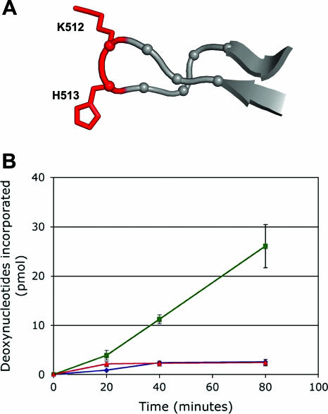
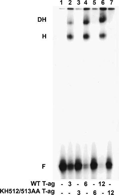
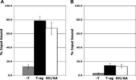
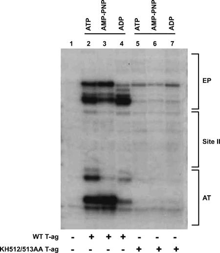
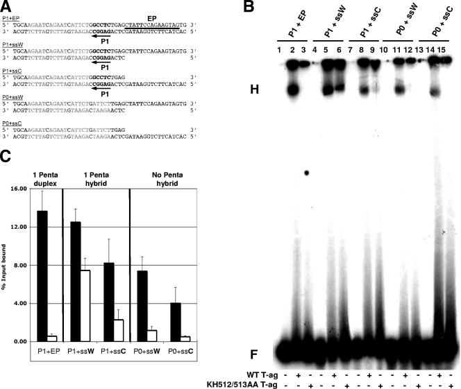
The interaction of simian virus 40 (SV40) T antigen (T-ag) with the viral origin has served as a model for studies of site-specific recognition of a eukaryotic replication origin and the mechanism of DNA unwinding. These studies have revealed that a motif termed the "beta-hairpin" is necessary for assembly of T-ag on the SV40 origin. Herein it is demonstrated that residues at the tip of the "beta-hairpin" are needed to melt the origin-flanking regions and that the T-ag helicase domain selectively assembles around one of the newly generated single strands in a manner that accounts for its 3'-to-5' helicase activity. Furthermore, T-ags mutated at the tip of the "beta-hairpin" are defective for oligomerization on duplex DNA; however, they can assemble on hybrid duplex DNA or single-stranded DNA (ssDNA) substrates provided the strand containing the 3' extension is present. Collectively, these experiments indicate that residues at the tip of the beta-hairpin generate ssDNA in the core origin and that the ssDNA is essential for subsequent oligomerization events.


Figures

FIG. 1.
FIG. 1.
Testing the ability of the KH512/513AA double mutant to catalyze initiation of DNA replication. (A) View of the “beta-hairpin” (residues 507 to 519) showing the positions of terminal residues K512 and H513 (in red), which were mutated to alanine. (B) SV40 in vitro replication assays (43, 80, 90) conducted for the indicated amounts of time with wt T-ag (green line) and the KH512/513AA double mutant (red line). As a negative control, the amount of T-ag-independent nonspecific incorporation was also determined (blue line).
FIG. 2.
FIG. 2.
Comparison of the abilities of T-ag and the KH512/513AA double mutant to oligomerize on the SV40 core origin. Band shift experiments, conducted with a 32P-labeled 64-bp duplex oligonucleotide containing the SV40 core origin and either 3, 6, or 12 pmol of T-ag, are shown in lanes 2, 4, and 6, respectively. Reactions conducted with the same amounts of the KH512/513AA double mutant are shown in lanes 3, 5, and 7. The control reaction in lane 1 was conducted in the absence of protein. The positions of T-ag hexamers (H), double hexamers (DH), and free DNA (F) are indicated.
FIG. 3.
FIG. 3.
Analysis of the relative abilities of T-ag and the KH512/513AA mutant to bind to duplex DNA substrates. Filter binding assays (11, 51) were used to establish the relative affinities of T-ag (black bars) and the KH512/513AA mutant (KH/AA; white bars) for either (A) a 64-bp oligonucleotide containing the SV40 core origin or (B) a 64-bp control oligonucleotide. As a control, filter binding assays were conducted in the absence of protein (−T; gray bars). The reactions were conducted under replication conditions (see Materials and Methods) in the presence of AMP-PNP and 6 pmol of either T-ag or the KH512/513AA mutant. The percentage of oligonucleotide bound was determined by nitrocellulose filter binding assays and scintillation counting.
FIG. 4.
FIG. 4.
Structural distortions in the SV40 origin are not catalyzed by the KH512/513AA mutant. T-ag (12 pmol) was incubated under replication conditions with pSV01ΔEP (0.27 pmol) in the presence of ATP (lane 2), AMP-PNP (lane 3), or ADP (lane 4). Additional reactions were conducted with 12 pmol of the KH512/513AA double mutant (lanes 5 to 7) in the presence of the identical nucleotides. As a control, the reaction in lane 1 was conducted in the absence of T-ag. After treatment with KMnO4, the locations of the oxidized bases were determined via primer extension reactions with a 32P-labeled oligonucleotide (see Materials and Methods). The products of the primer extension reactions were analyzed by electrophoresis on a 7% polyacrylamide gel containing 8 M urea. The locations of regions of the SV40 core origin, including the EP, site II, and the AT-rich region, are indicated to the right of the gel.
FIG. 5.
FIG. 5.
EMSA experiments conducted with the pentanucleotide 1-based set of oligonucleotides, T-ag, and the KH512/513AA mutant. (A) Pentanucleotide 1-based set of oligonucleotides used for EMSA experiments. The first three SV40 origin-derived oligonucleotides contained pentanucleotide 1 (P1; arrow). The 48-bp “P1+EP” oligonucleotide was entirely duplex; pentanucleotide 1 is indicated, the transition mutations that replaced the other pentanucleotides are shown in light gray, and the location of the EP region is indicated by a horizontal bar. The hybrid duplex/single-stranded “P1+ssW” and “P1+ssC” oligonucleotides contained pentanucleotide 1 and either a 3′ extension (Watson [W]) or a 5′ extension (Crick [C]) of the EP. The two control oligonucleotides, “P0+ssW” and “P0+ssC,” lacked pentanucleotide 1 but contained the same single-stranded extensions of the EP found in the “P1+ssW” and “P1+ssC” oligonucleotides. (B) Representative band shift experiment used to compare the abilities of T-ag (lanes 2, 5, 8, 11, and 14) and the KH512/513AA mutant (lanes 3, 6, 9, 12, and 15) to assemble on the indicated substrates (i.e., P1+EP, P1+ssW, P1+ssC, P0+ssW, and P0+ssC). The positions of the hexamers (H) and free DNA (F) are indicated. As controls, the reactions in lanes 1, 4, 7, 10, and 13 were conducted in the absence of protein. (C) Three separate EMSA experiments were quantitated with a Molecular Dynamics PhosphorImager and used to determine the percentages of input DNA present in the hexamers. Black bars symbolize experiments conducted with wt T-ag, while white bars symbolize those performed with the KH512/513AA mutant. It was apparent that the KH512/513AA mutant assembled at appreciable levels only on the P1+ssW substrate. Moreover, for the hybrid duplex/ssDNA substrates and either T-ag or the KH512/513AA mutant, the Watson strand was preferred over the Crick strand. Finally, for wt T-ag, the presence of a pentanucleotide in the hybrid duplex/ssDNA substrates led to a relatively minor increase (∼2-fold) in the level of assembly.
FIG. 6.
FIG. 6.
Support for the hypothesis that there exists an external path for ssDNA over the helicase domain. (A) Distribution of residues on the C-terminal surface of the helicase domain whose mutation selectively disrupted binding to ssDNA (blue residues, i.e., P429), helicase activity (green residues, i.e., G488, R498, D499, and P584), or both activities (cyan residues, i.e., D402, V404, P417, and K516). The six T-ag monomers in the T-ag hexamer (a to f) are indicated. Residues K512 and H513 are situated at the tip of the beta-hairpin, and their positions within the central channel are indicated (shown in red). (B) Residues on the lateral surface of the helicase domain which are defective, when mutated, for binding to ssDNA (blue residues, i.e., R349, R364, and D429), helicase activity (green residues, i.e., Y314, K315, H320, R357, W422, D466, G488, R498, and P584), or both activities (cyan residues, i.e., D402, V404, V413, and P417). The relative positions of the T-ag-obd (OBD) and the helicase subdomains (D2/D3 and D1) are indicated. (C) Electrostatic potential of the surface of the helicase domain. Blue surfaces indicate positive potential, along which negative ssDNA could transit, and red surfaces indicate negative potential (±10 kT/e). The relative positions of the T-ag domains are indicated as in panel B.
FIG. 7.
FIG. 7.
Models for beta-hairpin-dependent unwinding of the core origin and subsequent helicase activity. (A) The two images in panel A present a model for hexamer formation around a single strand of the SV40 origin. As shown in the upper figure, the SV40 core origin consists of a central region, containing four GAGGC pentanucleotides, and two flanking regions, the AT-rich region and the EP. Recognition of the SV40 origin by a T-ag monomer depends upon two interactions, namely, those of the A1 and B2 loops (green spheres) with a pentanucleotide (reviewed in reference 14) (pentanucleotide 1 is depicted by the pink arrow) and those between the beta-hairpin (shown in red) and the flanking sequences (65, 71). The lower figure depicts the subsequent assembly of the helicase domain (light blue spheres) on the 3′ extension of the EP, which is proposed to depend upon the selective coordination of the beta-hairpins around a single strand of origin DNA. The displaced strand is shown transiting the external surface of the helicase domain and then entering the T-ag-obd hexamer (dark blue spheres) via the recently described gap (54). For ease of viewing, two helicase domains have been removed. (B) Upon assembly of the double hexamer, it is proposed that DNA is routed through the complex as depicted (DNA is symbolized by green and yellow strands; DNA situated within the complex is depicted by dotted lines). The present studies indicate that the central channel of each T-ag hexamer contains the 3′ extension of the flanking sequence. Based on recent studies by Enemark and Joshua-Tor (25), ssDNA in the central channel is proposed to be pumped out of the helicase domain owing to beta-hairpin- and ATP-dependent interactions with the sugar-phosphate backbone. (The beta-hairpin is symbolized by red rectangles, and its position following ATP hydrolysis is depicted by red dotted lines). Finally, recent studies established that ssDNA selectively interacts with the T-ag-obd on the helicase proximal surface (64), one indication that upon entering the gap, ssDNA interacts with this surface.

References

    1. Abbate, E. A., J. M. Berger, and M. R. Botchan. 2004. The X-ray structure of the papillomavirus helicase in complex with its molecular matchmaker E2. Genes Dev. 18:1981-1996. - PMC - PubMed
    1. Ahnert, P., and S. S. Patel. 1997. Asymmetric interactions of hexameric bacteriophage T7 DNA helicase with the 5′- and 3′-tails of the forked DNA substrate. J. Biol. Chem. 272:32267-32273. - PubMed
    1. Arthur, A. K., A. Hoss, and E. Fanning. 1988. Expression of simian virus 40 T antigen in Escherichia coli: localization of T-antigen origin DNA-binding domain to within 129 amino acids. J. Virol. 62:1999-2006. - PMC - PubMed
    1. Arunkumar, A. I., V. Klimovich, X. Jiang, R. D. Ott, L. Mizoue, E. Fanning, and W. J. Chazin. 2005. Insights into hRPA32 C-terminal domain-mediated assembly of the simian virus 40 replisome. Nat. Struct. Mol. Biol. 12:332-339. - PMC - PubMed
    1. Auborn, K., M. Guo, and C. Prives. 1989. Helicase, DNA-binding, and immunological properties of replication-defective simian virus 40 mutant T antigens. J. Virol. 63:912-918. - PMC - PubMed

Publication types

MeSH terms

LinkOut - more resources