. 2007 Apr;81(8):3949-68.
doi: 10.1128/JVI.02333-06. Epub 2007 Feb 7.

Kaposi's sarcoma-associated herpesvirus induces sustained NF-kappaB activation during de novo infection of primary human dermal microvascular endothelial cells that is essential for viral gene expression

Affiliations

Kaposi's sarcoma-associated herpesvirus induces sustained NF-kappaB activation during de novo infection of primary human dermal microvascular endothelial cells that is essential for viral gene expression

Sathish Sadagopan et al. J Virol. 2007 Apr.

Abstract

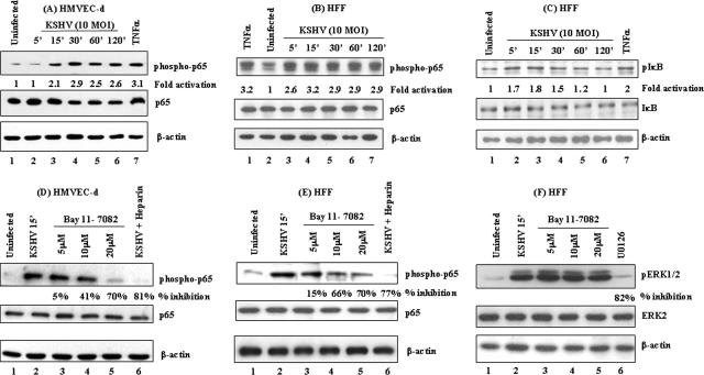
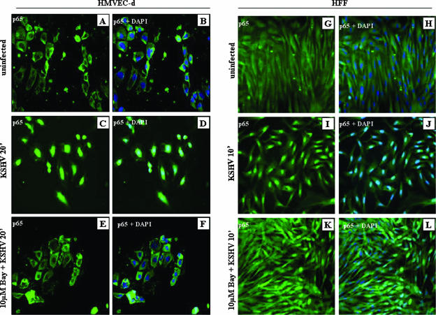
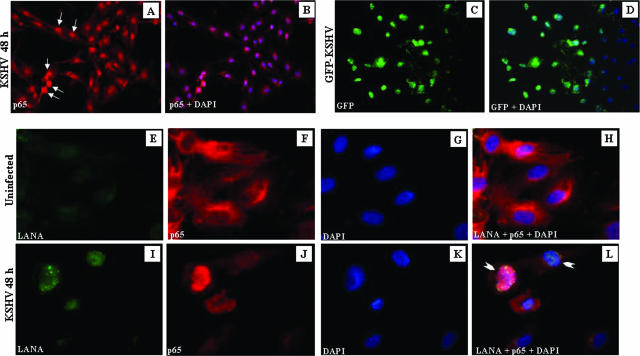
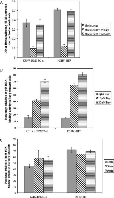
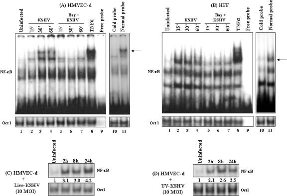
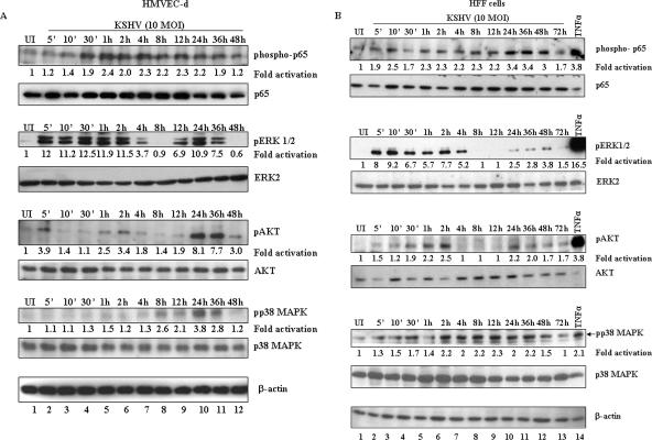
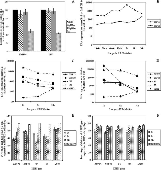
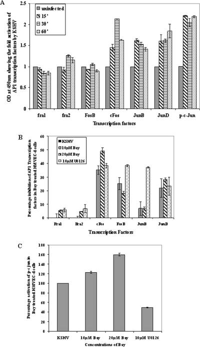
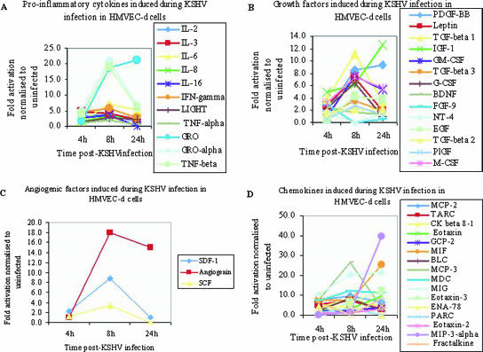
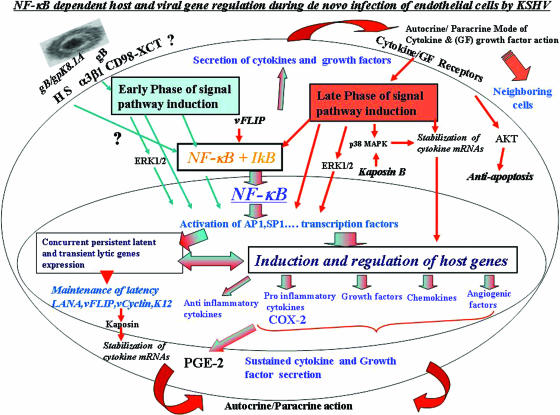
In vitro Kaposi's sarcoma-associated herpesvirus (KSHV) infection of primary human dermal microvascular endothelial (HMVEC-d) cells and human foreskin fibroblast (HFF) cells is characterized by the induction of preexisting host signal cascades, sustained expression of latency-associated genes, transient expression of a limited number of lytic genes, and induction of several cytokines, growth factors, and angiogenic factors. Since NF-kappaB is a key molecule involved in the regulation of several of these factors, here, we examined NF-kappaB induction during de novo infection of HMVEC-d and HFF cells. Activation of NF-kappaB was observed as early as 5 to 15 min postinfection by KSHV, and translocation of p65-NF-kappaB into nuclei was detected by immunofluorescence assay, electrophoretic mobility shift assay, and p65 enzyme-linked immunosorbent assay. IkappaB phosphorylation inhibitor (Bay11-7082) reduced this activation significantly. A sustained moderate level of NF-kappaB induction was seen during the observed 72 h of in vitro KSHV latency. In contrast, high levels of ERK1/2 activation at earlier time points and a moderate level of activation at later times were observed. p38 mitogen-activated protein kinase was activated only at later time points, and AKT was activated in a cyclic manner. Studies with UV-inactivated KSHV suggested a role for virus entry stages in NF-kappaB induction and a requirement for KSHV viral gene expression in sustained induction. Inhibition of NF-kappaB did not affect target cell entry by KSHV but significantly reduced the expression of viral latent open reading frame 73 and lytic genes. KSHV infection induced the activation of several host transcription factors, including AP-1 family members, as well as several cytokines, growth factors, and angiogenic factors, which were significantly affected by NF-kappaB inhibition. These results suggest that during de novo infection, KSHV induces sustained levels of NF-kappaB to regulate viral and host cell genes and thus possibly regulates the establishment of latent infection.


Figures

FIG. 1.
FIG. 1.
(A, B, and C) Detection of activated NF-κB in KSHV-infected HMVEC-d cells and HFF. HMVEC-d cells (A) and HFF (B) grown to 80% confluence were serum starved and infected with KSHV (10 DNA copies/cell), and p65 protein phosphorylation was monitored at the indicated time points. The cells were washed and lysed with RIPA lysis buffer, and the lysates were adjusted to equal amounts of protein, resolved on SDS-10% PAGE, and transferred to nitrocellulose membranes. The membranes were immunoblotted with monoclonal antibodies against phospho-p65 protein (top), total p65 protein (middle), or β-actin (bottom). (C) HFF that were either uninfected or infected with KSHV (10 DNA copies/cell) at various time points were Western blotted using phospho-IκB (top), total IκB (middle), and β-actin (bottom) antibodies. The level of phosphorylated p65 in uninfected cells was considered to be 1 for comparison. (D, E, and F) Specificity of NF-κB induction by KSHV and inhibition by Bay11-7082. Serum-starved HMVEC-d cells (D) and HFF (E and F), untreated or pretreated with 5, 10, or 20 μM Bay11-7082 (lanes 3, 4, and 5, respectively), were either uninfected (lane 1) or infected with 10 DNA copies/cell of KSHV for 15 min. For a control, serum-starved cells were infected for 30 min with virus preincubated with 100 μg/ml of heparin for 60 min at 37°C (lane 6). The cell lysates were reacted in Western blot reactions with anti-phospho-p65 antibodies (top). The membranes were stripped and reprobed with anti-p65 antibodies (middle) and β-actin antibodies (bottom). NF-κB induction with virus alone was considered 100%, and the data are presented as the percent inhibition of p65 phosphorylation. (F) Bay11-7082-pretreated HFF lysates were immunoblotted with phospho-ERK1/2 antibodies (top, lanes 1 to 5). ERK1/2 phosphorylation in virus-infected cells was measured in the presence of the MAPK inhibitor U0126 (top, lane 6). The blots were stripped and reprobed for total ERK2 (middle) and β-actin (bottom) levels. Each blot is representative of at least three independent experiments, and percent inhibition was calculated with respect to the phosphorylated levels of p65 in KSHV-infected cells without Bay11-7082 pretreatment.
FIG. 2.
FIG. 2.
Nuclear translocation of NF-κB-p65 in KSHV-infected cells. Serum-starved HMVEC-d cells and HFF grown in eight-well chamber slides were infected with KSHV (10 DNA copies/cell) for 20 min and 10 min, respectively; washed; fixed; permeabilized; and stained with anti-p65 polyclonal antibody. HMVEC-d cells and HFF were either uninfected (A, B, G, and H) or infected with KSHV (10 DNA copies/cell) (C, D, I, and J) or incubated with 10 μM Bay11-7082, followed by infection with KSHV (E, F, K, and L), and stained for NF-κB-p65. DAPI was used as a nuclear stain and merged with p65 staining.
FIG. 3.
FIG. 3.
Colocalization of NF-κB-p65 and ORF-73 (LANA-1) in KSHV-infected HMVEC-d cells. (A) Serum-starved HMVEC-d cells grown in eight-well chamber slides were infected with KSHV (10 DNA copies/cell) for 48 h, washed, fixed, permeabilized, and stained with anti-p65 polyclonal antibody. (B) Merged image of DAPI-stained nuclei with p65 staining. (C) HMVEC-d cells infected with GFP-KSHV for 48 h. (D) Merged image of DAPI-stained nuclei with GFP staining. (E to L) Colocalization of LANA-1 and p65. LANA (green in panels E and I) and p65 (red in panels F and J) in uninfected (E to H) and infected (I to L) HMVEC-d cells. (G and K) DAPI staining merged with LANA-1 and p65 (H and L). The arrows (A) indicate p65 nuclear staining, and the arrowheads (L) indicate cells positive for LANA-1 and p65 nuclear staining.
FIG. 4.
FIG. 4.
Detection of KSHV-induced nuclear translocation of NF-κB-p65 by ELISA. (A) Nuclear extracts from HMVEC-d cells and HFF infected with KSHV (10 DNA copies/cell) for 30 min were prepared and assayed for NF-κB DNA binding activity by ELISA. Plates immobilized with oligonucleotides specific for the κB site were incubated with nuclear extracts (5 μg/well), followed by ELISA with anti-p65 antibody. The competition experiment was done in a similar fashion but using plates coated with excess (20 pmol) NF-κB consensus site mutant or wt oligonucleotides. The data represent the averages ± standard deviations of three experiments. (B) HMVEC-d cells and HFF untreated or pretreated with various concentrations of Bay11-7082 for 1 h were infected with KSHV (10 DNA copies/cell) for 30 min, and nuclear extracts were prepared and assayed for NF-κB DNA binding activity. The percent nuclear translocation of NF-κB-p65 inhibition by Bay11-7082 pretreatment was calculated with respect to the DNA binding activities in untreated KSHV-infected cells. (C) Histograms depicting the kinetics of percent inhibition of DNA binding activity in nuclear extracts from HMVEC-d cells and HFF pretreated with 10 μM Bay11-7082 for 1 h and then infected with KSHV (10 DNA copies/cell) for different times. The data represent the averages ± standard deviations of three experiments.
FIG. 5.
FIG. 5.
EMSA detection of KSHV-induced NF-κB DNA binding activity. (A and B, top, lanes 1 to 7) HMVEC-d cells (A) and HFF (B), untreated or pretreated with 10 μM Bay11-7082 (1 h), were left uninfected or infected with KSHV (10 DNA copies/cell) for the indicated times. Nuclear extracts prepared from these cells were incubated with κB-specific probe. The specificity of DNA-protein interaction was assessed by induction with TNF-α (20 ng/ml) (lane 8), free probe (lane 9), normal probe (lane 11), or competitive EMSA using a 100× molar excess of unlabeled double-stranded oligonucleotide κB probe (cold probe; lane 10). Nuclear extracts from HMVEC-d cells infected with 10 DNA copies/cell of live KSHV (C) or UV-KSHV (D) for 2 h, 8 h, and 24 h were analyzed by EMSA. Each EMSA is representative of at least three independent experiments. (A and B, bottom, lanes 1 to 11, and C and D, bottom) Oct1 probe used as loading control and specificity control to demonstrate the binding of NF-κB to the specific probe. The arrows indicate the positions of the induced NF-κB complexes.
FIG. 6.
FIG. 6.
Sustained activation of NF-κB during de novo infection of target cells by KSHV. Proteins prepared from HMVEC-d cells (A) and HFF (B) that were uninfected (UI) or infected with KSHV (10 DNA copies/cell) for 5 min, 10 min, 30 min, 1 h, 2 h, 4 h, 8 h, 12 h, 24 h, 36 h, 48 h, and 72 h were resolved by SDS-PAGE and transferred onto nitrocellulose membranes. Phosphorylated and total p65, ERK 1/2, AKT, and p38 MAPK proteins were detected with the respective antibodies. Each blot is representative of at least three independent experiments. The phosphorylation levels in the uninfected cells were considered to be 1 for comparison. For a control, cells were induced with TNF-α for 20 min.
FIG. 7.
FIG. 7.
Effect of Bay11-7082 on KSHV entry and KSHV ORF 73, ORF 50, K5, K8, and vIRF2 gene expression. (A) HMVEC-d cells and HFF, untreated or incubated with different concentrations of inhibitors for 1 h at 37°C, were infected with KSHV at 10 DNA copies/cell. For a control, cells were preincubated with 50 μM LY294002 (LY) for 1 h at 37°C before virus was added. After 2 h of incubation, the cells were washed twice with PBS to remove the unbound virus, treated with trypsin-EDTA for 5 min at 37°C to remove the bound but noninternalized virus, and washed, and total DNA was isolated. This was normalized, and the numbers of KSHV genome copies were estimated by real-time DNA PCR for ORF 73. The cycle threshold values were used to plot the standard graph and to calculate the relative copy numbers of viral DNA in the samples. The data are presented as the percent entry of KSHV DNA internalization obtained when the cells were incubated with virus alone. Each reaction was done in duplicate, and each bar represents the mean and standard deviation of three experiments. (B to F) Untreated HMVEC-d cells and HFF or cells pretreated with 10 μM Bay11-7082 for 1 h were infected with KSHV (10 DNA copies/cell) for 2 h, 8 h, and 24 h, and RNA was isolated and treated with DNase I for 1 h. A total of 250 ng of DNase-treated RNA was subjected to real-time RT-PCR with ORF 73, ORF 50, K5, K8, and vIRF2 gene-specific primers and TaqMan probes. Standard graphs generated using known concentrations of DNase-treated in vitro-transcribed ORF 73, ORF 50, K5, K8, and vIRF2 transcripts were used to calculate the relative copy numbers of viral transcripts and were normalized with GAPDH. Each reaction was done in duplicate, and each point represents the average ± standard deviation of three independent experiments. (B) Kinetics of ORF 73 and 50 gene expression in HMVEC-d cells. (C and D) Comparative kinetics of ORFs 73 and 50, K5, K8, and vIRF2 in HMVEC-d cells and HFF, respectively. (E and F) Histograms depicting the percent inhibition of KSHV ORF 73 and 50, K5, K8, and vIRF2 expression in the presence of Bay11-7082 in HMEVC-d cells and HFF, respectively.
FIG. 8.
FIG. 8.
Effect of NF-κB inhibition on AP-1 transcription factor activation. (A) Nuclear extracts prepared from uninfected HMVEC-d cells or HMVEC-d cells infected with KSHV for 15 min, 30 min, and 60 min were tested for the activation of AP-1-regulated transcription factors by incubating the nuclear extracts with the plate-immobilized oligonucleotides containing the AP-1 transcription factor-specific site, followed by ELISA with antibodies to the respective transcription factors. The histogram represents the activation levels of phospho-c-Jun, JunB, JunD, Fra1, Fra2, Fos-B, and c-Fos in the nuclear extracts from KSHV-infected HMVEC-d cells. The data represent the averages and standard deviations of three experiments, and the values shown here are after normalization with uninfected cells. (B) Histogram depicting the percent inhibition of DNA binding of AP-1 transcription factors in nuclear extracts from HMVEC-d cells pretreated with two different concentrations of Bay11-7082 and 10 μM U0126, followed by infection with KSHV. (C) Histogram depicting the percent activation of DNA binding of the phospho-c-Jun transcription factor in HMVEC-d nuclear extracts. Percent inhibition and percent activation were calculated with respect to the DNA binding activities in KSHV-infected HMVEC-d cells without Bay11-7082 pretreatment. The data represent the averages ± standard deviations of three experiments.
FIG. 9.
FIG. 9.
Up regulation of proinflammatory cytokines, growth factors, angiogenic factors, and chemokines in HMVEC-d cells by KSHV. Densitometric analysis of cytokine array blots was carried out to determine the difference in the release of human cytokines from serum-starved, untreated HMVEC-d cells and KSHV-infected cells at three different time points. The values were normalized to identical background levels using the Ray Bio Human Cytokine antibody array V analysis tool. The increases in the cytokine levels were calculated by dividing the respective values obtained from infected-cell supernatants with the values obtained from uninfected-cell supernatants and cytokines showing significant change represented in a line graph format. (A) Proinflammatory cytokines, (B) growth factors, (C) angiogenic factors, and (D) chemokines that showed significant changes with respect to uninfected cells are represented in the graph.
FIG. 10.
FIG. 10.
Schematic representation depicting the early and late induction phases of NF-κB during in vitro KSHV infection of HMVEC-d cells and their potential roles in transcription factor regulation, establishment and maintenance of KSHV infection, and cytokine secretion. In the early phase of NF-κB induction (blue arrows), virus binding and entry lead to signal pathway induction, such as FAK, Src, PI 3-K, AKT, PKC-ζ, MAPK-ERK1/2, and NF-κB signal molecules. Activated NF-κB translocates into the nucleus, which coincides with viral-DNA entry into the infected-cell nuclei, concurrent transient expression of limited viral lytic genes, and persistent latent gene expression. Overlapping with these events, a limited number of cytokines and growth factors are induced, which is initiated by transcription factors, like AP-1 (induced by ERK1/2 and NF-κB). Early activation of NF-κB and ERK1/2 also leads to the activation and release of NF-κB-inducible host factors, which act in autocrine and paracrine fashions on the infected, as well as neighboring, cells. The autocrine action of these factors, along with viral gene expression, probably contributes to the second, or late, phase of signal pathway activation (red arrows), including sustained NF-κB activation and phosphorylation of p38 MAPK, ERK1/2, and AKT required for the maintenance of latency. The blue and red arrows together indicate pathways induced during both early and late phases of KSHV infection.

Similar articles

Cited by

References

    1. Akula, S. M., N. P. Pramod, F. Z. Wang, and B. Chandran. 2001. Human herpesvirus 8 envelope-associated glycoprotein B interacts with heparan sulfate-like moieties. Virology 284:235-249. - PubMed
    1. Akula, S. M., F. Z. Wang, J. Vieira, and B. Chandran. 2001. Human herpesvirus 8 interaction with target cells involves heparan sulfate. Virology 282:245-255. - PubMed
    1. An, J., A. K. Lichtenstein, G. Brent, and M. B. Rettig. 2002. The Kaposi sarcoma-associated herpesvirus (KSHV) induces cellular interleukin 6 expression: role of the KSHV latency-associated nuclear antigen and the AP1 response element. Blood 99:649-654. - PubMed
    1. An, J., Y. Sun, R. Sun, and M. B. Rettig. 2003. Kaposi's sarcoma-associated herpesvirus encoded vFLIP induces cellular IL-6 expression: the role of the NF-κB and JNK/AP1 pathways. Oncogene 22:3371-3385. - PubMed
    1. Baeuerle, P. A., and D. Baltimore. 1996. NF-kappa B: ten years after. Cell 87:13-20. - PubMed

Publication types

MeSH terms

LinkOut - more resources