. 2007;35(6):e38.
doi: 10.1093/nar/gkm017. Epub 2007 Feb 7.

Motif programming: a microgene-based method for creating synthetic proteins containing multiple functional motifs

Affiliations

Motif programming: a microgene-based method for creating synthetic proteins containing multiple functional motifs

Hirohide Saito et al. Nucleic Acids Res. 2007.

Abstract

The presence of peptide motifs within the proteins provides the synthetic biologist with the opportunity to fabricate novel proteins through the programming of these motifs. Here we describe a method that enables one to combine multiple peptide motifs to generate a combinatorial protein library. With this method, a set of sense and antisense oligonucleotide primers were prepared. These primers were mixed and polymerized, so that the resultant DNA consisted of combinatorial polymers of multiple microgenes created from the stochastic assembly of the sense and antisense primers. With this motif-mixing method, we prepared a protein library from the BH1-4 motifs shared among Bcl-2 family proteins. Among the 41 clones created, 70% of clones had a stable, presumably folded expression product in human cells, which was detectable by immunohistochemistry and western blot. The proteins obtained varied with respect to both the number and the order of the four motifs. The method enables homology-independent polymerization of DNA blocks that coded motif sequences, and the frequency of each motif within a library can be adjusted in a tailor-made manner. This motif programming has a potential for creating a library with a large proportion of folded/functional proteins.


Figures

Figure 1.
Figure 1.
Single microgene-based method. A single microgene block has motif A (red) and motif B (cyan) embedded in different reading frames. The microgene (shown by the double helix) is polymerized in tandem using a microgene polymerization reaction (MPR) in which two overlapping MPR primer pairs having a 3′ mismatched base (shown by red letters with dots) are used. During MPR, insertion and/or deletion mutations randomly occur at junctions between microgene blocks (shown by wavy lines labeled ‘FS’) causing the reading frame to randomly shift, leading to combinatorial polymerization of motifs A and B.
Figure 2.
Figure 2.
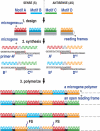
Schematic diagram of a motif-mixing protocol used in this study. Initially, we designed DNA sequences for microgenescore that each encode a peptide motif to be mixed in their first reading frames, after which sense and antisense MPR primers were synthesized based on these microgenescore. These primers share 3′ sequences that enable base-pair formation between the sense and antisense primers, but contain mismatched bases at their 3′-OH ends (shown by red letters with dots). In the polymerization step, motifs can be embedded either in the sense or antisense primer. In the figure, motifs A and B are embedded in the sense primers, producing primers AS and BS, while motifs C and D are in the antisense primers, producing primers CAS and DAS. The thermal cycle reaction is carried out in the presence of these MPR primers, a thermostable, a DNA polymerase and dNTP. The resultant high molecular weight DNAs are combinatorial polymers of multiple microgenes created by stochastic base paring of the MPR primers. In some clones, nucleotide insertions or deletions allow frame shift mutations (denoted by FS), so that peptide sequences encoded by the second and third reading frames appear in the translated products.
Figure 3.
Figure 3.
Mixing four short DNAs using the new protocol. (A) MPR primers AS (sense), BAS, CAS and DAS (all antisense) were designed so that they contained a restriction endonuclease recognition site (red, orange, green or cyan). The sense and antisense primers overlap at the 3′-region, forming six base pairs (shown by black bars). All of the primers had a mismatched adenosine residue at the 3′-OH end (represented by a purple A with asterisks). (B) Polymerization of four microgenes. The four MPR primers described in (A) were polymerized to yield a large DNA fragment; note that a pair of MPR primers (both sense and antisense) was necessary for the polymerization (compare lane 1 with lanes 2–6). The sense primer AS (0.4 µM) and antisense primers (BAS, CAS and DAS; 0.4 µM total) were mixed as shown in the figure. (C) The MPR products obtained were digested with the indicated restriction enzymes and analyzed by gel electrophoresis (1% agarose). The primers used and their concentrations were same as in (B): lanes 1–4, AS (0.4 µM) + BAS (0.4 µM); lanes 5–8, AS (0.4 µM) + CAS (0.4 µM); lanes 9–12, AS (0.4 µM) + DAS (0.4 µM); lanes 13–16, AS (0.4 µM) + BAS (0.2 µM) + CAS (0.2 µM); lanes 17–20, AS (0.4 µM) + BAS (0.134 µM) + CAS (0.134 µM) + DAS (0.134 µM); lane M, size standards.
Figure 4.
Figure 4.
Mixing four BH motifs. (A) Structure of human Bcl-xL, an anti-apoptotic multidomain protein (based on the [1R2D]). The core regions of the BH1, BH2, BH3 and BH4 motifs focused on in this study are shown in cyan, orange, red and green, respectively, though we used the BH3 motif from Noxa instead of Bcl-xL. (B) Core microgenes that encode BH peptides in their first reading frames; the color scheme is the same as in (A). (C) MPR primers used in this study. CACC tetranucleotides that facilitated directional cloning into pcDNA were added at the 5′ end of the sense primers (BH3S and BH4S). Six base pairs were formed in the 3′-regions between the sense and antisense primers. They also have mismatched adenosines at their 3′-OH ends (shown in purple). Derivatives of antisense primers (BH1AS+, BH2AS+ and BH3AS+) had an extra CC at their 5′ termini so that the reconstituted microgenes would have lengths that were multiples of three. To monitor incorporation of the corresponding blocks into polymers, the recognition sequences for SalI, XhoI, HindIII, BglII and EcoRI were introduced into BH4S, BH3S, BH3AS(+), BH2AS(+) and BH1AS(+), respectively (shown by italics). (D) The designed microgenes used in this study. They generated the BH1core–BH4 core peptide motifs, which consisted of arranged blocks. (E) Microgenes polymers prepared from combinations of the primers shown in Table 1. The DNA polymers obtained from pools a–d were digested with the motif-specific restriction enzymes and electrophoresed through 1% agarose.
Figure 5.
Figure 5.
(A)The ratio of BH1core–BH4core in pool c or pool d was determined from the sequences of 41 randomly selected clones. (B) Primary structure of artificial protein, a8, which contained the two BH4core and two BH3core motifs. (C) Localization of the a8 protein. The a8 plasmids were transfected into MCF-7 cells using lipofectamine 2000 (Invitrogen) according to the manufacturer's instructions. The cells were incubated for 24 h and then fixed in methanol for 10 min. The localizations of synthetic proteins were analyzed using the penta-his antibody with Alexa Fluor 488 dyes (Alexa-His, QIAGEN). The mitochondria (Mito) and nucleus were probed by Mitotracker Orange (Molecular Probes) and DAPI, respectively. Stained samples were analyzed using a confocal laser scanning microscope.
Figure 6.
Figure 6.
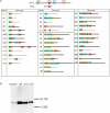
(A) Primary structure of synthetic proteins generated from four libraries (pools a–d) prepared under different conditions (see also Table 1). Forty-one clones were sequenced, and their expression was investigated in MCF-7 cells. Transient protein expression in MCF-7 cells was observed for 28 of the 41 clones (68%, shown by bold black bars), which was detected immunohistochemically using anti-c-myc antibody; all of the synthetic proteins contained a myc epitope (EQKLISEEDL) and a poly-histidine tag at their C-terminus. The lengths of the bars correspond to the relative number of amino acids (e.g. d29 has 221 amino acid residues). The order and arrangement of BH1–BH4core in each protein are shown by squares (the color coding of each motif is the same as in Figure 4). In pools-a and -b, MPR primers were designed so that the reconstituted microgenes would have lengths that were not multiples of three. The reading frame of the microgene polymers was altered at every junction between the microgene units, unless the junction contained insertion/deletion mutations. In contrast, the microgenes in pools-c and -d were designed to maintain the reading frame throughout the polymers. Reflecting this design, clones obtained from pools-c and -d contained fewer frameshifts than those from pools-a and -b. (B) MCF-7 cells were transfected with DNA encoding a8, a10, a12 or empty vector (control), and after 24 h, the clones were analyzed by western blotting using anti-myc antibody. Predicted sizes of synthetic proteins were detected.

Similar articles

Cited by

References

    1. Benner SA. Synthetic biology: act natural. Nature. 2003;421:118. - PubMed
    1. Szostak JW. In vitro genetics. Trends Biochem. Sci. 1992;17:89–93. - PubMed
    1. Saito H, Kourouklis D, Suga H. An in vitro evolved precursor tRNA with aminoacylation activity. EMBO J. 2001;20:1797–1806. - PMC - PubMed
    1. Lee DH, Granja JR, Martinez JA, Severin K, Ghadri MR. A self-replicating peptide. Nature. 1996;382:525–528. - PubMed
    1. Elowitz MB, Leibler S. A synthetic oscillatory network of transcriptional regulators. Nature. 2000;403:335–338. - PubMed

Publication types