. 2007 Feb 13;104(7):2199-204.
doi: 10.1073/pnas.0611217104. Epub 2007 Feb 7.

Global proteomic profiling of phosphopeptides using electron transfer dissociation tandem mass spectrometry

Affiliations

Global proteomic profiling of phosphopeptides using electron transfer dissociation tandem mass spectrometry

Henrik Molina et al. Proc Natl Acad Sci U S A. .

Abstract

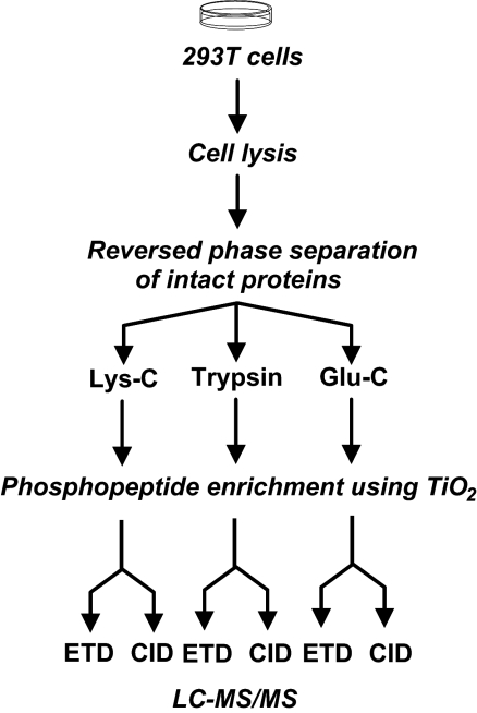
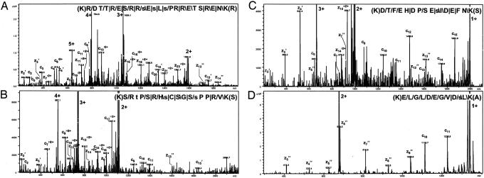
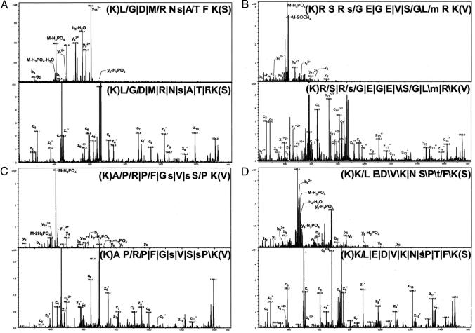
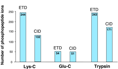
Electron transfer dissociation (ETD) is a recently introduced mass spectrometric technique that provides a more comprehensive coverage of peptide sequences and posttranslational modifications. Here, we evaluated the use of ETD for a global phosphoproteome analysis. In all, we identified a total of 1,435 phosphorylation sites from human embryonic kidney 293T cells, of which 1,141 ( approximately 80%) were not previously described. A detailed comparison of ETD and collision-induced dissociation (CID) modes showed that ETD identified 60% more phosphopeptides than CID, with an average of 40% more fragment ions that facilitated localization of phosphorylation sites. Although our data indicate that ETD is superior to CID for phosphorylation analysis, the two methods can be effectively combined in alternating ETD and CID modes for a more comprehensive analysis. Combining ETD and CID, from this single study, we were able to identify 80% of the known phosphorylation sites in >1,000 phosphorylated peptides analyzed. A hierarchical clustering of the identified phosphorylation sites allowed us to discover 15 phosphorylation motifs that have not been reported previously. Overall, ETD is an excellent method for localization of phosphorylation sites and should be an integral component of any strategy for comprehensive phosphorylation analysis.


Conflict of interest statement

Conflict of interest statement: D.M.H. and N.T. are employees of Agilent Technologies. All other authors have declared no conflict of interest.

Figures

Fig. 1.
Fig. 1.
A schematic depicting the strategy for analysis of phosphopeptides carried out in this study. All fractions were analyzed by LC-MS/MS. A total of 30 Lys-C digested fractions were analyzed by LC-MS/MS (ETD); 12 of the selected Lys-C fractions (1/3 of each sample was used) were subjected to a replicate analysis, with one part of a sample analyzed by ETD and the other by CID; the remaining 2/3 of the 12 protein fractions were digested by using trypsin or Glu-C, respectively, and analyzed in replicate by LC-MS/MS (CID) and LC-MS/MS (ETD). A fraction (7%) of the digested samples before enrichment of phosphopeptides was also analyzed by CID.
Fig. 2.
Fig. 2.
MS/MS spectra of four phosphorylated Lys-C peptides identified by ETD. Phosphopeptides with charge states of + 5 (A), +4 (B), +3 (C) and +2 (D) are shown. The four peptides originate from splicing factor, arginine/serine-rich 2 interacting protein, splicing coactivator subunit SRm300, Bcl2-associated transcription factor 1 and DnaJ homolog, subfamily C, member 9, respectively. The peptide sequence with the fragmentation pattern is shown in each panel. The signs: “/”, “ /”, and “|” designate that the C-terminal type fragments, N-terminal fragments, or both types of fragments, respectively, were identified. For all spectra, except A, the intensity axis has been enlarged by a factor of ≈5. Fragment ions resulting from charge stripping of the precursor ion are assigned with charge states (in bold). Small letters indicate phosphorylated residues.
Fig. 3.
Fig. 3.
Phosphopeptide identifications from ETD and CID experiments. (A) The overlap among phosphopeptides identified from Lys-C digested samples. (B) The corresponding data for nonphosphorylated peptides from the same samples.
Fig. 4.
Fig. 4.
Direct comparison of phosphopeptides subjected to alternating CID and ETD experiments. The CID and ETD experiments identified the exact same sites for the two phosphopeptides show in A and B (from tumor protein D52-like 2 isoform E and tripartite motif-containing 28 protein, respectively) whereas C and D show phosphopeptides with identical amino acid sequence but different assignments of the phosphorylated residues (PDZ and LIM domain 5 isoform a and tumor protein D52 isoform 1, respectively). The peptide sequence with the fragmentation pattern is shown in each panel. The signs: “/”, “ /”, and “|” designate that the C-terminal type fragments, N-terminal fragments, or both type of fragments, respectively, were identified. All intensity axes have been enlarged ≈5 times.
Fig. 5.
Fig. 5.
A summary of phosphopeptide analysis by CID and ETD. Identical samples digested with three proteases were analyzed by CID and ETD. The number of phosphopeptide ions identified in each of the sample sets digested with trypsin, Glu-C, and Lys-C is shown.

References

    1. Haynes PA, Aebersold R. Anal Chem. 2000;72:5402–5410. - PubMed
    1. Greis KD, Hayes BK, Comer FI, Kirk M, Barnes S, Lowary TL, Hart GW. Anal Biochem. 1996;234:38–49. - PubMed
    1. Chalkley RJ, Burlingame AL. J Am Soc Mass Spectrom. 2001;12:1106–1113. - PubMed
    1. Zubarev RA, Kelleher NL, McLafferty FW. J Am Chem Soc. 1998;120:3265–3266.
    1. Syka JE, Coon JJ, Schroeder MJ, Shabanowitz J, Hunt DF. Proc Natl Acad Sci USA. 2004;101:9528–9533. - PMC - PubMed

Publication types

Substances

LinkOut - more resources