. 2007 Feb 13;104(7):2448-53.
doi: 10.1073/pnas.0611134104. Epub 2007 Feb 7.

Spinal cholinergic interneurons regulate the excitability of motoneurons during locomotion

Affiliations

Spinal cholinergic interneurons regulate the excitability of motoneurons during locomotion

Gareth B Miles et al. Proc Natl Acad Sci U S A. .

Abstract

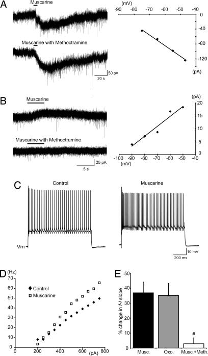
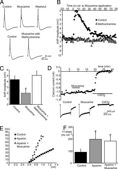
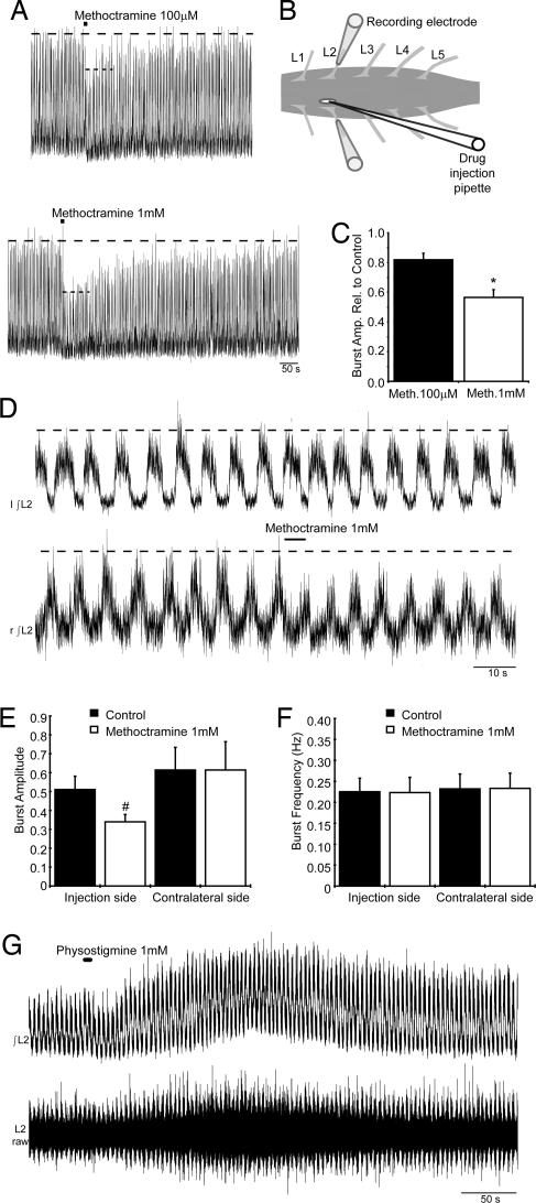
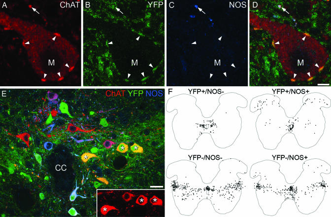
To effect movement, motoneurons must respond appropriately to motor commands. Their responsiveness to these inputs, or excitability, is regulated by neuromodulators. Possible sources of modulation include the abundant cholinergic "C boutons" that surround motoneuron somata. In the present study, recordings from motoneurons in spinal cord slices demonstrated that cholinergic activation of m2-type muscarinic receptors increases excitability by reducing the action potential afterhyperpolarization. Analyses of isolated spinal cord preparations in which fictive locomotion was elicited demonstrated that endogenous cholinergic inputs increase motoneuron excitability during locomotion. Anatomical data indicate that C boutons originate from a discrete group of interneurons lateral to the central canal, the medial partition neurons. These results highlight a unique component of spinal motor networks that is critical in ensuring that sufficient output is generated by motoneurons to drive motor behavior.


Conflict of interest statement

The authors declare no conflict of interest.

Figures

Fig. 1.
Fig. 1.
Muscarinic receptor activation increases spinal MN excitability. (A and B) Voltage-clamp traces (Left) and the current–voltage relationships (Right) of methoctramine-insensitive inward currents (A) and methoctramine-sensitive outward currents (B) induced by muscarine (100 μM, locally applied) in MNs held at −60 mV. The methoctramine was bath-applied (10 μM). (C) Increased MN firing in response to the same amplitude of current injection in the presence of muscarine (50 μM, bath-applied) compared with control. (D) Increased steady-state f–I slope for an MN in the presence of muscarine (50 μM, bath-applied). (E) Pooled data showing changes in the slopes of f–I plots in response to bath applications of muscarine (50 μM, n = 19), oxotremorine (100 μM, n = 6), or muscarine (50 μM) with methoctramine (10 μM, n = 8). #, significantly different to muscarine alone.
Fig. 2.
Fig. 2.
Muscarinic receptor activation increases spinal MN excitability via a reduction in the AHP. (A) Single action potentials (truncated) and AHPs elicited in an MN showing the methoctramine-sensitive (10 μM, bath-applied) effects of muscarine (100 μM, locally applied). (B) Plots of AHP amplitude versus time relative to the local application of muscarine (100 μM) in control and in the presence of methoctramine (10 μM). (C) Average AHP amplitude in MNs in control conditions (n = 10), during bath application of muscarine (50 μM, n = 10) and during bath application of both muscarine (50 μM) and methoctramine (10 μM, n = 8). (D) Unchanged amplitude of calcium currents (measured at the end of voltage steps, −60 mV to a test potential of 0 mV, 500-ms duration, delivered every 30 sec) plotted versus time during bath application of muscarine (50 μM). CdCl2 (500 μM) eliminated the currents, confirming the specificity of our protocol for voltage-activated Ca2+ currents (n = 2). Traces at bottom show examples of currents elicited in control, muscarine, and CdCl2. (E and F) Steady-state f–I plots for one MN (E) and average MN f–I slopes (F; n = 6) in control, apamin (100 nM), and both apamin and muscarine (50 μM). #, significantly different to control.
Fig. 3.
Fig. 3.
Endogenous cholinergic activity increases MN output during fictive locomotion. (A) Local methoctramine application (100 μM and 1 mM, 10-sec duration) decreased locomotor output as seen in rectified/integrated L2 ventral root recordings. (B) Schematic demonstrating positioning of the drug injection pipette through a slit made in the pia matter above the motor column. (C) Average reduction by methoctramine (100 μM and 1 mM) of locomotor-related burst amplitude reported relative to control (n = 4). (D) Local methoctramine application (1 mM, 5-sec duration) during fictive locomotion did not affect contralateral activity. (E and F) Average amplitude and frequency of locomotor-related bursts recorded before and during methoctramine application (1 mM, 1- to 5-sec duration, n = 8). (G) Rectified/integrated (Upper) and raw (Lower) traces of locomotor output recorded from the L2 root during the local application of physostigmine (1 mM, 5-sec duration). ∗, significantly different to 100 μM methoctramine. #, significantly different to control.
Fig. 4.
Fig. 4.
C boutons likely originate from interneurons located lateral to the central canal (lamina X/medial lamina VII). (A–D) High-magnification confocal images of a single optical section through an MN (M) from a Dbx1-YFP mouse. Staining for ChAT, YFP, and NOS indicates that C boutons (arrowheads) are YFP+ but lack NOS, whereas some other ChAT+ terminals are NOS+ (arrow). (E) A single optical section through the region surrounding the central canal (CC) from a Dbx1-YFP mouse. Asterisks mark ChAT+/YFP+/NOS neurons. (Inset) Strong ChAT immunoreactivity in these cells. (F) Plots of all ChAT+ neurons (apart from those in the motor nuclei) from 10 60-μm upper lumbar spinal cord sections of a Dbx1-YFP mouse (gray matter outlined). Cells have been distinguished based on the presence or absence of YFP and NOS. (Scale bars: A–D, 5 μm; E, 20 μm.)

References

    1. Brownstone RM. Prog Neurobiol. 2006;78:156–172. - PMC - PubMed
    1. Rossignol S. Curr Opin Neurobiol. 2000;10:708–716. - PubMed
    1. Conradi S, Skoglund S. Acta Physiol Scand Suppl. 1969;333:5–52. - PubMed
    1. Nagy JI, Yamamoto T, Jordan LM. Synapse. 1993;15:17–32. - PubMed
    1. Li W, Ochalski PA, Brimijoin S, Jordan LM, Nagy JI. Neuroscience. 1995;65:879–891. - PubMed

Publication types