. 2007 Feb 13;104(7):2548-53.
doi: 10.1073/pnas.0609337104. Epub 2007 Feb 7.

The PsbQ protein defines cyanobacterial Photosystem II complexes with highest activity and stability

Affiliations

The PsbQ protein defines cyanobacterial Photosystem II complexes with highest activity and stability

Johnna L Roose et al. Proc Natl Acad Sci U S A. .

Abstract

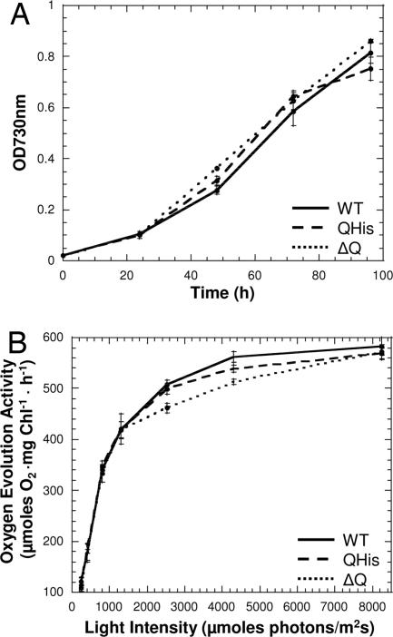
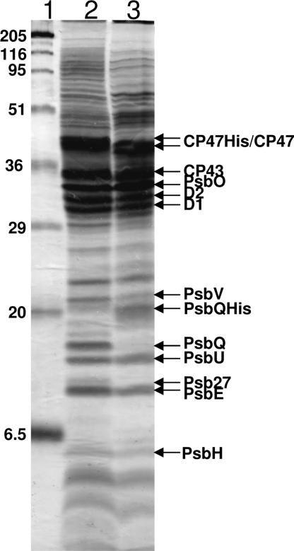
Light-induced conversion of water to molecular oxygen by Photosystem II (PSII) is one of the most important enzymatic reactions in the biosphere. PSII is a multisubunit membrane protein complex with numerous associated cofactors, but it continually undergoes assembly and disassembly due to frequent light-mediated damage as a result of its normal function. Thus, at any instant, there is heterogeneity in the subunit compositions of PSII complexes within the cell. In particular, cyanobacterial PSII complexes have five associated extrinsic proteins, PsbO, PsbP, PsbQ, PsbU, and PsbV. However, little is known about the interactions of the more recently identified PsbQ protein with other components in cyanobacterial PSII. Here we show that PSII complexes can be isolated from the cyanobacterium Synechocystis sp. PCC 6803 on the basis of the presence of a polyhistidine-tagged PsbQ protein. Purification of PSII complexes using a tagged extrinsic protein has not been previously described, and this work conclusively demonstrates that PsbQ is present in combination with the PsbO, PsbU, and PsbV proteins in cyanobacterial PSII. Moreover, PsbQ-associated PSII complexes have higher activity and stability relative to those isolated using histidine-tagged CP47, an integral membrane protein. Therefore, we conclude that the presence of PsbQ defines the fully assembled and optimally active form of the enzyme.


Conflict of interest statement

The authors declare no conflict of interest.

Figures

Fig. 1.
Fig. 1.
Construction of the QHis strain and segregation analysis. (A) psbQ locus of the QHis strain. The psbQ gene, which contains additional sequence at its 3′ end encoding an octa-histidine tag (gray), is followed by a downstream gentamycin resistance marker and the next downstream gene, ureD. The arrows show the location of the primers used for segregation analysis. (B) PCR segregation analysis of WT and QHis cells. The sizes of the products in base pairs (bp) are shown on the right. (C) Immunodetection of the PsbQ protein in WT and QHis cells. The sizes and positions of the molecular mass standards are shown on the left.
Fig. 2.
Fig. 2.
Photosynthetic activity of the QHis strain. (A) Photoautotrophic growth of WT, Qhis, and ΔpsbQ cells in BG11 medium. Error bars represent the standard error of the mean (n = 3). (B) Oxygen-evolution activities of WT, Qhis, and ΔpsbQ (ΔQ) cells in BG11 medium. Error bars represent the standard error of the mean (n = 3).
Fig. 3.
Fig. 3.
Isolation of PSII from QHis cells. (A) Chromatogram of PsbQ-tagged PSII isolation. The first arrow designates the start of sample injection, and the second arrow indicates the start of sample elution. The pooled peak fractions are enclosed by a rectangle. (B) Fluorescence emission spectrum at 77 K for PsbQ-tagged PSII. Peaks are located at 683 and 691 nm.
Fig. 4.
Fig. 4.
Polypeptide compositions of isolated PSII. Each lane contained 5 μg of Chl-containing sample: (1) molecular mass standards, (2) CP47-tagged PSII, and (3) PsbQ-tagged PSII.
Fig. 5.
Fig. 5.
Oxygen-evolution activity of CP47- and PsbQ-tagged PSII complexes as a function of light intensity. Error bars represent the standard error of the mean (n = 3).
Fig. 6.
Fig. 6.
Model of PSII biogenesis and repair. The PSII subunits are colored as follows: D1 (light green); D2, Cyt b559, CP43 (dark green); CP47 (cyan); Psb27 (pink); PsbO (teal); PsbV (dark blue); PsbU (light blue); PsbQ (purple); and damaged D1 (yellow). The top half of the cycle represents steps in the synthesis half of the pathway resulting in fully assembled dimers on the far right, and the bottom half of the cycle shows the disassembly of the complex and removal of the damaged D1 protein. Refer to Discussion for further details.

References

    1. Ferreira KN, Iverson TM, Maghlaoui K, Barber J, Iwata S. Science. 2004;303:1831–1838. - PubMed
    1. Kamiya N, Shen J-R. Proc Natl Acad Sci USA. 2003;100:98–103. - PMC - PubMed
    1. Loll B, Kern J, Saenger W, Zouni A, Biesiadka J. Nature. 2005;438:1040–1044. - PubMed
    1. Zouni A, Witt HT, Kern J, Fromme P, Krauss N, Saenger W, Orth P. Nature. 2001;409:739–743. - PubMed
    1. Aro EM, Suorsa M, Rokka A, Allahverdiyeva Y, Paakkarinen V, Saleem A, Battchikova N, Rintamaki E. J Exp Bot. 2005;56:347–356. - PubMed

Publication types

MeSH terms

LinkOut - more resources