. 2007 Feb 13;104(7):2193-8.
doi: 10.1073/pnas.0607084104. Epub 2007 Feb 7.

Analysis of phosphorylation sites on proteins from Saccharomyces cerevisiae by electron transfer dissociation (ETD) mass spectrometry

Affiliations

Analysis of phosphorylation sites on proteins from Saccharomyces cerevisiae by electron transfer dissociation (ETD) mass spectrometry

An Chi et al. Proc Natl Acad Sci U S A. .

Abstract

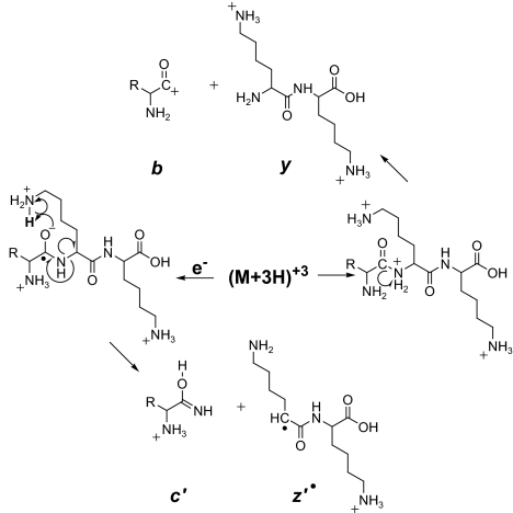
We present a strategy for the analysis of the yeast phosphoproteome that uses endo-Lys C as the proteolytic enzyme, immobilized metal affinity chromatography for phosphopeptide enrichment, a 90-min nanoflow-HPLC/electrospray-ionization MS/MS experiment for phosphopeptide fractionation and detection, gas phase ion/ion chemistry, electron transfer dissociation for peptide fragmentation, and the Open Mass Spectrometry Search Algorithm for phosphoprotein identification and assignment of phosphorylation sites. From a 30-microg (approximately 600 pmol) sample of total yeast protein, we identify 1,252 phosphorylation sites on 629 proteins. Identified phosphoproteins have expression levels that range from <50 to 1,200,000 copies per cell and are encoded by genes involved in a wide variety of cellular processes. We identify a consensus site that likely represents a motif for one or more uncharacterized kinases and show that yeast kinases, themselves, contain a disproportionately large number of phosphorylation sites. Detection of a pHis containing peptide from the yeast protein, Cdc10, suggests an unexpected role for histidine phosphorylation in septin biology. From diverse functional genomics data, we show that phosphoproteins have a higher number of interactions than an average protein and interact with each other more than with a random protein. They are also likely to be conserved across large evolutionary distances.


Conflict of interest statement

The authors declare no conflict of interest.

Figures

Fig. 1.
Fig. 1.
Peptide fragmentation. Scheme for generating type b and y ions by CAD and type c and z· ions by ETD.
Fig. 2.
Fig. 2.
Phosphopeptide chromatogram and mass spectra. (A) Base peak, ion chromatogram for a 90 min separation of yeast phosphopeptides enriched from a 30-μg aliquot of yeast total cell protein. (B and C) ETD mass spectra recorded on (M + 3H)+3 ions from peptides eluting under peaks I and II, respectively, in A. Observed c and z· ions are indicated on the peptide sequence by ⌉ and ⌊, respectively.
Fig. 3.
Fig. 3.
Phosphopeptide mass spectra. ETD mass spectra recorded on (M + 6H)+6 ions at m/z 683.3 for a 35 residue phosphopeptide of MW 4,093 (A), and (M + 3H)+3 ions from a pHis containing peptide at the C terminus of the septin protein, Cdc10 (B). Observed c and z· ions are indicated on the peptide sequence by ⌉ and ⌊, respectively. Observed doubly charged c and z· ions are indicated by an additional label, circle and asterisk, respectively.
Fig. 4.
Fig. 4.
Phosphoprotein and amino acid frequencies. (A) Comparison of GO Slim (33) term frequencies between the whole genome and the phosphoproteins. (B) Comparison of protein abundances between the entire genome and the phosphoproteins. The distribution of phosphoprotein abundances is comparable to that of the genomic background. (C–E) Log2 ratios of per-site amino acid frequencies relative to the genomic background. (C) Sites identified as acidophilic by SCANSITE. (D) Sites identified as basophilic by SCANSITE. (E) Sites not recognized by SCANSITE.
Fig. 5.
Fig. 5.
Phosphoprotein interactions and conservation. (A) A subset of the KEGG sce04110 cell cycle pathway (34). Proteins phosphorylated in this study appear as bold nodes. Known physical interactions are represented by blue edges, and known genetic interactions are shown as red edges. (B) A comparison of phosphoprotein interactions to those of random genomic samples. Clique interactions represent genetic or physical interactions between phosphoproteins (or within random subsamples), and total interactions contain all known genetic or physical interactions between phosphoproteins/sampled proteins and the yeast genome. (C) A representation of the number of model organisms (A. gossypi, C. elegans, D. melanogaster, H. sapiens, and A. thaliana) across which yeast proteins are conserved with significant BLASTP hits. Phosphoproteins are much more likely than a random yeast protein to be conserved (leftmost bars), and conserved phosphoproteins are much more likely to be conserved in all five genomes examined (rightmost bars). Conservation in just one genome is largely explained by the data from the closest organism to S. cerevisiae, A. Gossypi (overlay in darker colors). Error bars represent ± 1 standard deviation.

References

    1. Ficarro SB, McLeland ML, Burke DJ, Ross MM, Shabanowitz J, Hunt DF, White FM. Nat Biotechnol. 2002;20:301–305. - PubMed
    1. Syka JEP, Coon JJ, Schroeder MJ, Shabanowitz J, Hunt DF. Proc Natl Acad Sci USA. 2004;101:9528–9533. - PMC - PubMed
    1. Coon JJ, Ueberheide B, Syka JEP, Dryhurst DD, Ausio J, Shabanowitz J, Hunt DF. Proc Natl Acad Sci USA. 2005;102:9463–9468. - PMC - PubMed
    1. Ghaemmaghami S, Huh W-K, Bower K, Howson RW, Belle A, Dephoure N, O'Shea EK, Weissman JS. Nature. 2003;425:737–741. - PubMed
    1. West AH, Stock AM. Trends Biochem Sci. 2001;26:369–376. - PubMed

Publication types

MeSH terms