. 2007 Apr;18(4):1397-409.
doi: 10.1091/mbc.e06-08-0693. Epub 2007 Feb 7.

Muscle satellite cells and endothelial cells: close neighbors and privileged partners

Affiliations

Muscle satellite cells and endothelial cells: close neighbors and privileged partners

Christo Christov et al. Mol Biol Cell. 2007 Apr.

Abstract

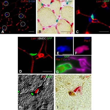
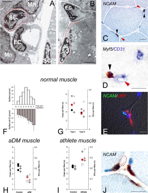
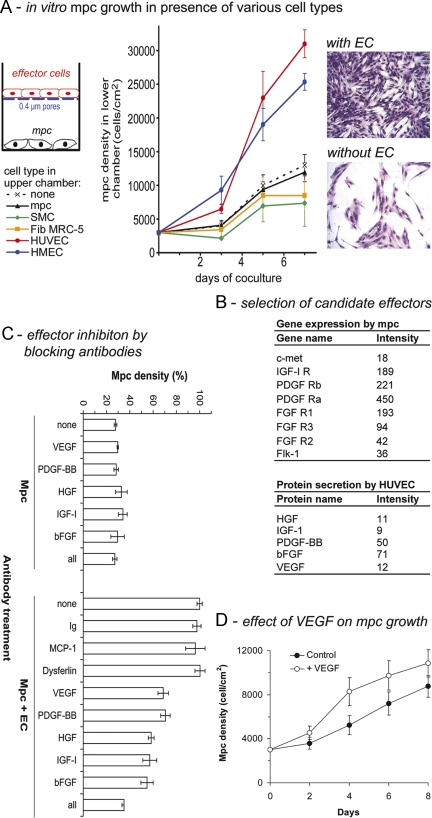
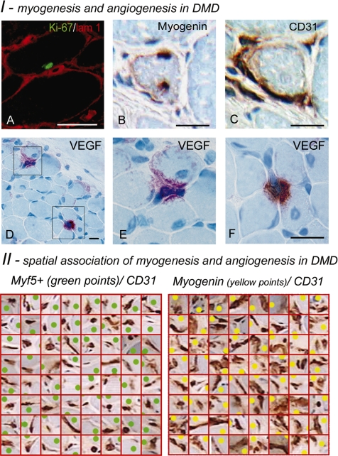
Genetically engineered mice (Myf5nLacZ/+, Myf5GFP-P/+) allowing direct muscle satellite cell (SC) visualization indicate that, in addition to being located beneath myofiber basal laminae, SCs are strikingly close to capillaries. After GFP(+) bone marrow transplantation, blood-borne cells occupying SC niches previously depleted by irradiation were similarly detected near vessels, thereby corroborating the anatomical stability of juxtavascular SC niches. Bromodeoxyuridine pulse-chase experiments also localize quiescent and less quiescent SCs near vessels. SCs, and to a lesser extent myonuclei, were nonrandomly associated with capillaries in humans. Significantly, they were correlated with capillarization of myofibers, regardless to their type, in normal muscle. They also varied in paradigmatic physiological and pathological situations associated with variations of capillary density, including amyopathic dermatomyositis, a unique condition in which muscle capillary loss occurs without myofiber damage, and in athletes in whom capillaries increase in number. Endothelial cell (EC) cultures specifically enhanced SC growth, through IGF-1, HGF, bFGF, PDGF-BB, and VEGF, and, accordingly, cycling SCs remained mainly juxtavascular. Conversely, differentiating myogenic cells were both proangiogenic in vitro and spatiotemporally associated with neoangiogenesis in muscular dystrophy. Thus, SCs are largely juxtavascular and reciprocally interact with ECs during differentiation to support angio-myogenesis.


Figures

Figure 1.
Figure 1.
SC proximity to capillaries in mouse models. (A) TA muscle cryosections from Myf5nlacZ/+ mouse in which SC nuclei expressing nuclear β-Gal activity (blue pseudocolor) are observed close to microvessels whose thick basal lamina is visualized by enhanced collagen IV immunoreactivity (red); bar, 10 μm. (B) TA muscle cryosections from Myf5nlacZ/+ mouse in which most SC nuclei expressing nuclear β-Gal activity (blue) are found close to capillaries expressing alkaline phosphatase activity (red); bar, 10 μm. (C) TA muscle cryosections from Myf5GFP-P/+ mouse in which one SC–expressing cytoplasmic GFP (green) is closely associated with a capillary. Laminin 1 is labeled in red. Bar, 10 μm. (D) TA muscle cryosections from a WT mouse transplanted with GFP+ BM-derived cells (BMDC) showing a blood-borne GFP+ cell housed in a juxtavascular SC niche. Bar, 10 μm. (E) The nucleus (blue) that expresses the satellite cell marker Pax7 (red) (F) belongs to a blood-borne GFP+ cell (green) (G). Collagen IV immunostaining (purple) performed as a second step indicates that the cell is sublaminal and juxtavascular (G). (H and I) TA muscle cryosections from a Myf5nlacZ/+ mouse perinatally infused with BrdU showing one label-retaining SC located close to a capillary: BrdU immunostaining appears in fluorescent green in both dark field (H) and merge with DIC (H), microvascular alkaline phosphatase activity appears in red at both fluorescent (H) and photonic (I) microscopy; SC nuclear β-Gal antigen is revealed in brown by immunoperoxidase (I). Bar, 10 μm.
Figure 2.
Figure 2.
Topological relations between SCs and capillaries. (A and B) Transmission electron microscopy of rat SCs. Both apposition of SC body to EC (A) and apposition of a cytoplasmic process elongated from a distant SC toward EC (B) are observed. For clarity, SC plasma membrane was underlined by red dots. Please note the absence of direct SC-to-EC contact. Bars, 2 μm. (C–E) Chromogenic or fluorescent immunostainings of human deltoid muscle showing extreme proximity of SCs and capillaries. In C, SCs (black arrowheads) are immunostained for NCAM and capillaries (red arrowheads) are identified without immunostaining. In D, a SC nucleus is immunostained for Myf 5 (black arrowhead) and capillaries (red arrowheads) are immunostained for CD31. (E) NCAM of SC plasma membrane is green, and the EC marker vWF is red. Bar, 10 μm. (F and G) Correlations between SCs and capillaries in normal muscle. Left graph (F), histogram of myofiber capillarization frequency (top) is close to Gaussian, whereas SC frequency (bottom) increases linearly with myofiber capillarization (data pooled from 3 normal deltoid muscles). Right graph (G), numbers of both capillaries (●) and SCs (+) are different and largely proportionate in type I and type II myofibers (individual results from 3 normal deltoid muscles identified by different colors). (H) Correlations between SCs and capillaries in aDM muscle. A proportionate loss of muscle capillaries (●) and SCs (+) was found in 3 patients with aDM (in red, gray, and white, respectively) compared with 3 age, sex, and muscle-matched normal controls (in black). (I and J) Heavy-trained muscle. (I) compared with their controls (in black), 3 athletes (in red, gray and white, respectively) show an increase of both muscle capillaries (●) and SCs (+). (J) SC clusters are formed by accumulations of SCs (NCAM+, brown) belonging to different myofibers, often adjacent to a myonucleus (hematoxylin nuclear counterstaining in blue) and surrounding the same capillary (red dots). Bar, 10 μm.
Figure 3.
Figure 3.
ECs stimulate SC growth. (A) In vitro mpc growth in presence of various cell types. Mpc cocultures were done using a bicameral insert avoiding direct cell–cell contacts between cell types. Coculture with either HUVECs or HMECs markedly increased mpc growth, whereas other cell types, including SMC, MRC-5 fibroblasts, or heterologous mpc, had no effect. Photos show representative mpc cultures with and without HUVECs at day 5 of coculture. (B) Selection of candidate effectors. DNA macroarray and protein array showed expression of 5 growth factor receptor mRNAs and their ligands by human mpc and HUVECs, respectively, assessed by signals above internal negative controls. Signal intensity is conventionally expressed in arbitrary densitometric units. (C) Functional involvement of the candidate effectors. Mpc grown alone and incubated with blocking antibodies against IGF-1, HGF, bFGF, PDGF-BB, and VEGF showed no significant change in their growth. In contrast, in cocultures, the same antibodies induced reduction of HUVEC-induced growth effect. This was not observed with nonrelevant antibodies (whole Igs, anti-MCP-1, or anti-Dysferlin antibodies) assessing implication of the five effectors in EC paracrine effects on mpc. (D) Incubation of mpc cultures with recombinant VEGF. Human mpc cultured with VEGF (25 ng/ml) showed significant enhancement of their growth compared with mpc cultured without VEGF (p < 0.05). Results are the mean ± SEM of 5 experiments run in duplicate.
Figure 4.
Figure 4.
Myogenic cells are proangiogenic. (I) In vitro mpc proangiogenic activity. Compared with control (top), mpc (day 14) conditioned-medium (bottom) stimulated the formation of tubular-like structures by HUVECs in matrigel (24-h exposure, phase contrast). Proangiogenic activity significantly increased with mpc differentiation, assessed by Myogenin immunoblotting. (II) In vivo localization of VEGF. (A) Normal human muscle show weak microvascular immunostaining. (B) Amyopathic dermatomyositis shows virtually no signal. (C) Athlete muscle shows VEGF immunostaining in SCs with elongated processes. (D–F) Immunofluorescence shows a myofiber undergoing postnecrotic regeneration enclosing marginally located NCAM/CD56+ myogenic cells (green) coexpressing VEGF (red). Bar, 5 μm (except C where magnification is about fourfold higher).
Figure 5.
Figure 5.
Combined myogenesis and angiogenesis in DMD. (I) Myogenesis and angiogenesis in DMD. (A) Double immunostaining for nuclear Ki-67 antigen (green) and laminin 1 (red) shows a sublaminal SC undergoing cell cycling close to a capillary. (B and C) Alternate sections immunostained for Myogenin and CD31, showing differentiating myogenic cells directly adjacent to transversely oriented neovessels. (D–F) VEGF immunostaining showing two discrete foci of intense positivity visualizing VEGF, sequestered next to myofibers with a central nucleus, directing transverse angiogenic sprouting. (II) Spatial association of myogenesis and angiogenesis in DMD. Selected squares enclosing both CD31+ ECs (brown) and either MYF5+ (green points) or myogenin+ (yellow points) cells from quadrat test applied to a DMD muscle. Myogenin+ cells undergoing late myogenic differentiation were associated with larger EC cytoplasmic areas than undifferentiated MYF5+-positive SCs (these illustrations were built from alternate sections immunostained for MYF5, CD31, and Myogenin, myogenic cell positions being manually reported in the graphic plane of image reconstruction of CD31 immunostaining).

References

    1. Ameln H., Gustafsson T., Sundberg C. J., Okamoto K., Jansson E., Poellinger L., Makino Y. Physiological activation of hypoxia inducible factor-1 in human skeletal muscle. FASEB J. 2005;19:1009–1011. - PubMed
    1. Asakura A., Komaki M., Rudnicki M. Muscle satellite cells are multipotential stem cells that exhibit myogenic, osteogenic, and adipogenic differentiation. Differentiation. 2001;68:245–253. - PubMed
    1. Arsic N., Zacchigna S., Zentilin L., Ramirez-Correa G., Pattarini L., Salvi A., Sinagra G., Giacca M. Vascular endothelial growth factor stimulates skeletal muscle regeneration in vivo. Mol. Ther. 2004;10:844–854. - PubMed
    1. Beauchamp J. R., Heslop L., Yu D. S., Tajbakhsh S., Kelly R. G., Wernig A., Buckingham M. E., Partridge T. A., Zammit P. S. Expression of CD34 and myf5 defines the majority of quiescent adult skeletal muscle satellite cells. J. Cell Biol. 2000;151:1221–1234. - PMC - PubMed
    1. Bischoff R., Franzini-Armstrong C. Satellite and stem cells in muscle regeneration. In: Engel A. G., Franzini-Armstrong C., editors. Myology. New-York: McGraw Hill; 2004. pp. 66–86.

MeSH terms

Substances

LinkOut - more resources