Recurrent circuits in layer II of medial entorhinal cortex in a model of temporal lobe epilepsy
- PMID: 17287497
- PMCID: PMC6673582
- DOI: 10.1523/JNEUROSCI.3182-06.2007
Recurrent circuits in layer II of medial entorhinal cortex in a model of temporal lobe epilepsy
Abstract
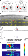
Patients and laboratory animal models of temporal lobe epilepsy display loss of layer III pyramidal neurons in medial entorhinal cortex and hyperexcitability and hypersynchrony of less vulnerable layer II stellate cells. We sought to test the hypothesis that loss of layer III pyramidal neurons triggers synaptic reorganization and formation of recurrent, excitatory synapses among layer II stellate cells in epileptic pilocarpine-treated rats. Laser-scanning photo-uncaging of glutamate focally activated neurons in layer II while excitatory synaptic responses were recorded in stellate cells. Photostimulation revealed previously unidentified, functional, recurrent, excitatory synapses between layer II stellate cells in control animals. Contrary to the hypothesis, however, control and epileptic rats displayed similar levels of recurrent excitation. Recently, hyperexcitability of layer II stellate cells has been attributed, at least in part, to loss of GABAergic interneurons and inhibitory synaptic input. To evaluate recurrent inhibitory circuits in layer II, we focally photostimulated interneurons while recording inhibitory synaptic responses in stellate cells. IPSCs were evoked more than five times more frequently in slices from control versus epileptic animals. These findings suggest that in this model of temporal lobe epilepsy, reduced recurrent inhibition contributes to layer II stellate cell hyperexcitability and hypersynchrony, but increased recurrent excitation does not.
Figures


Similar articles
-
Hyperexcitability, interneurons, and loss of GABAergic synapses in entorhinal cortex in a model of temporal lobe epilepsy.J Neurosci. 2006 Apr 26;26(17):4613-23. doi: 10.1523/JNEUROSCI.0064-06.2006. J Neurosci. 2006. PMID: 16641241 Free PMC article.
-
Reduced inhibition and increased output of layer II neurons in the medial entorhinal cortex in a model of temporal lobe epilepsy.J Neurosci. 2003 Sep 17;23(24):8471-9. doi: 10.1523/JNEUROSCI.23-24-08471.2003. J Neurosci. 2003. PMID: 13679415 Free PMC article.
-
Diversity and excitability of deep-layer entorhinal cortical neurons in a model of temporal lobe epilepsy.J Neurophysiol. 2012 Sep;108(6):1724-38. doi: 10.1152/jn.00364.2012. Epub 2012 Jun 27. J Neurophysiol. 2012. PMID: 22745466
-
Interaction between superficial layers of the entorhinal cortex and the hippocampus in normal and epileptic temporal lobe.Epilepsy Res. 1998 Sep;32(1-2):183-93. doi: 10.1016/s0920-1211(98)00050-3. Epilepsy Res. 1998. PMID: 9761319 Review.
-
Interneurones are not so dormant in temporal lobe epilepsy: a critical reappraisal of the dormant basket cell hypothesis.Epilepsy Res. 1998 Sep;32(1-2):93-103. doi: 10.1016/s0920-1211(98)00043-6. Epilepsy Res. 1998. PMID: 9761312 Review.
Cited by
-
Increased excitatory synaptic input to granule cells from hilar and CA3 regions in a rat model of temporal lobe epilepsy.J Neurosci. 2012 Jan 25;32(4):1183-96. doi: 10.1523/JNEUROSCI.5342-11.2012. J Neurosci. 2012. PMID: 22279204 Free PMC article.
-
Grid cells in pre- and parasubiculum.Nat Neurosci. 2010 Aug;13(8):987-94. doi: 10.1038/nn.2602. Epub 2010 Jul 25. Nat Neurosci. 2010. PMID: 20657591
-
The mechanism of abrupt transition between theta and hyper-excitable spiking activity in medial entorhinal cortex layer II stellate cells.PLoS One. 2010 Nov 4;5(11):e13697. doi: 10.1371/journal.pone.0013697. PLoS One. 2010. PMID: 21079802 Free PMC article.
-
Layer-specific modulation of entorhinal cortical excitability by presubiculum in a rat model of temporal lobe epilepsy.J Neurophysiol. 2015 Nov;114(5):2854-66. doi: 10.1152/jn.00823.2015. Epub 2015 Sep 16. J Neurophysiol. 2015. PMID: 26378210 Free PMC article.
-
Neural Circuitry Polarization in the Spinal Dorsal Horn (SDH): A Novel Form of Dysregulated Circuitry Plasticity during Pain Pathogenesis.Cells. 2024 Feb 25;13(5):398. doi: 10.3390/cells13050398. Cells. 2024. PMID: 38474361 Free PMC article. Review.
References
-
- Agrawal N, Alonso A, Ragsdale DS. Increased persistent sodium currents in rat entorhinal cortex layer V neurons in a post-status epilepticus model of temporal lobe epilepsy. Epilepsia. 2003;44:1601–1604. - PubMed
-
- Bailey SJ, Dhillon A, Woodhall GL, Jones RSG. Lamina-specific differences in GABAB autoreceptor-mediated regulation of spontaneous GABA release in rat entorhinal cortex. Neuropharmacology. 2004;46:31–42. - PubMed
-
- Bartolomei F, Khalil M, Wendling F, Sontheimer A, Régis J, Ranjeva J-P, Guye M, Chauvel P. Entorhinal cortex involvement in human mesial temporal lobe epilepsy: an electrophysiologic and volumetric study. Epilepsia. 2005;46:677–687. - PubMed
Publication types
MeSH terms
Substances
LinkOut - more resources
Full Text Sources
