. 2007 Feb 7;27(6):1247-54.
doi: 10.1523/JNEUROSCI.5320-06.2007.

The inhalation anesthetic isoflurane induces a vicious cycle of apoptosis and amyloid beta-protein accumulation

Affiliations

The inhalation anesthetic isoflurane induces a vicious cycle of apoptosis and amyloid beta-protein accumulation

Zhongcong Xie et al. J Neurosci. .

Abstract

The anesthetic isoflurane has been reported to induce apoptosis and increase Abeta generation and aggregation. However, the molecular mechanism underlying these effects remains unknown. We therefore set out to assess whether the effects of isoflurane on apoptosis are linked to amyloid beta-protein (Abeta) generation and aggregation. For this purpose, we assessed the effects of isoflurane on beta-site amyloid beta precursor protein (APP)-cleaving enzyme (BACE) and gamma-secretase, the proteases responsible for Abeta generation. We also tested the effects of inhibitors of Abeta aggregation (iAbeta5, a beta-sheet breaker peptide; clioquinol, a copper-zinc chelator) on the ability of isoflurane to induce apoptosis. All of these studies were performed on naive human H4 neuroglioma cells as well as those overexpressing APP (H4-APP cells). Isoflurane increased the levels of BACE and gamma-secretase and secreted Abeta in the H4-APP cells. Isoflurane-induced Abeta generation could be blocked by the broad-based caspase inhibitor Z-VAD. The Abeta aggregation inhibitors, iAbeta5 and clioquinol, selectively attenuated caspase-3 activation induced by isoflurane. However, isoflurane was able to induce caspase-3 activation in the absence of any detectable alterations of Abeta generation in naive H4 cells. Finally, Abeta potentiated the isoflurane-induced caspase-3 activation in naive H4 cells. Collectively, these findings suggest that isoflurane can induce apoptosis, which, in turn, increases BACE and gamma-secretase levels and Abeta secretion. Isoflurane also promotes Abeta aggregation. Accumulation of aggregated Abeta in the media can then promote apoptosis. The result is a vicious cycle of isoflurane-induced apoptosis, Abeta generation and aggregation, and additional rounds of apoptosis, leading to cell death.


Figures

Figure 1.
Figure 1.
Treatment with 2% isoflurane induces caspase-3 activation and decreases cell viability without detectable changes in APP processing and Aβ generation in naive H4 cells. A, The 2% isoflurane treatment (lanes 1–3) induces caspase-3 cleavage (activation) compared with control conditions (lanes 4–6) in naive H4 cells. There is no significant difference in the amounts of β-actin between the control- or 2% isoflurane-treated naive H4 cells. B, Caspase-3 activation assessed by quantifying the ratio of caspase-3 fragment to caspase-3-FL in the Western blots. Quantification of the Western blot shows that the 2% isoflurane treatment (black bar) increases caspase-3 activation compared with control conditions (white bar), normalized to β-actin levels. C, Treatment with 2% isoflurane (black bar) decreases cell viability compared with control conditions (white bar) in naive H4 cells. D, Treatment with 2% isoflurane (lanes 3, 4) does not alter the levels of APP-FL and APP-CTFs compared with control conditions (lanes 1, 2). There is no significant difference in the amounts of β-actin in the control- or 2% isoflurane-treated naive H4 cells. E, Quantification of the Western blot shows that the 2% isoflurane treatment (black bar) does not alter the protein levels of APP-FL compared with control conditions (white bar) in H4 naive cells, normalized to β-actin levels. F, Quantification of the Western blot shows that the 2% isoflurane treatment (black bar) does not alter the protein levels of APP-CTFs compared with control conditions (white bar) in H4 naive cells, normalized to β-actin levels. G, Treatment with 2% isoflurane (black bar) does not increase the generation of Aβ40 and Aβ42 compared with control conditions (white bar). Data are means ± SD; n = 9–10 for each experimental group. t test was used to compare the difference between control condition and the 2% isoflurane treatment condition (*p < 0.05; **p < 0.01).
Figure 2.
Figure 2.
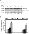
The caspase inhibitor Z-VAD inhibits the caspase-3 activation and the increases in Aβ generation induced by 2% isoflurane in H4-APP cells. A, Treatment with 2% isoflurane (lanes 5, 6) induces caspase-3 cleavage (activation) compared with control conditions (lanes 1, 2) or Z-VAD (100 μm) treatment (lanes 3, 4). The Z-VAD treatment inhibits the caspase-3 cleavage induced by 2% isoflurane (lane 7, 8). There is no significant difference in the amounts of β-actin in the H4-APP cells with the above treatments. B, Quantitation of the Western blot shows that the 2% isoflurane treatment (black bar) increases caspase-3 activation compared with control conditions (white bar) or the Z-VAD (100 μm) treatment (gray bar), normalized to β-actin levels. The isoflurane-induced caspase-3 activation is inhibited by the Z-VAD treatment (striped bar). C, Z-VAD inhibits the isoflurane-induced changes in APP processing in H4-APP cells. Treatment with 2% isoflurane (lanes 5, 6) decreases the protein levels of APP-FL and APP-CTFs compared with control conditions (lanes 1, 2) or Z-VAD (100 μm) treatment (lanes 3, 4). The Z-VAD treatment (lanes 7, 8) inhibits the isoflurane-induced decreases in the protein levels of APP-FL and APP-CTFs. There is no significant difference in the amounts of β-actin in the H4-APP cells with all of the above treatments. D, Quantification of the Western blot shows that 2% isoflurane treatment (black bar) decreases the protein levels of APP-FL compared with the control condition (white bar) or Z-VAD treatment (gray bar), normalized to β-actin levels. The isoflurane-induced decrease in the protein levels of APP-FL is inhibited by the Z-VAD treatment (striped bar). E, Quantification of the Western blot also shows that 2% isoflurane treatment (black bar) decreases the protein levels of APP-CTFs compared with the control condition (white bar) or Z-VAD treatment (gray bar), normalized to β-actin levels. The isoflurane-induced decrease in the protein levels of APP-CTFs is also inhibited by the Z-VAD treatment (striped bar). F, Z-VAD inhibits the isoflurane-induced increases in Aβ generation. Treatment with 2% isoflurane (black bar) increases the levels of Aβ40 compared with the control condition (white bar). Z-VAD treatment alone (gray bar) does not change the levels of Aβ40; however, Z-VAD treatment inhibits the isoflurane-induced increases in the levels of Aβ40. Data are means ± SD; n = 6 for each experimental group. t test is used to compare the difference between control conditions and 2% isoflurane treatment (*p < 0.05; **p < 0.01) and the difference between 2% isoflurane plus DMSO treatment and 2% isoflurane plus Z-VAD treatment (# p < 0.05; ## p < 0.01).
Figure 3.
Figure 3.
Isoflurane increases levels of BACE and nicastrin in H4-APP cells. A, Treatment with 2% isoflurane (lane 1) induces caspase-3 cleavage (17 kDa; activation), increases the level of BACE (65 kDa), and decreases the APP-FL level (110 kDa) compared with the control condition (lane 2). There is no significant difference in the amounts of β-actin between the control condition- or 2% isoflurane-treated H4-APP cells. B, Quantification of the Western blot shows that 2% isoflurane treatment (black bar) induces caspase-3 activation, increases BACE levels, and decreases APP-FL levels, normalized to β-actin levels. C, Treatment with 2% isoflurane (lanes 4–6) increases the levels of immature and mature nicastrin compared with the control condition (lanes 1–3). There is no significant difference in the amounts of β-actin between the control- or 2% isoflurane-treated H4-APP cells. D, Quantification of the Western blot shows that 2% isoflurane treatment (black bar) increases the levels of both immature and mature nicastrin, normalized to β-actin levels. Data are means ± SD; n = 3 for each experimental group. t test was used to compare the difference between control condition and 2% isoflurane treatment (*p < 0.05; **p < 0.01).
Figure 4.
Figure 4.
iAβ5 and clioquinol specifically attenuate isoflurane-induced caspase-3 activation in H4-APP cells. A, iAβ5 plus 2% isoflurane treatment (lane 2) results in a lower degree of caspase-3 cleavage than 2% isoflurane treatment alone (lane 1). There is no significant difference in the amounts of β-actin between the 2% isoflurane- and iAβ5 plus 2% isoflurane-treated H4-APP cells. B, Quantification of the Western blot, based on the ratio of caspase-3 fragment to caspase-3 FL, shows that iAβ5 treatment (black bar) reduces the isoflurane-induced caspase-3 activation (white bar), normalized to β-actin levels. C, STS treatment (lane 3), but not iAβ5 treatment (lane 2), causes caspase-3 activation compared with control condition (lane 1) in H4-APP cells. iAβ5 plus STS treatment (lane 4) leads to a degree of caspase-3 activation similar to STS treatment alone (lane 3). There is no significant difference in the amounts of β-actin in the control-, iAβ5-, STS-, or iAβ5 plus STS-treated H4-APP cells. D, Quantification of the Western blot shows that iAβ5 (striped bar) does not reduce the STS-induced (black bar) caspase-3 activation, normalized to β-actin levels. E, Two percent isoflurane (lane 3) or STS (lane 5) treatment causes caspase-3 activation compared with control condition (lane 1) or clioquinol treatment (lane 2) in H4-APP cells. Clioquinol plus 2% isoflurane treatment (lane 4) leads to a lower degree of caspase-3 activation than 2% isoflurane treatment alone (lane 3). Clioquinol plus STS treatment (lane 6) leads to a degree of caspase-3 activation similar to STS treatment alone (lane 5). There is no significant difference in the amounts of β-actin in all of the above treatments in H4-APP cells. F, Quantification of the Western blot shows that clioquinol reduces the isoflurane-induced (gray bar vs black bar), but not STS-induced (dotted bar vs striped bar), caspase-3 activation, normalized to β-actin levels. Data are means ± SD; n = 3 for each experimental group. t test was used to compare the difference of caspase-3 activation between control condition and STS or 2% isoflurane treatment (*p < 0.05; **p < 0.01) and between saline treatment and iAβ5 or clioquinol treatment (# p < 0.05).
Figure 5.
Figure 5.
Aβ potentiates the isoflurane-induced caspase-3 activation in H4 naive cells. A, Treatments with 2% isoflurane (lanes 3, 7, 11) cause caspase-3 cleavage (activation) compared with control conditions (lanes 1, 5, 9) in H4 naive cells. Aβ plus 2% isoflurane treatment (lanes 4, 8, 12) leads to a greater degree of caspase-3 cleavage than 2% isoflurane treatment alone (lanes 3, 7, 11) in a dose-dependent manner. Aβ treatments alone (lanes 2, 6, 10) also cause caspase-3 activation compared with saline treatments (lanes 1, 5, 9) in a dose-dependent manner. There is no significant difference in the amounts of β-actin between the control- or 2% isoflurane-treated H4-APP cells. B, Quantification of the Western blot shows that Aβ [0 (white bar), 2.5 (gray bar), 5 (black bar), and 7.5 μm (striped bar)] induces caspase-3 activation and potentiates the isoflurane-induced caspase-3 activation in a dose-dependent manner, normalized to β-actin levels. Data are means ± SD; n = 3 for each experimental group. t test was used to compare the difference of caspase-3 activation between saline and Aβ treatment in 2% isoflurane-treated cells (*p < 0.05; **p < 0.01) and in cells with the control condition (# p < 0.05; ## p < 0.01).
Figure 6.
Figure 6.
Hypothetical pathway by which isoflurane induces a vicious cycle of apoptosis and Aβ generation and aggregation. Isoflurane induces caspase-3 activation/apoptosis. Caspase activation, in turn, increases the activities of both BACE and γ-secretase, which serve to increase Aβ generation/accumulation. Isoflurane also enhances Aβ aggregation, which further induces caspase-3 activation and apoptosis. Elevated Aβ generation/accumulation and Aβ aggregation then further induce apoptosis, leading to a vicious cycle of isoflurane-induced apoptosis and Aβ generation/accumulation and aggregation.

Similar articles

Cited by

References

    1. Cherny RA, Atwood CS, Xilinas ME, Gray DN, Jones WD, McLean CA, Barnham KJ, Volitakis I, Fraser FW, Kim Y, Huang X, Goldstein LE, Moir RD, Lim JT, Beyreuther K, Zheng H, Tanzi RE, Masters CL, Bush AI. Treatment with a copper-zinc chelator markedly and rapidly inhibits beta-amyloid accumulation in Alzheimer's disease transgenic mice. Neuron. 2001;30:665–676. - PubMed
    1. De Felice FG, Vieira MN, Saraiva LM, Figueroa-Villar JD, Garcia-Abreu J, Liu R, Chang L, Klein WL, Ferreira ST. Targeting the neurotoxic species in Alzheimer's disease: inhibitors of Abeta oligomerization. FASEB J. 2004;18:1366–1372. - PubMed
    1. Eckenhoff RG, Johansson JS, Wei H, Carnini A, Kang B, Wei W, Pidikiti R, Keller JM, Eckenhoff MF. Inhaled anesthetic enhancement of amyloid-beta oligomerization and cytotoxicity. Anesthesiology. 2004;101:703–709. - PubMed
    1. Florent S, Malaplate-Armand C, Youssef I, Kriem B, Koziel V, Escanye MC, Fifre A, Sponne I, Leininger-Muller B, Olivier JL, Pillot T, Oster T. Docosahexaenoic acid prevents neuronal apoptosis induced by soluble amyloid-beta oligomers. J Neurochem. 2006;96:385–395. - PubMed
    1. Francis R, McGrath G, Zhang J, Ruddy DA, Sym M, Apfeld J, Nicoll M, Maxwell M, Hai B, Ellis MC, Parks AL, Xu W, Li J, Gurney M, Myers RL, Himes CS, Hiebsch R, Ruble C, Nye JS, Curtis D. aph-1 and pen-2 are required for Notch pathway signaling, gamma-secretase cleavage of betaAPP, and presenilin protein accumulation. Dev Cell. 2002;3:85–97. - PubMed

Publication types

MeSH terms