Comparative Study
. 2007 Feb 7;27(6):1396-404.
doi: 10.1523/JNEUROSCI.5188-06.2007.

Differential spatial representation of taste modalities in the rat gustatory cortex

Affiliations
Comparative Study

Differential spatial representation of taste modalities in the rat gustatory cortex

Riccardo Accolla et al. J Neurosci. .

Abstract

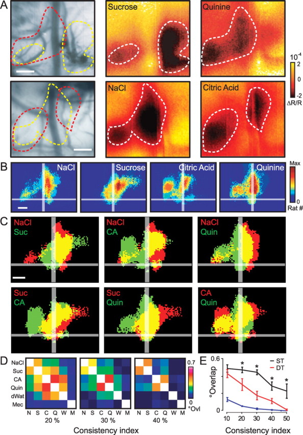
Discrimination between foods is crucial for the nutrition and survival of animals. Remarkable progress has been made through molecular and genetic manipulations in the understanding of the coding of taste at the receptor level. However, much less is known about the cortical processing of taste sensation and the organizing principles of the gustatory cortex (GC). Using genetic tracing, it has recently been shown that sweet and bitter taste are processed through segregated neuronal circuitries along the gustatory pathway up to the cortical level. This is in disagreement with the evidence that GC neurons recorded in both anesthetized and behaving animals responded to multiple taste modalities (including sweet and bitter). To investigate the functional architecture of the GC in regard to taste modalities, we used in vivo intrinsic optical imaging, a technique that has been successfully applied to explore the organization of other neocortical regions. We found that four of the primary taste modalities (sweet, bitter, salty, and sour) are represented by distinctive spatial patterns but that no region was specific to a single modality. In addition, we found that two tastants of similar hedonic value (pleasant or unpleasant) activated areas with more common regions than two tastants with opposite hedonic value. In summary, we propose that these specific cortical patterns can be used to discriminate among various tastants.


Figures

Figure 1.
Figure 1.
Localization of the primary gustatory cortex within the rat insular cortexA, Approximate size and location of the primary GC with respect to anatomical landmarks (blood vessels) and other sensory areas of the brain. mca, Middle cerebral artery; rhv, rhinal veins; S1BF, primary somatosensory area barrel field. Scale bar, 2 mm. B, Coronal section showing location of the GC within the insular cortex (IC), between piriform cortex (Pir) and primary somatosensory cortex (S1). lot, Lateral olfactory tract; rs, rhinal sulcus. Scale bar, 2 mm.
Figure 2.
Figure 2.
In vivo imaging of taste evoked response in the gustatory cortex. A, Left, Vascular pattern taken with a 546 nm illumination filter. The following images represent intrinsic responses averaged over 36 presentations of control (no stimulus), distilled water, and tastants of different modalities. Frames are averaged over a period of 2 s, 1 s after stimulus onset. Tastant-activated area is outlined and reported in all images. Scale bar, 500 μm. B, Interanimal variability analysis of the cortical regions activated by tastants. Population maps (n = 27) of statistically responsive pixels to distilled water and to the average of all tastants responses are shown. The color scale gives a measure of the consistency of the response across animals (high values meaning that the same region was activated in many rats). Scale bar, 1 mm. C, Anatomical localization of the responsive area. Injection of a red dye in the taste-activated cortex before perfusion and sectioning of rat brain. The red spot in the coronal section counterstained with fluorescent Nissl stain (green) is localized within the insular cortex. rrhv, Rostral rhinal vein. See Figure 1 for other abbreviations. Scale bar, 1 mm.
Figure 3.
Figure 3.
Signals imaged in the gustatory cortex are taste specific. A1, Schematic drawing of the taste stimulation system. A2, Intrinsic response averaged over all control and tastant presentations (144 and 24, respectively). Frames are averaged over a period of 2 s from response onset. B1, Schematic drawing of the mechanical stimulation system. B2, Response after mechanical stimulation of the tongue (120 averaged presentations; frames averaged 1.5 s from response onset) in the same animal as in A. Outlined (in red and yellow) over the vessel pattern are the two nonoverlapping regions activated by taste and mechanical stimulation. C, Interanimal variability analysis of cortical regions activated by mechanical stimulation of the tongue (n = 5). The white outline (reported from Fig. 2 B and rescaled) shows that the separation between gustatory and somatosensory responsive areas is consistent across animals. Scale bar, 1 mm. stim., Stimulation; Dors, dorsal; Ventr, ventral; Caud, caudal; Rostr, rostral; Mech., mechanical. See Figure 1 for other abbreviations.
Figure 4.
Figure 4.
Analysis of concentration-dependent response to NaCl. A, Example of intrinsic response to three concentrations of NaCl (average of 6 presentations each). Frames are averaged over a period of 2 s from response onset. The white outline is traced with respect to the 500 mm response. Scale bar, 500 μm. B, Time course of responses to control, dH2O, and NaCl averaged across animals (n = 7). The black bar above the curves indicates the duration of stimulus application. C, Superposition of the dose curves represented in B at an expanded timescale. D, Histograms across animals representing from left to right: decreasing latencies (response time onset), decreasing average time to reach response peak, and increasing maximum amplitude of the response with increasing concentrations. Values (B–D) are means ± SEM. E, Population maps (n = 7) of statistically responsive pixels. Note the stereotyped regions activated between animals at several concentrations. Scale bar, 500 μm.
Figure 5.
Figure 5.
Differential activation patterns of tastant modalities. A, Two different examples of activation maps after sucrose and quinine stimulation (top) and NaCl and citric acid stimulation (bottom). Twenty-eight presentations of each stimulus are averaged. Note that the some regions activated are clearly different. B, Population maps for the four basic taste modalities, NaCl (n = 18), sucrose (n = 15), citric acid (n = 8), and quinine (n = 8). Imaging was done on a total of 27 animals, testing at least two tastants chosen randomly among the four. Color scale “Max” represents 60% of total number of animals. C, Comparison of activated areas between tastants. The yellow areas represent overlapping regions. Single stimulus surfaces are derived from B considering pixels responding with a consistency index of 20%. Scale bars, 500 μm. D, Matrices showing the relative degree of overlap (°Ovl) between stimuli. The three matrices are computed from different consistency indices (20, 30, and 40%). Color-coded values are the result of the ratio between the overlap area (yellow in C) and the total activated area. The value for the white squares is 1. N, NaCl; Suc, S, sucrose; CA, C, citric acid; Quin, Q, quinine; dWat, W, distilled water; Mec, M, mechanical. E, Overlap curves as a function of the consistency index. Mean overlap values among same tastants pairs (ST) (black), different tastants pairs (DT) (red), and between mechanical stimulation and tastants (blue). Difference between black and red line is statistically significant using ANOVA (*Newman–Keuls post hoc test, at least p < 0.0004).
Figure 6.
Figure 6.
Behavioral assessment of hedonic value associated to different tastants. A, Organization of a taste preference trial. The rat's head first enters the sampling port, breaks an infrared (IR) beam and begins to lick the sampling spout. After 1 s, the taste solution is delivered for 500 ms. The frequency of licking is monitored throughout the trial. The next trial is initialized at least 2.5 s after the end of the delivery of the last stimulus, and the tastants are presented in a random manner with at least one water trial between two different taste stimuli. B, Behavioral preference to the four compounds used at the same concentrations as in the imaging experiment. The histograms represent the relative average licking pattern across subjects (n = 6) obtained from the responses of the single rat across five experimental sessions (250 trials for each session). The result is obtained as a ratio between stimulus and water response (dotted line indicates a ratio of 1). We averaged across a time window of 1 s, right after the stimulus presentation. Note the significant increase of the licking rate with respect to the baseline (1 s period before stimulus application) for 500 mm sucrose and 500 mm NaCl and the decrease for 20 mm quinine and 10 mm citric acid (*at least p < 0.005, all statistics with paired t test analysis). Error bars indicate SEM.

Similar articles

Cited by

References

    1. Adolphs R, Tranel D, Koenigs M, Damasio AR. Preferring one taste over another without recognizing either. Nat Neurosci. 2005;8:860–861. - PubMed
    1. Bakin JS, Kwon MC, Masino SA, Weinberger NM, Frostig RD. Suprathreshold auditory cortex activation visualized by intrinsic signal optical imaging. Cereb Cortex. 1996;6:120–130. - PubMed
    1. Barnett EM, Evans GD, Sun N, Perlman S, Cassell MD. Anterograde tracing of trigeminal afferent pathways from the murine tooth pulp to cortex using herpes simplex virus type 1. J Neurosci. 1995;15:2972–2984. - PMC - PubMed
    1. Bartoshuk LM. NaCl thresholds in man: thresholds for water taste or NaCl taste. J Comp Physiol Psychol. 1974;87:310–325. - PubMed
    1. Bonhoeffer T, Grinvald A. Iso-orientation domains in cat visual cortex are arranged in pinwheel-like patterns. Nature. 1991;353:429–431. - PubMed

Publication types

MeSH terms