. 2007 Feb 7;27(6):1405-10.
doi: 10.1523/JNEUROSCI.4564-06.2007.

Presynaptic alpha-synuclein aggregates, not Lewy bodies, cause neurodegeneration in dementia with Lewy bodies

Affiliations

Presynaptic alpha-synuclein aggregates, not Lewy bodies, cause neurodegeneration in dementia with Lewy bodies

Michael L Kramer et al. J Neurosci. .

Abstract

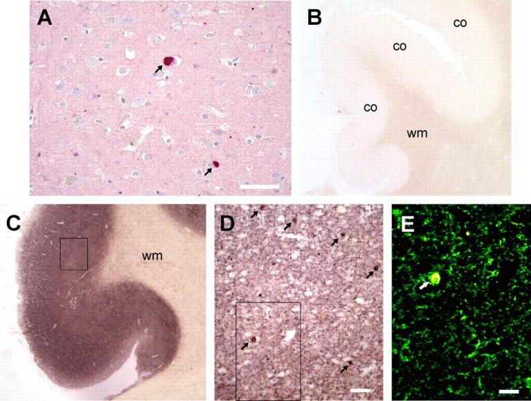
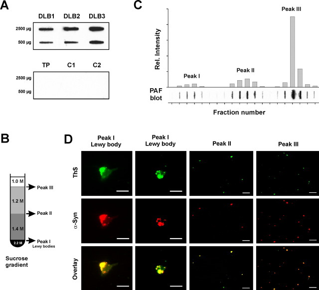
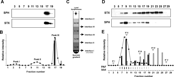
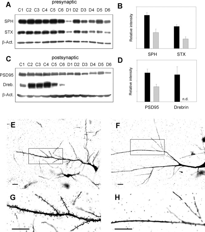
Lewy bodies, the pathological hallmark of dementia with Lewy bodies (DLB), are large juxtanuclear inclusions of aggregated alpha-synuclein. However, the small number of cortical Lewy bodies relative to the total neuron count does not correlate with the extent of cognitive impairment. In contrast to dopaminergic neurons in Parkinson's disease, nerve cell loss is usually less prevalent in the cortex of DLB, suggesting a different mechanism of neurodegeneration. Because antibodies used for immunodetection per se do not generally differentiate the aggregated from the physiological and monomeric isoform of alpha-synuclein, we developed the paraffin-embedded tissue (PET) blot and the protein aggregate filtration (PAF) assay for the sensitive and selective detection of alpha-synuclein aggregates in tissue slides and brain homogenates, respectively. In contrast to common immunohistochemistry, the PET blot detected an enormous number of small alpha-synuclein aggregates, which, in contrast to the few Lewy bodies, may explain the cognitive impairment in DLB. Using the PAF assay, we demonstrate that the absolute majority of alpha-synuclein aggregates are located at presynaptic terminals, suggesting a severe pathological impact on synaptic function. Indeed, parallel to the massive presynaptic accumulation of alpha-synuclein aggregates, we observed significant synaptic pathology with almost complete loss of dendritic spines at the postsynaptic area. Our results provide strong evidence for a novel concept of neurodegeneration for DLB in which synaptic dysfunction is caused by presynaptic accumulation of alpha-synuclein aggregates. This concept may also be valid for Parkinson's disease.


Figures

Figure 1.
Figure 1.
Revealing previously undetectable, small granular α-synuclein aggregates with the PET blot. A, Common immunohistochemistry (LB509) identifies only α-synuclein deposits (dark red) as Lewy bodies (arrows) and few Lewy neurites (dots or threads). B, C, Compared with a control (B), the PET blot method detects diffuse α-synuclein aggregates in the frontal cortex (co) of the gyrus cinguli but not in the white matter (wm). The enormous number of α-synuclein aggregates is visible as a dark brown color reaction throughout the cortex in DLB. D, The magnification (see box in C) shows that the majority of α-synuclein aggregates other than Lewy bodies (arrows) form fine granular deposits. E, The virtual fluorescence image of the PET blot section (box in D) created by digital processing clearly demonstrates that the fine granular α-synuclein aggregates are much smaller compared with Lewy bodies (arrow). Scale bars: A, 50 μm; D, 20 μm; E, 10 μm.
Figure 2.
Figure 2.
Small granular α-synuclein deposits represent the absolute majority of α-synuclein aggregates in DLB. A, The new developed PAF assay specifically detects α-synuclein aggregates in brain homogenates of DLB compared with a case with tau pathology (TP) and non-dementia controls (C1 and C2). B, Sucrose gradient with the indicated molar concentrations for the separation of α-synuclein aggregates in brain homogenates by ultracentrifugation, which was originally used for purification of Lewy bodies (Iwatsubo et al., 1996). C, PAF assay of the sucrose gradient fraction with the plot of its densitometric analysis. D, Identification of small granular α-synuclein (α-Syn) aggregates in the three sucrose gradient peaks by immunofluorescence (LB509; red) and thioflavin S (ThS) fluorescence (green). Scale bars, 20 μm.
Figure 3.
Figure 3.
Small granular α-synuclein aggregates are trapped in synaptosomes, indicating a presynaptic localization. A, Location of synaptosomes by Western blot analysis of sucrose gradient fractions for syntaxin (STX; HPC-1) as the synaptic plasma membrane marker and synaptophysin (SPH; SY38) as the synaptic vesicle marker. B, Plot of the relative densitometric intensities for α-synuclein aggregates (filled circles), syntaxin (gray bars), and synaptophysin (black bars). C, Synaptosomes of peak III were subjected to hypotonic shock and applied to a more differentiated sucrose gradient. D, Western blot analysis of the gradient in C for synaptophysin and syntaxin. E, PAF blot analysis of the gradient in C for α-synuclein aggregates and their densitometric quantification (filled circles) together with synaptophysin (black bars) and syntaxin (gray bars). The fractions corresponding to the IFs of the gradient in C are marked by inverted triangles.
Figure 4.
Figure 4.
Pathophysiological changes at synapses in DLB. A–D, Brain homogenates of six confirmed DLB cases (D1–D6) are compared with age-matched non-dementia controls (C1–C6). A, C, Western blot analysis for presynaptic markers (A) synaptophysin (SPH; SY38) and syntaxin (STX; HPC-1) and postsynaptic markers (C) postsynaptic density protein (PSD95, rabbit anti-PSD95) and drebrin (Dreb.; M2F6), including the loading control β-actin (β-Act.). B, D, Plot of the β-actin normalized average intensities for presynaptic marker (B) and postynaptic marker (D) proteins of non-dementia controls (black bars) and DLB cases (gray bars) ± SE. E–H, Golgi–Cox–Davenport staining of brain slices for visualization of dendrites and their spines in non-dementia control (E, G) and DLB (F, H). G and H show the magnification of the black boxes in E and F. Scale bars, 50 μm.

Similar articles

Cited by

References

    1. Burn DJ. Cortical Lewy body disease. J Neurol Neurosurg Psychiatry. 2004;75:175–178. - PMC - PubMed
    1. Chandra S, Gallardo G, Fernandez-Chacon R, Schluter OM, Sudhof TC. Alpha-synuclein cooperates with CSPalpha in preventing neurodegeneration. Cell. 2005;123:383–396. - PubMed
    1. Dodd PR, Watson WE, Morrison MM, Johnston GA, Bird ED, Cowburn RF, Hardy JA. Uptake of gamma-aminobutyric acid and l-glutamic acid by synaptosomes from postmortem human cerebral cortex: multiple sites, sodium dependence and effect of tissue preparation. Brain Res. 1989;490:320–331. - PubMed
    1. Engert F, Bonhoeffer T. Dendritic spine changes associated with hippocampal long-term synaptic plasticity. Nature. 1999;399:66–70. - PubMed
    1. Fujiwara H, Hasegawa M, Dohmae N, Kawashima A, Masliah E, Goldberg MS, Shen J, Takio K, Iwatsubo T. Alpha-synuclein is phosphorylated in synucleinopathy lesions. Nat Cell Biol. 2002;4:160–164. - PubMed

Publication types

Substances