

G alpha(q)-coupled muscarinic acetylcholine receptors enhance nicotinic acetylcholine receptor signaling in Caenorhabditis elegans mating behavior
- PMID: 17287516
- PMCID: PMC6673585
- DOI: 10.1523/JNEUROSCI.4320-06.2007
G alpha(q)-coupled muscarinic acetylcholine receptors enhance nicotinic acetylcholine receptor signaling in Caenorhabditis elegans mating behavior
Abstract
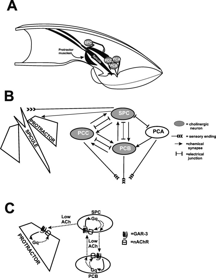
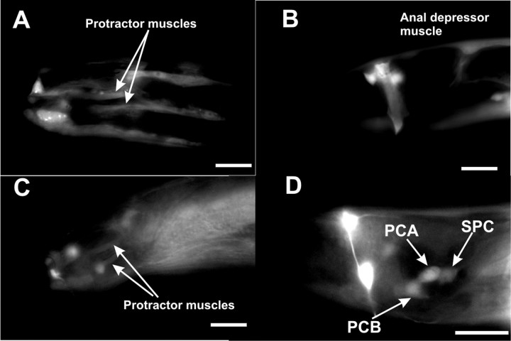
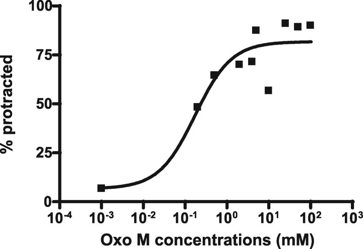
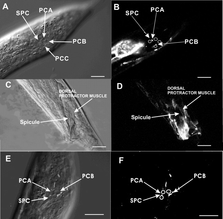
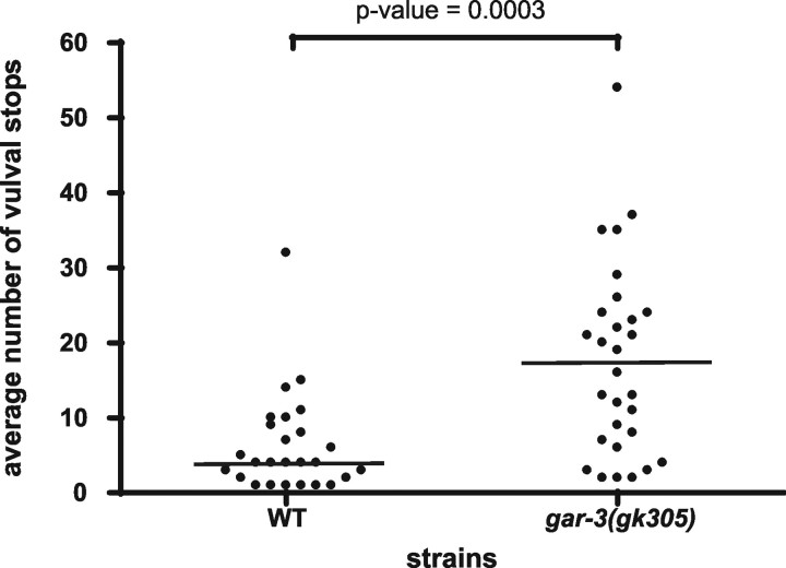
In this study, we address why metabotropic and ionotropic cholinergic signaling pathways are used to facilitate motor behaviors. We demonstrate that a G alpha(q)-coupled muscarinic acetylcholine receptor (mAChR) signaling pathway enhances nicotinic acetylcholine receptor (nAChR) signaling to facilitate the insertion of the Caenorhabditis elegans male copulatory spicules into the hermaphrodite during mating. Previous studies showed that ACh (acetylcholine) activates nAChRs on the spicule protractor muscles to induce the attached spicules to extend from the tail. Using the mAChR agonist Oxo M (oxotremorine M), we identified a GAR-3(mAChR)-G alpha(q) pathway that promotes protractor muscle contraction by upregulating nAChR signaling before mating. GAR-3(mAChR) is expressed in the protractor muscles and in the spicule-associated SPC and PCB cholinergic neurons. However, ablation of these neurons or impairing cholinergic transmission reduces drug-induced spicule protraction, suggesting that drug-stimulated neurons directly activate muscle contraction. Behavioral analysis of gar-3 mutants indicates that, in wild-type males, GAR-3(mAChR) expression in the SPC and PCB neurons is required for the male to sustain rhythmic spicule muscle contractions during attempts to breach the vulva. We propose that the GAR-3(mAChR)/G alpha(q) pathway sensitizes the spicule neurons and muscles before and during mating so that the male can respond to hermaphrodite vulva efficiently.
Figures

Similar articles
-
Coordination of opposing sex-specific and core muscle groups regulates male tail posture during Caenorhabditis elegans male mating behavior.BMC Biol. 2009 Jun 22;7:33. doi: 10.1186/1741-7007-7-33. BMC Biol. 2009. PMID: 19545405 Free PMC article.
-
Caenorhabditis elegans UNC-103 ERG-like potassium channel regulates contractile behaviors of sex muscles in males before and during mating.J Neurosci. 2003 Apr 1;23(7):2696-705. doi: 10.1523/JNEUROSCI.23-07-02696.2003. J Neurosci. 2003. PMID: 12684455 Free PMC article.
-
Regulation of distinct muscle behaviors controls the C. elegans male's copulatory spicules during mating.Cell. 2001 Dec 14;107(6):777-88. doi: 10.1016/s0092-8674(01)00600-6. Cell. 2001. PMID: 11747813
-
Regulation of muscarinic acetylcholine receptor signaling.Pharmacol Ther. 2003 May;98(2):197-220. doi: 10.1016/s0163-7258(03)00032-9. Pharmacol Ther. 2003. PMID: 12725869 Review.
-
Acetylcholine.WormBook. 2007 Jan 30:1-21. doi: 10.1895/wormbook.1.131.1. WormBook. 2007. PMID: 18050502 Free PMC article. Review.
Cited by
-
STR-33, a novel G protein-coupled receptor that regulates locomotion and egg laying in Caenorhabditis elegans.J Biol Chem. 2011 Nov 18;286(46):39860-70. doi: 10.1074/jbc.M111.241000. Epub 2011 Sep 21. J Biol Chem. 2011. PMID: 21937442 Free PMC article.
-
Coordination of opposing sex-specific and core muscle groups regulates male tail posture during Caenorhabditis elegans male mating behavior.BMC Biol. 2009 Jun 22;7:33. doi: 10.1186/1741-7007-7-33. BMC Biol. 2009. PMID: 19545405 Free PMC article.
-
A cholinergic-regulated circuit coordinates the maintenance and bi-stable states of a sensory-motor behavior during Caenorhabditis elegans male copulation.PLoS Genet. 2011 Mar;7(3):e1001326. doi: 10.1371/journal.pgen.1001326. Epub 2011 Mar 10. PLoS Genet. 2011. PMID: 21423722 Free PMC article.
-
Behavioral decay in aging male C. elegans correlates with increased cell excitability.Neurobiol Aging. 2012 Jul;33(7):1483.e5-23. doi: 10.1016/j.neurobiolaging.2011.12.016. Epub 2012 Jan 27. Neurobiol Aging. 2012. PMID: 22285759 Free PMC article.
-
Deciphering and modulating G protein signalling in C. elegans using the DREADD technology.Sci Rep. 2016 Jul 27;6:28901. doi: 10.1038/srep28901. Sci Rep. 2016. PMID: 27461895 Free PMC article.
References
-
- Alfonso A, Grundahl K, Duerr JS, Han HP, Rand JB. The Caenorhabditis elegans unc-17 gene: a putative vesicular acetylcholine transporter. Science. 1993;261:617–619. - PubMed
Publication types
MeSH terms
Substances
Grants and funding
LinkOut - more resources
Full Text Sources
Molecular Biology Databases