. 2007 Feb 7;27(6):1422-33.
doi: 10.1523/JNEUROSCI.2382-06.2007.

Estrogen regulates Bcl-w and Bim expression: role in protection against beta-amyloid peptide-induced neuronal death

Affiliations

Estrogen regulates Bcl-w and Bim expression: role in protection against beta-amyloid peptide-induced neuronal death

Mingzhong Yao et al. J Neurosci. .

Abstract

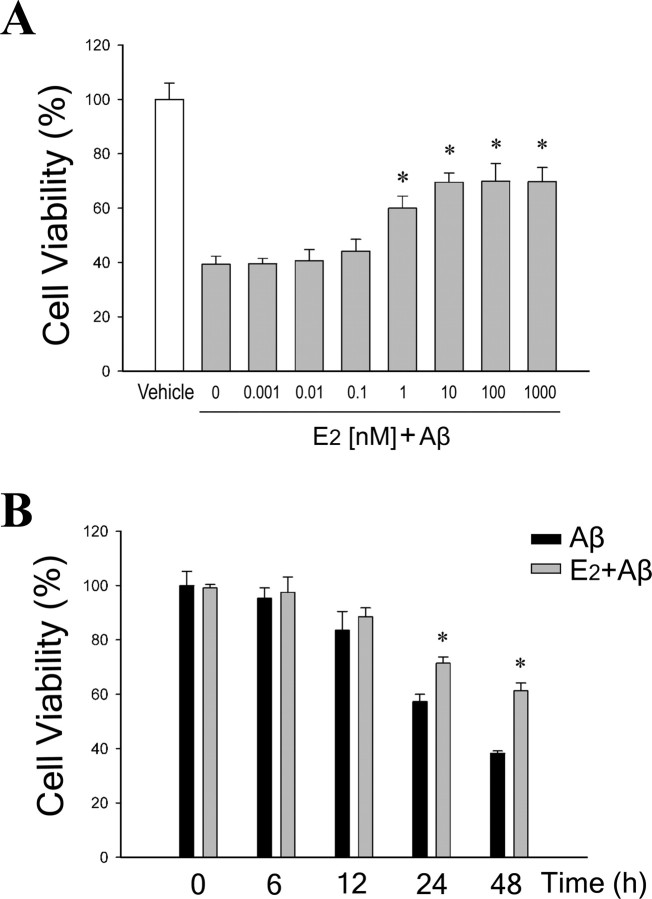
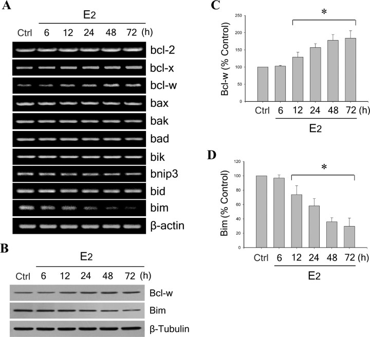
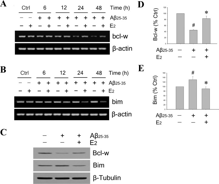
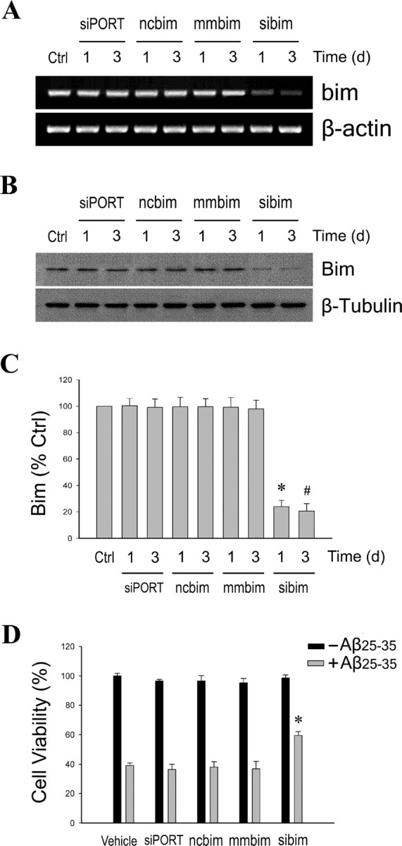
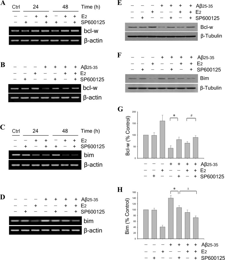
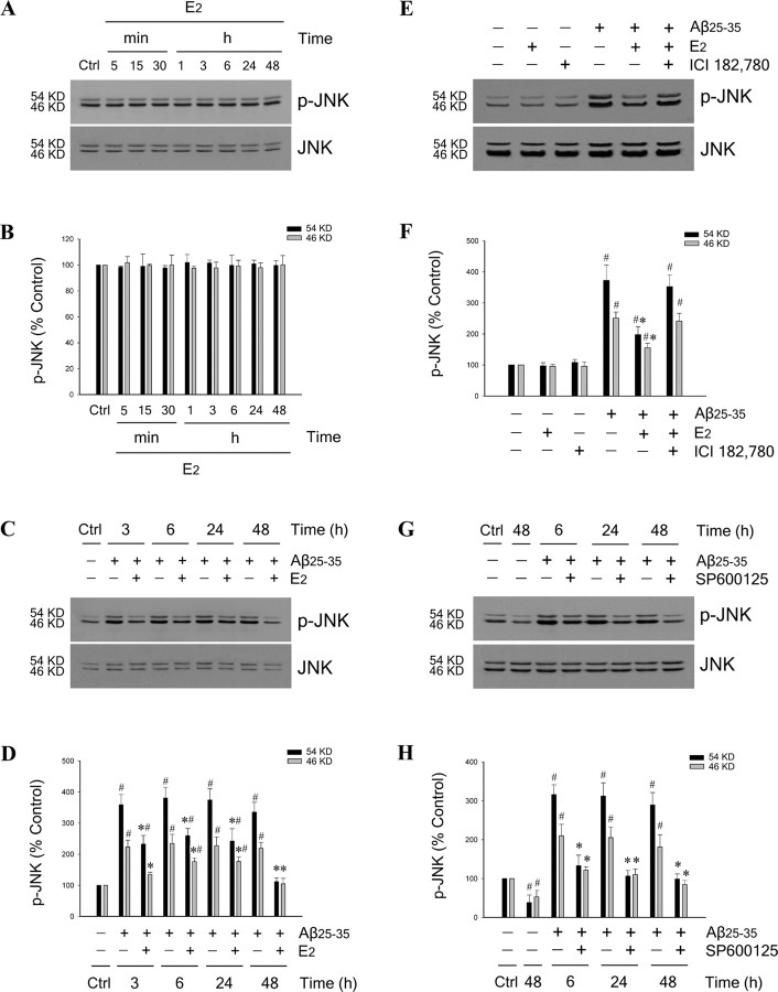
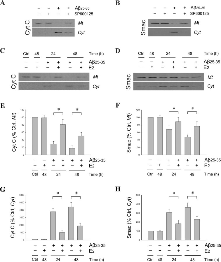
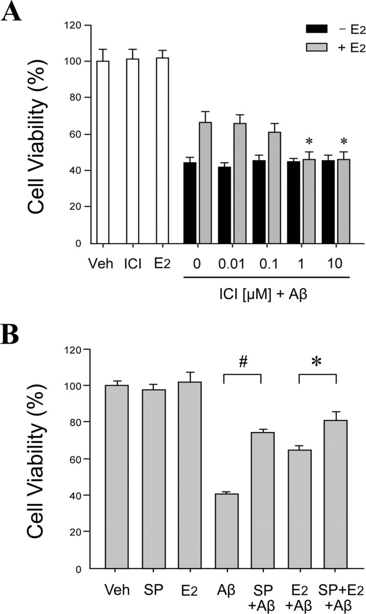
Estrogen is neuroprotective against a variety of insults, including beta-amyloid peptide (Abeta); however, the underlying mechanism(s) is not fully understood. Here, we report that 17beta-estradiol (E2) selectively regulates neuronal expression of the Bcl-2 family (bcl-2, bcl-x, bcl-w, bax, bak, bad, bik, bnip3, bid, and bim). In primary cerebrocortical neuron cultures under basal conditions, we observe that E2 upregulates expression of antiapoptotic Bcl-w and downregulates expression of proapoptotic Bim in an estrogen receptor (ER)-dependent manner. In the presence of toxic levels of Abeta, we observe that E2 attenuates indices of neuronal apoptosis: c-Jun N-terminal kinase (JNK)-dependent downregulation of Bcl-w and upregulation of Bim, mitochondrial release of cytochrome c and Smac, and cell death. These neuroprotective effects of E2 against Abeta-induced apoptosis are mimicked by the JNK inhibitor SP600125 (anthra[1,9-cd]pyrazol-6(2H)-one). In addition, E2 attenuates Abeta-induced JNK phosphorylation in an ER-dependent manner, but does not affect basal levels of JNK phosphorylation. These results suggest that E2 may reduce Abeta-induced neuronal apoptosis at least in part by two complementary pathways: (1) ER-dependent, JNK-independent upregulation of Bcl-w and downregulation of Bim under basal conditions, and (2) ER-dependent inhibition of Abeta-induced JNK activation and subsequent JNK-dependent downregulation of Bcl-w and upregulation of Bim, resulting in mitochondrial release of cytochrome c and Smac and eventual cell death. These data provide new understanding into the mechanisms contributing to estrogen neuroprotection, a neural function with potential therapeutic relevance to Alzheimer's disease.


Figures

Figure 1.
Figure 1.
E2 reduces Aβ-induced neuronal death in a dose- and time-dependent manner. A, Neuron cultures were pretreated with increasing concentrations (0.001–1000 nm) of E2 for 60 min, followed by exposure to 25 μm25–35 for 48 h, and then assayed for cell viability. Data show mean cell viability (+SEM) from a representative experiment (n = 6). Significance is defined as follows: *p < 0.01 compared with Aβ25–35 condition. B, Neuron cultures were pretreated with 0 nm (black bars) or 10 nm (gray bars) E2 for 60 min, followed by exposure to 25 μm25–35 for the indicated times, and then assayed for cell viability. Data show mean cell viability (+SEM) from a representative experiment (n = 6). Significance is defined as follows: *p < 0.01 compared with Aβ treatment at the matched time point.
Figure 2.
Figure 2.
Estrogen upregulates Bcl-w and downregulates Bim expression. A, E2 induces a time-dependent increase of bcl-w and decrease of bim. Neuron cultures were treated with 10 nm E2 for 6–72 h, and mRNA expression levels of Bcl-2 family members (bcl-2, bcl-x, bcl-w, bax, bak, bad, bik, bnip3, bid, and bim) were detected by RT-PCR, followed by agarose gel electrophoresis. β-actin served as an internal control. B, Representative Western blots show E2 regulation of Bcl-w (top panel) and Bim (middle panel) protein levels; β-tubulin (bottom panel) was used as an internal control. C, D, Relative amounts of Bcl-w (C) and Bim (D) protein levels were determined by densitometric scanning of Western blots from three to five independent experiments. Data are represented as a mean (+SEM) percentage of control values. *p < 0.01 relative to vehicle-treated control group (Ctrl).
Figure 3.
Figure 3.
Estrogen attenuates Aβ-induced Bcl-w downregulation and Bim upregulation. A, B, Neuron cultures were pretreated with 10 nm E2 for 60 min, followed by exposure to 25 μm25–35 for indicated times, and analysis of bcl-w (A) and bim (B) mRNA expression by RT-PCR. β-actin served as an internal control. C, Neuron cultures were pretreated with 10 nm E2 for 60 min followed by exposure to 25 μm25–35 for 48 h. A representative Western blot of culture lysates shows protein levels of Bcl-w (top panel) and Bim (middle panel); β-tubulin (bottom panel) was used as a control. D, E, Relative amounts of Bcl-w (D) and Bim (E) protein levels were determined by densitometric scanning of Western blots from three to four independent experiments. Data are represented as a mean (+SEM) percentage of control values. #p < 0.01 compared with vehicle-treated control group; *p < 0.01 relative to Aβ25–35-treated condition.
Figure 4.
Figure 4.
Suppression of endogenous Bim expression decreases Aβ-induced neuronal death. Primary neuron cultures were transfected for 1 or 3 d with bim-specific siRNA (sibim), scrambled siRNA (ncbim), mismatched siRNA (mmbim), or siPORT amine vehicle (siPORT), or were untreated (Ctrl). A, Representative agarose gels of RT-PCR products show endogenous bim mRNA expression decreased only by specific bim siRNA (top) with no change in levels of β-actin, an internal control (bottom). B, Protein levels of Bim were similarly affected by the siRNA treatments, as determined by Western blot (top), with no change in β-tubulin levels (bottom). C, Relative amounts of Bim were determined by densitometric scanning of Western blots from three independent experiments. Data are represented as a mean (+SEM) percentage of vehicle-treated control (Ctrl) values. *p < 0.01 relative to 1 d ncbim control group; #p < 0.01 relative to 3 d ncbim control group. D, Neuron cultures were exposed for 48 h to 25 μm25–35 24 h after transfection with siRNA. Data show mean (+SEM) cell viability from a representative experiment (n = 4). *p < 0.01 relative to respective ncbim condition.
Figure 5.
Figure 5.
Estrogen regulation of Bcl-w and Bim expression is ER dependent. A, C, Representative agarose gels of RT-PCR products using bcl-w (A) or bim (C) primers show that the ER antagonist ICI 182,780 blocks E2 regulation of bcl-w and bim under basal conditions. Neuron cultures were pretreated with 1 μm ICI 182,780 for 60 min, followed by 10 nm E2 for 24 and 48 h, respectively. β-actin served as an internal control. B, D, Representative agarose gels of RT-PCR products using bcl-w (B) or bim (D) primers show that the ER antagonist ICI 182,780 blocks E2 inhibition of Aβ-induced changes in bcl-w and bim expression. Neuron cultures were pretreated with 1 μm ICI 182,780 for 60 min, followed by 10 nm E2 for 60 min, and then exposed to 25 μm25–35 for 24 and 48 h. β-actin served as an internal control. E, ER dependence of E2 bcl-w and bim regulation was extended to protein expression using Western blots. A representative Western blot shows Bcl-w (top panel), Bim (middle panel), and β-tubulin (bottom panel) expression in lysates from neuron cultures that were pretreated with 1 μm ICI 182,780, followed 60 min later with 10 nm E2 treatment, and then 60 min later exposed to 25 μm25–35 for 48 h. F, G, Relative protein levels of Bcl-w (F) and Bim (G) were determined by densitometric scanning of Western blots from four independent experiments. Data show mean (+SEM) percentages of control values. *p < 0.01 relative to E2-treated condition; #p < 0.01 compared with E2 plus Aβ25–35 treatment.
Figure 6.
Figure 6.
Estrogen regulation of Bcl-w and Bim expression is JNK independent under basal conditions but involves JNK signaling under Aβ challenge. A, C, Representative agarose gels of RT-PCR products using bcl-w (A) or bim (C) primers show that, under basal conditions, the JNK inhibitor SP600125 neither blocks E2 regulation of bcl-w and bim nor independently affects expression of bcl-w and bim. Neuron cultures were pretreated with 100 nm SP600125 for 60 min, followed by 10 nm E2 for 24 and 48 h. β-actin served as an internal control. B, D, Representative agarose gels of RT-PCR products using bcl-w (B) or bim (D) primers show that the JNK inhibitor SP600125 both independently and additively with E2 blocks Aβ-induced changes in bcl-w and bim expression. Neuron cultures were pretreated with 100 nm SP600125 for 60 min, followed by 10 nm E2 for 60 min, and then exposed to 25 μm25–35 for 24 and 48 h. β-actin served as an internal control. Effects of JNK inhibition on E2 regulation of bcl-w and bim were extended to protein expression using Western blots. E, F, Representative blots show Bcl-w (E) and Bim (F) and expression in lysates from neuron cultures that were pretreated with 100 nm SP600125, followed 60 min later with 10 nm E2 treatment, and then 60 min later exposed to 25 μm25–35 for 48 h. G, H, Relative protein levels of Bcl-w (G) and Bim (H) were determined by densitometric scanning of Western blots from three independent experiments. *p < 0.01 relative to Aβ25–35 condition; #p < 0.01 compared with E2 plus Aβ25–35 treatment; Δp < 0.01 relative to Aβ25–35 plus SP600125 condition.
Figure 7.
Figure 7.
Estrogen reduces Aβ-induced JNK activation. A, Neuron cultures were treated with 10 nm E2 for the indicated times and then assessed by Western blot with phospho-JNK (p-JNK) (top panel) and pan-JNK (bottom panel) antibodies. B, Blots were quantified by band densitometry for both the 54 kDa (black bars) and 46 kDa (gray bars) JNK forms. C, D, E2 reduces Aβ-induced JNK activation, as shown by both representative Western blots (C) and densitometric quantification of blots (D), in neuron cultures that were pretreated with 10 nm E2 for 60 min, followed by exposure to 25 μm25–35 for the indicated times. E, F, Representative Western blots probed with phospho-JNK (top panel) and pan-JNK (bottom panel) antibodies (E) and densitometric quantification of blots (F) show that pretreatment of neuron cultures with 1 μm ICI 182,780 for 60 min attenuates the inhibitory effect of 10 nm E2 treatment on JNK phosphorylation induced by exposure to 25 μm25–35 for 6 h. G, H, The JNK inhibitor SP600125 reduces Aβ-induced JNK activation, as shown by representative Western blots (G) and densitometric quantification of blots (H) from lysates of neuron cultures that were pretreated with 100 nm SP600125 for 60 min, followed by exposure to 25 μm25–35 for the indicated times. All experiments were repeated in three or more independent culture preparations. Data show mean phospho-JNK:total JNK ratios, normalized to the vehicle control condition. *p < 0.01 relative to matched Aβ25–35 condition at the same time point; #p < 0.01 relative to vehicle control (Ctrl) condition.
Figure 8.
Figure 8.
Estrogen reduces JNK-dependent mitochondrial cytochrome c and Smac release induced by Aβ. A, B, Neuron cultures were pretreated with 100 nm JNK inhibitor SP600125 for 60 min, followed by exposure to 25 μm25–35 for 48 h, and then analyzed for cytochrome c (Cyt C) (A) and Smac (B) content in mitochondrial (Mt) and cytosolic (Cyt) extracts by Western blot. Neuron cultures were pretreated with 10 nm E2 for 60 min, followed by exposure to 25 μm25–35 for 24 and 48 h. C, D, Levels of cytochrome c (C) and Smac (D) in mitochondrial and cytosolic extracts were analyzed by Western blot. EH, Relative protein levels of cytochrome c, in the mitochondrial fraction (E) and cytosolic fraction (G), and Smac, in the mitochondrial fraction (F) and cytosolic fraction (H), were determined by densitometric scanning of Western blots from three independent experiments. Data are presented as mean (+SEM) percentages of vehicle-treated control (Ctrl) values. *p < 0.01 relative to Aβ25–35 condition at 24 h; #p < 0.01 relative to Aβ25–35 condition at 48 h.
Figure 9.
Figure 9.
Aβ toxicity is JNK dependent and attenuated by ER-dependent estrogen actions. A, E2-induced neuroprotection against Aβ is ER dependent. Primary neuron cultures were pretreated with ICI 182,780 (ICI) at concentrations ranging from 0.01 to 10 μm for 60 min, followed by treatment with 0 nm (black bars) or 10 nm (gray bars) E2 for 60 min, and then exposed to 25 μm25–35 for 48 h. Controls groups with vehicle treatment (Veh), 10 nm E2 alone, and 1 μm ICI alone are shown in white bars. Data show mean cell viability (+SEM) from a representative experiment (n = 4). *p < 0.01 compared with E2 plus Aβ25–35 and 0 μm ICI treatments. B, Inhibition of JNK signaling reduces Aβ toxicity and increases estrogen neuroprotection. Neuron cultures were pretreated with 100 nm JNK inhibitor SP600125 (SP) for 60 min, followed by 10 nm E2 for 60 min, and then exposure to 25 μm25–35 for 48 h. Data show mean cell viability (+SEM) from a representative experiment (n = 4). Significance is defined as follows: #p < 0.01 compared with Aβ treatment; *p < 0.01 compared with E2 plus Aβ25–35 treatment.
Figure 10.
Figure 10.
Estrogen neuroprotection may involve two complementary pathways. Under basal conditions (solid lines), E2 increases expression of antiapoptotic Bcl-w and decreases expression of proapoptotic Bim, events that counteract the mitochondrial pathway of apoptosis. This may represent an estrogen maintenance pathway of neuron survival. Under toxic challenges such as Aβ exposure (dashed lines), E2 can inhibit activation of JNK, functionally mimicking the pharmacological JNK inhibitor SP600125. JNK activation induces the mitochondrial pathway of apoptosis involving decreased expression of Bcl-w and increased expression of Bim, followed by mitochondrial release of cytochrome c and Smac, and eventually neuron death. Because E2 does not alter basal JNK activity, such a protective mechanism may represent an estrogen response pathway that is activated after injury.

Similar articles

Cited by

References

    1. Agostinho P, Oliveira CR. Involvement of calcineurin in the neurotoxic effects induced by amyloid-beta and prion peptides. Eur J Neurosci. 2003;17:1189–1196. - PubMed
    1. Ahlbom E, Prins GS, Ceccatelli S. Testosterone protects cerebellar granule cells from oxidative stress-induced cell death through a receptor mediated mechanism. Brain Res. 2001;892:255–262. - PubMed
    1. Alkayed NJ, Goto S, Sugo N, Joh HD, Klaus J, Crain BJ, Bernard O, Traystman RJ, Hurn PD. Estrogen and Bcl-2: gene induction and effect of transgene in experimental stroke. J Neurosci. 2001;21:7543–7550. - PMC - PubMed
    1. Antonsson B, Martinou JC. The Bcl-2 protein family. Exp Cell Res. 2000;256:50–57. - PubMed
    1. Bae MA, Song BJ. Critical role of c-Jun N-terminal protein kinase activation in troglitazone-induced apoptosis of human HepG2 hepatoma cells. Mol Pharmacol. 2003;63:401–408. - PubMed

Publication types

MeSH terms

Substances

LinkOut - more resources