The neural code of auditory phantom perception
- PMID: 17287523
- PMCID: PMC6673575
- DOI: 10.1523/JNEUROSCI.3711-06.2007
The neural code of auditory phantom perception
Abstract
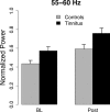
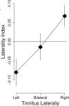
Tinnitus is defined by an auditory perception in the absence of an external source of sound. This condition provides the distinctive possibility of extracting neural coding of perceptual representation. Previously, we had established that tinnitus is characterized by enhanced magnetic slow-wave activity (approximately 4 Hz) in perisylvian or putatively auditory regions. Because of works linking high-frequency oscillations to conscious sensory perception and positive symptoms in a variety of disorders, we examined gamma band activity during brief periods of marked enhancement of slow-wave activity. These periods were extracted from 5 min of resting spontaneous magnetoencephalography activity in 26 tinnitus and 21 control subjects. Results revealed the following, particularly within a frequency range of 50-60 Hz: (1) Both groups showed significant increases in gamma band activity after onset of slow waves. (2) Gamma is more prominent in tinnitus subjects than in controls. (3) Activity at approximately 55 Hz determines the laterality of the tinnitus perception. Based on present and previous results, we have concluded that cochlear damage, or similar types of deafferentation from peripheral input, triggers reorganization in the central auditory system. This produces permanent alterations in the ongoing oscillatory dynamics at the higher layers of the auditory hierarchical stream. The change results in enhanced slow-wave activity reflecting altered corticothalamic and corticolimbic interplay. Such enhancement facilitates and sustains gamma activity as a neural code of phantom perception, in this case auditory.
Figures


References
-
- Berg P, Scherg M. A multiple source approach to the correction of eye artifacts. Electroencephalogr Clin Neurophysiol. 1994;90:229–241. - PubMed
-
- Calford MB, Rajan R, Irvine DR. Rapid changes in the frequency tuning of neurons in cat auditory cortex resulting from pure-tone-induced temporary threshold shift. Neuroscience. 1993;55:953–964. - PubMed
-
- Delorme A, Makeig S. EEGLAB: an open source toolbox for analysis of single-trial EEG dynamics including independent component analysis. J Neurosci Methods. 2004;134:9–21. - PubMed
-
- Eggermont JJ, Roberts LE. The neuroscience of tinnitus. Trends Neurosci. 2004;27:676–682. - PubMed
Publication types
MeSH terms
LinkOut - more resources
Full Text Sources
Other Literature Sources
Medical
