. 2007 Feb 7;27(6):1485-97.
doi: 10.1523/JNEUROSCI.4720-06.2007.

Xenopus vocalizations are controlled by a sexually differentiated hindbrain central pattern generator

Affiliations

Xenopus vocalizations are controlled by a sexually differentiated hindbrain central pattern generator

Heather J Rhodes et al. J Neurosci. .

Abstract

Male and female African clawed frogs (Xenopus laevis) produce rhythmic, sexually distinct vocalizations as part of courtship and mating. We found that Xenopus vocal behavior is governed by a sexually dimorphic central pattern generator (CPG) and that fictive vocalizations can be elicited from an in vitro brain preparation by application of serotonin or by electrical stimulation of a premotor nucleus. Male brains produced fictive vocal patterns representing two calls commonly produced by males in vivo (advertisement and amplectant call), as well as one call pattern (release call) that is common for juvenile males and females in vivo but rare for adult males. Female brains also produced fictive release call. The production of male calls is androgen dependent in Xenopus; to test the effects of androgens on the CPG, we examined fictive calling in the brains of testosterone-treated females. Both fictive male advertisement call and release call were produced. This suggests that all Xenopus possess a sexually undifferentiated pattern generator for release call. Androgen exposure leads to a gain-of-function, allowing the production of male-specific call types without prohibiting the production of the undifferentiated call pattern. We also demonstrate that the CPG is located in the brainstem and seems to rely on the same nuclei in both males and females. Finally, we identified endogenous serotonergic inputs to both the premotor and motor nuclei in the brainstem that may regulate vocal activity in vivo.


Figures

Figure 1.
Figure 1.
Male fictive vocal patterns resemble nerve recordings of in vivo advertisement call. A, In vitro nerve recording before and after 5-HT application shows the onset of fictive advertisement call ∼35 s after 5-HT was added to the bath (gray bar). The only other activity apparent in the recording is respiratory activity (arrow). A, Inset shows bracketed region in A with an enlarged timescale, demonstrating that each of the large-amplitude bursts on the right is a bout of fictive advertisement call. B, Fictive (left) and in vivo (right) nerve recordings of male advertisement call showing slow, fast, and loud–slow (l–s) trills, as well as transition (trn) CAPs between slow and fast trills. B, Inset, Simultaneous recordings of nerve activity and sound production in a freely behaving frog. Traces show a portion of fast and loud–slow trill; each CAP in the nerve recording (N; top) is followed by the production of a click of sound (S; bottom trace) several milliseconds later. Scale bar, 50 ms. C, D, Fictive (C1) and in vivo (D1) nerve recordings of glottal bursts associated with respiration; enlarged traces of slow (left) and fast (right) trill CAPs for fictive (C2) and in vivo (D2) recordings in B; five traces are overlaid for each. Timescales are the same for C1 and D1, as well as for C2 and D2. E, Frequency histogram showing the bimodal distribution of instantaneous CAP rates for one individual fit with two Gaussian curves. CAPs within 2 SD of the peak of the lower distribution were designated as slow trill, and those within 2 SD of the upper distribution were fast trill (ranges marked with arrows). Values falling between those ranges were transitional clicks; for additional detail on CAP designations, see Materials and Methods. F, Instantaneous CAP frequency versus CAP order for one bout of in vivo (top) and one bout of fictive (bottom) advertisement call, showing CAP designation as slow, transition, fast, or loud–slow trill.
Figure 2.
Figure 2.
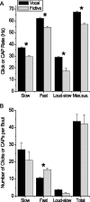
A comparison of fictive and in vivo male advertisement call. A, Instantaneous CAP rates for slow, fast, and loud–slow trills, as well as the maximum sustained CAP rate, for fictive advertisement call are lower than click rates produced by the same animals in vivo. B, Number of clicks and CAPs per bout for each trill type are compared for the in vivo and in vitro conditions. Total includes slow, fast, loud–slow, and transition. n = 10 animals for each bar; error bars are SEM. Compared with Wilcoxon's signed-rank test; *p < 0.0125.
Figure 3.
Figure 3.
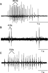
Several male calls were produced as fictive vocalizations. A, An example of fictive slow trill without fast trill. B, An example of fictive amplectant call. C, An example of fictive male release call. Average CAP rates are given for a short series of CAPs from each trace.
Figure 4.
Figure 4.
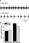
Female brains produce sex-specific fictive vocalizations. A, Fictive release call (top) compared with an audio recording of a release call in vivo (bottom). B, CAP rates for female fictive calls are similar to those observed in vivo. Vocal data are from Potter et al. (2005). Bar graph shows means, and error bars are SEM.
Figure 5.
Figure 5.
T-females produce male-like vocal patterns in vivo and in vitro. A, Fictive (top) and vocal (bottom) recordings are from one animal after 8 weeks of testosterone exposure. Slow and fast trill segments are marked. The fictive call resembles male advertisement call more closely than the sound produced in vivo, which shows an abnormal pause between slow and fast trill. B, Fictive advertisement calls produced by three T-females are compared with fictive calls from male brains and in vivo calls by T-females. Dots are means CAP rates for the three T-females that produced advertisement call in this study. Gray and black solid lines are means for male fictive and T-female vocalizations, respectively; dashed lines are ±1 SD. T-female vocal data from Potter et al. (2005); male data are the same as shown previously (Fig. 2).
Figure 6.
Figure 6.
Transection experiments show that DTAM is necessary for 5-HT-induced fictive vocalizations in both sexes. A, Examples of male advertisement call (middle column) and female release call (right column) in vivo, in vitro, and after a series of transections. Drawings (left column) show the condition of the brain during each recording. The location of DTAM is marked with black triangles; nucleus IX–X is marked with black ovals. All traces in each column are from a single animal, except the in vivo female recording. A1, Sound recordings in vivo. A2, Nerve recordings of fictive calls in the whole brain. A3, Fictive call after the telencephalon and diencephalon were removed revealed lengthened fast trill for males and normal call for females. A4, Fictive call after bisection of the midbrain and rostral brainstem resulted in reduced fast trill amplitude and rate in the male (slow and fast trill segments are marked) but no change in the female call. A5, Removal of the rostral brainstem (containing DTAM) resulted in the loss of all fictive vocalizations in both males and females. B, Plots showing CAP rate (top row), number of CAPs per bout (middle row), and CAP area (bottom row) for male fast trill (left column), male slow trill (middle column), and female release call (right column) for the conditions illustrated in A1–A4. n = 4 for all. Analyzed with repeated-measures ANOVA followed by Tukey–Kramer post hoc test. *p < 0.05 on Tukey–Kramer comparing marked condition with whole-brain fictive condition (A2).
Figure 7.
Figure 7.
Electrical stimulation of DTAM can induce fictive advertisement call. A, A sample trace: continuous stimulation to DTAM at 20 Hz produced two bouts of fictive advertisement call. Stimulus artifact is gray, and nerve response is black. B, Enlargement of gray boxed region in A showing characteristic CAP shapes for slow and fast trill. C, A cresyl violet-stained horizontal section shows the lesion (arrow on left) at the rostral end of DTAM (location marked with dashed line on right). Fourth V, Fourth ventricle; CBL, cerebellum; Isthmi, nucleus isthmi; OT, optic tectum; V, nerve V; VIII, nerve VIII. D, Average CAP rate during slow and fast trill in response to three stimulus frequencies in two brains. Slow trill rate increases with increasing stimulus frequency, whereas fast trill does not change. Error bars are SEM. E, Frequency histograms show the distribution of CAP latencies from preceding stimuli for fast (top row) and slow (bottom row) trill, in response to stimuli at 40 Hz (left column) and 20 Hz (right column). Only slow trill with 20 Hz stimulation shows a strong relationship between stimulus and CAP timing.
Figure 8.
Figure 8.
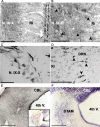
Endogenous serotonergic inputs to motor and premotor nuclei. A, B, Serotonergic neurons (black) are located along the midline of the brainstem in the dorsal raphe nucleus (DRN) and project laterally through the reticular formation (Ri) and into the laryngeal motor nucleus (N. IX-X). Arrowheads in B indicate varicose serotonergic fibers in the laryngeal motor nucleus. Sections in A–D are horizontal, medial is to the right, and rostral is to the top. Scale bars: A, 500 μm; B, 250 μm. C, Backfill of nerve IX–X with the transneuronal tracer WGA-HRP (punctate black stain) resulted in filled motoneuron axons (top left) and cell bodies. Counterstained with cresyl violet (light gray). Scale bar, 100 μm. D, Secondary label with transneuronal tracer was found in the DRN medial to laryngeal motor nucleus (arrowheads). Scale bar, 100 μm. E, Serotonergic fibers are found in the region of DTAM in the midbrain, lateral to the fourth ventricle (fourth V.) and ventral to the cerebellum (CBL). F, The location of DTAM was confirmed with Nissl stain of an adjacent tissue section of E and further by back-labeling DTAM neurons with rhodamine dextran/and overlaying with the adjacent Nissl-stained section (E, inset). Scale bars: E, F, 500 μm.

Similar articles

Cited by

References

    1. Bass AH, Baker R. Sexual dimorphisms in the vocal control system of a teleost fish: morphology of physiologically identified neurons. J Neurobiol. 1990;21:1155–1168. - PubMed
    1. Bass AH, Baker R. Phenotypic specification of hindbrain rhombomeres and the origins of rhythmic circuits in vertebrates. Brain Behav Evol. 1997;50(Suppl 1):3–16. - PMC - PubMed
    1. Birinyi A, Antal M, Wolf E, Szekely G. The extent of the dendritic tree and the number of synapses in the frog motoneuron. Eur J Neurosci. 1992;4:1003–1012. - PubMed
    1. Brahic CJ, Kelley DB. Vocal circuitry in Xenopus laevis: telencephalon to laryngeal motor neurons. J Comp Neurol. 2003;464:115–130. - PMC - PubMed
    1. Cowley KC, Schmidt BJ. A comparison of motor patterns induced by N-methyl-d-aspartate, acetylcholine and serotonin in the in vitro neonatal rat spinal cord. Neurosci Lett. 1994;171:147–150. - PubMed

Publication types

MeSH terms

LinkOut - more resources