. 2007 Apr;175(4):1695-705.
doi: 10.1534/genetics.107.070946. Epub 2007 Feb 7.

Valproic acid affects membrane trafficking and cell-wall integrity in fission yeast

Affiliations

Valproic acid affects membrane trafficking and cell-wall integrity in fission yeast

Makoto Miyatake et al. Genetics. 2007 Apr.

Abstract

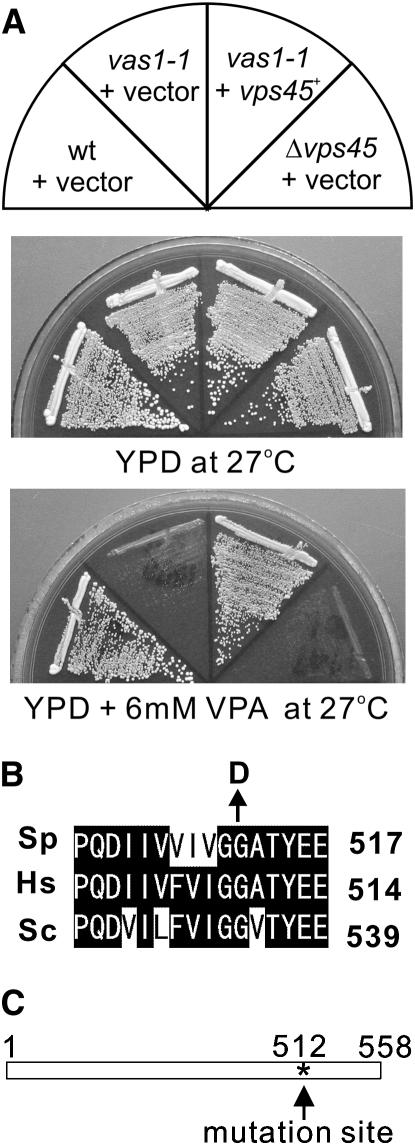
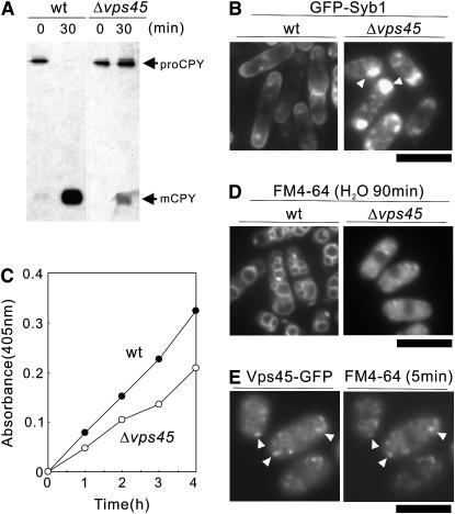
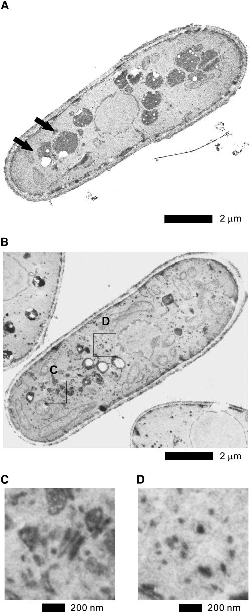
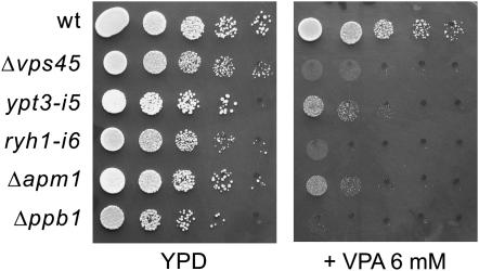
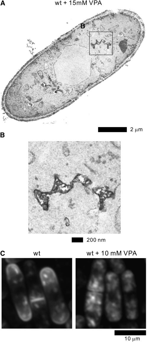
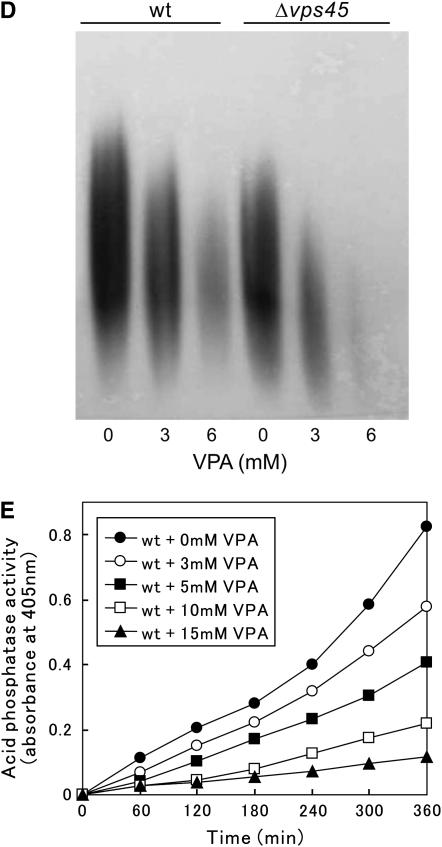
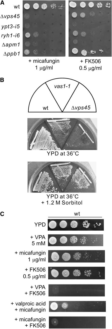
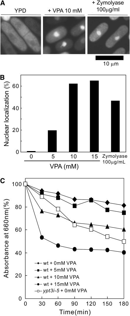
Valproic acid (VPA) is widely used to treat epilepsy and manic-depressive illness. Although VPA has been reported to exert a variety of biochemical effects, the exact mechanisms underlying its therapeutic effects remain elusive. To gain further insights into the molecular mechanisms of VPA action, a genetic screen for fission yeast mutants that show hypersensitivity to VPA was performed. One of the genes that we identified was vps45+, which encodes a member of the Sec1/Munc18 family that is implicated in membrane trafficking. Notably, several mutations affecting membrane trafficking also resulted in hypersensitivity to VPA. These include ypt3+ and ryh1+, both encoding a Rab family protein, and apm1+, encoding the mu1 subunit of the adaptor protein complex AP-1. More importantly, VPA caused vacuolar fragmentation and inhibited the glycosylation and the secretion of acid phosphatase in wild-type cells, suggesting that VPA affects membrane trafficking. Interestingly, the cell-wall-damaging agents such as micafungin or the inhibition of calcineurin dramatically enhanced the sensitivity of wild-type cells to VPA. Consistently, VPA treatment of wild-type cells enhanced their sensitivity to the cell-wall-digesting enzymes. Altogether, our results suggest that VPA affects membrane trafficking, which leads to the enhanced sensitivity to cell-wall damage in fission yeast.


Figures

F<sc>igure</sc> 1.—
Figure 1.—
Mutation in the vas1+/vps45+ gene causes VPA-sensitive phenotype. (A) The VPA sensitivities of the vas1-v1/vps45-v1 mutant cells. Cells transformed with the multicopy vector pDB248 or the vector containing the vps45+ gene were streaked onto each plate containing YPD or YPD plus 6 mm VPA and then incubated for 4 days at 27°. (B) Alignment of partial protein sequences of S. pombe (Sp) Vps45 with related proteins from human (Hs) and S. cerevisiae (Sc). Sequence alignment was performed using the Clustal W program. Solid background indicates identical amino acids. Arrow indicates the highly conserved glycine 512, which, when mutated to aspartic acid, resulted in VPA-sensitive function in Vps45. (C) Linear representation of the structure of Vps45 and where the vas1-v1 mutation resides. Asterisk indicates the mutation site.
F<sc>igure</sc> 2.—
Figure 2.—
Defects in membrane trafficking in the Δvps45 mutant cells. (A) Processing of CPY in vivo. Wild-type strain and Δvps45 mutant cells were pulse labeled with Express-35S-label for 10 min at 27° and chased. The immunoprecipitates were separated on an SDS–10% polyacrylamide gel. The autoradiograms of the fixed dried gels are shown. proCPY, precursor form of CPY; mCPY, mature form of CPY. (B) Intracellular localization of GFP-Syb1 in Δvps45 mutant cells. Wild-type strain and Δvps45 mutant cells expressing chromosome-borne GFP-Syb1 were cultured in YPD medium at 27°. The localization of GFP-Syb1 was examined by fluorescence microscopy. Bar, 10 μm. (C) Defective secretion of acid phosphatase in Δvps45 cells. Wild-type strain and Δvps45 cells were grown to an optical density at 660 nm of 0.3 and assayed for secreted acid phosphatase activity. The data shown are representative of multiple experiments. (D) Δvps45 mutant cells are defective in vacuole fusion. Wild-type (wt) strain and Δvps45 mutant cells were grown in YPD medium at 27°. Cells were collected, labeled with FM 4-64 fluorescent dye (see materials and methods), resuspended in water, and examined by fluorescence microscope. Photographs were taken after 90 min. Bar, 10 μm. (E) Vps45-GFP is concentrated at the Golgi/endosomal compartments. Wild-type cells, expressing chromosome-borne Vps45-GFP cultured in EMM in the absence of thiamine for 12 hr, were incubated with the FM4-64 dye for 5 min to visualize early endosomes. Arrowheads indicate the dot-like structures of Vps45-GFP and early endosomes stained with FM4-64, respectively. Bar, 10 μm.
F<sc>igure</sc> 3.—
Figure 3.—
Electron microscopic analysis of Δvps45 mutant cells and the effect of VPA treatment on wild-type cells. (A) The wild-type cells and (B) the Δvps45 mutant cells grown at 27° were analyzed by electron microscopy. Representative thin sections are shown. Arrows in A indicate vacuoles in wild-type cells. The boxed regions C and D in B are enlarged. Bar, 2 μm. (C) The enlargement of Golgi structures of Δvps45 mutant cells. Bar, 200 nm. (D) The enlargement of the accumulated vesicle structures in Δvps45 mutant cells. Bar, 200 nm.
F<sc>igure</sc> 4.—
Figure 4.—
The Δvps45 mutant cells and various membrane trafficking mutants displayed hypersensitivity to VPA. Wild-type cells and the various mutant cells were spotted onto each plate containing YPD or YPD plus 6 mm VPA and then incubated for 3 days at 27°. Cells were spotted in serial 10-fold dilutions starting with OD660 = 0.3 of log-phase cells (5 μl).
F<sc>igure</sc> 5.—
Figure 5.—
Effect of VPA on membrane trafficking of wild-type cells. (A) Electron microscopy of wild-type cells treated with 15 mm VPA for 12 hr. The boxed region B in A is enlarged. Bar, 2 μm. (B) The enlargement of the fragmented vacuoles in wild-type cells treated with VPA. Bar, 200 nm. (C) Effect of VPA on the intracellular localization of GFP-Syb1 in wild-type cells. Wild-type strain expressing the chromosome-borne GFP-Syb1 was cultured in YPD medium and treated with 10 mm VPA for 10 hr at 27°. The localization of GFP-Syb1 was examined by fluorescence microscopy. Bar, 10 μm. (D) VPA-induced defects in glycosylation. Acid phosphatase glycosylation in the wild-type strain and the Δvps45 mutant cells. Effects of the addition of 3 or 6 mm VPA to the medium on these cellular processes were also examined. Immunoblot analysis and acid phosphatase staining were performed as described in materials and methods. (E) VPA treatment caused the defective secretion of acid phosphatase in wild-type cells. Wild-type strains treated with indicated concentrations of VPA were grown to an optical density at 660 nm of 0.3 and assayed for the secreted acid phosphatase activity as indicated. The data presented are representative of three independent experiments.
F<sc>igure</sc> 6.—
Figure 6.—
The synergistic effect of VPA and cell-wall damage in fission yeast. (A) The Δvps45 mutant cells and various membrane trafficking mutants displayed sensitivity to the cell-wall-damaging agent micafungin and the immunosuppressant FK506. Wild-type cells and the various mutant cells as indicated were spotted onto each plate containing YPD plus 1 μg/ml micafungin or YPD plus 0.5 μg/ml FK506 and then incubated for 3 days at 27°. Cells were spotted in serial 10-fold dilutions starting with OD660 = 0.3 of log-phase cells (5 μl). (B) The temperature sensitivities of the vps45 mutants. Wild-type cells, vas1-1 cells, or Δvps45 cells were streaked onto each plate containing YPD or YPD plus 1.2 m sorbitol and then incubated for 3 days at 36°. (C) Synergism between VPA and cell-wall perturbation. Wild-type cells were spotted onto the plates with or without 5 mm VPA, 0.5 μg/ml FK506, or 1.0 μg/ml micafungin, individually or in combination, and then incubated for 4 days at 27°.
F<sc>igure</sc> 7.—
Figure 7.—
VPA activates calcineurin signaling. (A) VPA and glucanase treatment stimulates translocation of GFP-Prz1 from the cytosol to the nucleus. The cellular localization of Prz1 was examined by immunofluorescence microscopy in wild-type cells carrying the chromosomally tagged GFP-Prz1 with or without the addition of 10 mm VPA for 6 hr or with the addition of 100 μg/ml of zymolyase 20T for 3 hr. Bar, 10 μm. (B) Quantification of the nuclear accumulation of GFP-Prz1 in wild-type cells with or without the addition of various concentrations of VPA for 6 hr or with the addition of 100 μg/ml of zymolyase for 3 hr to the medium. Data represent the average of four experiments. (C) Effect of VPA on cell-wall digestion by β-glucanase. Wild-type cells exponentially growing in YPD medium were treated with the indicated concentrations of VPA for 8 hr and incubated with 100 μg/ml of β-glucanase (zymolyase 20T) at 30° with vigorous shaking. Cell lysis was monitored by measuring optical density at 660 nm (the value before adding the enzyme was taken as 100%). The data shown are representative of multiple experiments.

Similar articles

Cited by

References

    1. Achachi, A., A. Florins, N. Gillet, C. Debacq, P. Urbain et al., 2005. Valproate activates bovine leukemia virus gene expression, triggers apoptosis, and induces leukemia/lymphoma regression in vivo. Proc. Natl. Acad. Sci. USA 102: 10309–10314. - PMC - PubMed
    1. Babst, M., G. Odorizzi, E. J. Estepa and S. D. Emr, 2000. Mammalian tumor susceptibility gene 101 (TSG101) and the yeast homologue, Vps23p, both function in late endosomal trafficking. Traffic 1: 248–258. - PubMed
    1. Bankaitis, V. A., l. M. Johnson, and S. D. Emr, 1986. Isolation of yeast mutants defective in protein targeting to the vacuole. Proc. Natl. Acad. Sci. USA 83: 9075–9079. - PMC - PubMed
    1. Basi, G., E. Schmid and K. Maundrell, 1993. TATA box mutations in the Schizosaccharomyces pombe nmt1 promoter affect transcription efficiency but not the transcription start point or thiamine repressibility. Gene 123: 131–136. - PubMed
    1. Bates, S., D. M. MacCallum, G. Bertram, C. A. Munro, H. B. Hughes et al., 2005. Candida albicans Pmr1p, a secretory pathway P-type Ca2+/Mn2+-ATPase, is required for glycosylation and virulence. J. Biol. Chem. 280: 23408–23415. - PubMed

Publication types

MeSH terms