. 2007 May 15;404(1):71-80.
doi: 10.1042/BJ20070005.

Mapping binding sites for the PDE4D5 cAMP-specific phosphodiesterase to the N- and C-domains of beta-arrestin using spot-immobilized peptide arrays

Affiliations

Mapping binding sites for the PDE4D5 cAMP-specific phosphodiesterase to the N- and C-domains of beta-arrestin using spot-immobilized peptide arrays

George S Baillie et al. Biochem J. .

Abstract

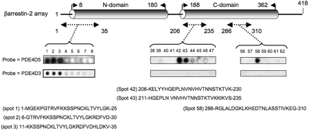
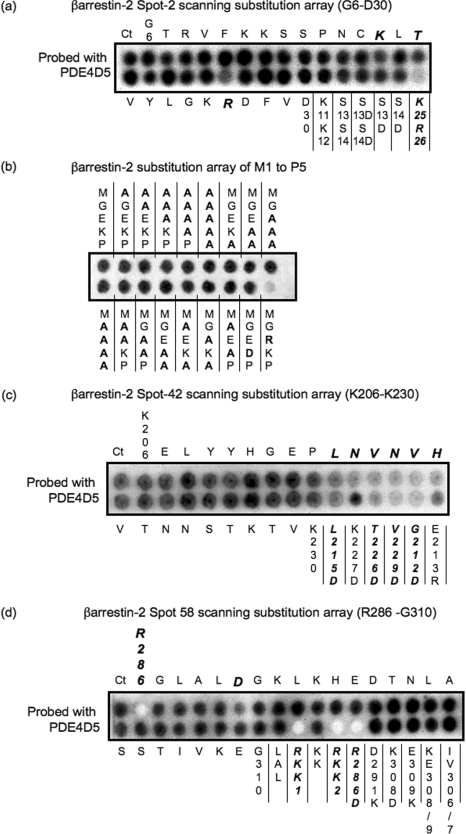
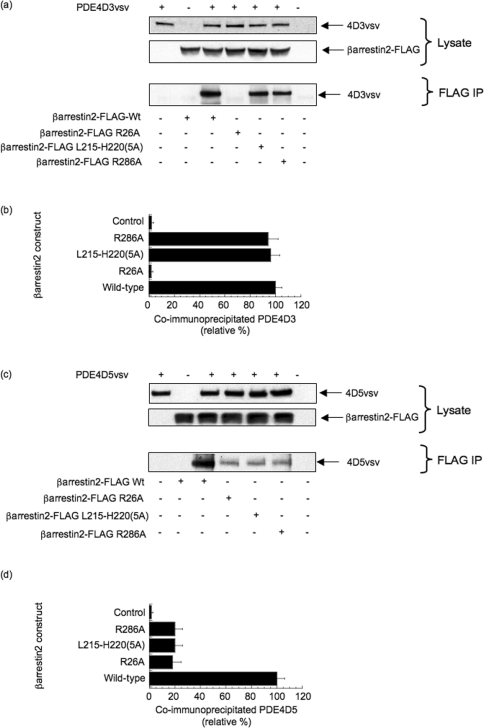
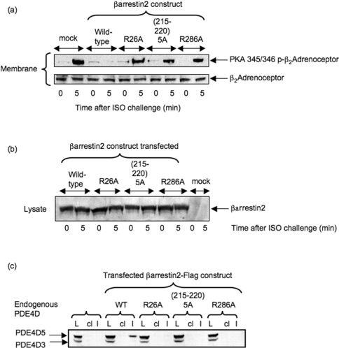
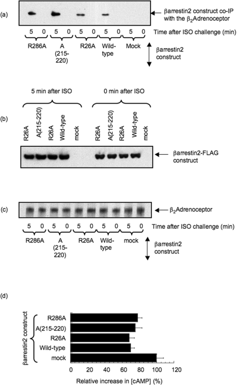
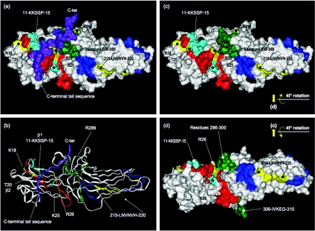
Beta2-ARs (beta2-adrenoceptors) become desensitized rapidly upon recruitment of cytosolic beta-arrestin. PDE4D5 (family 4 cAMP-specific phosphodiesterase, subfamily D, isoform 5) can be recruited in complex with beta-arrestin, whereupon it regulates PKA (cAMP-dependent protein kinase) phosphorylation of the beta2-AR. In the present study, we have used novel technology, employing a library of overlapping peptides (25-mers) immobilized on cellulose membranes that scan the entire sequence of beta-arrestin 2, to define the interaction sites on beta-arrestin 2 for binding of PDE4D5 and the cognate long isoform, PDE4D3. We have identified a binding site in the beta-arrestin 2 N-domain for the common PDE4D catalytic unit and two regions in the beta-arrestin 2 C-domain that confer specificity for PDE4D5 binding. Alanine-scanning peptide array analysis of the N-domain binding region identified severely reduced interaction with PDE4D5 upon R26A substitution, and reduced interaction upon either K18A or T20A substitution. Similar analysis of the beta-arrestin 2 C-domain identified Arg286 and Asp291, together with the Leu215-His220 region, as being important for binding PDE4D5, but not PDE4D3. Transfection with wild-type beta-arrestin 2 profoundly decreased isoprenaline-stimulated PKA phosphorylation of the beta2-AR in MEFs (mouse embryo fibroblasts) lacking both beta-arrestin 1 and beta-arrestin 2. This effect was negated using either the R26A or the R286A mutant form of beta-arrestin 2 or a mutant with substitution of an alanine cassette for Leu215-His220, which showed little or no PDE4D5 binding, but was still recruited to the beta2-AR upon isoprenaline challenge. These data show that the interaction of PDE4D5 with both the N- and C-domains of beta-arrestin 2 are essential for beta2-AR regulation.


Figures

Figure 1
Figure 1. Probing a β-arrestin 2 peptide array with PDE4D5–GST and PDE4D3–GST
β-Arrestin 2 is shown schematically with its N- and C-domains. Results show immobilized peptide ‘spots’ of overlapping 25-mer peptides each shifted along by five amino acids in the entire β-arrestin 2 sequence probed for interaction with either PDE4D5–GST or PDE4D3–GST and detection by immunoblotting. Positively interacting peptides generate dark spots, while those that do not interact leave white (blank) spots. In all other sections of the array, spots were blank with either probe. Spot numbers relate to peptides in the scanned array and whose sequence is given as indicated. Arrays probed with purified GST did not yield any positively interacting spots. These data are typical of experiments performed three times.
Figure 2
Figure 2. Alanine-scanning substitution analysis to probe the binding sites for PDE4D5–GST in the N- and C-domains of β-arrestin 2
β-Arrestin 2 peptide arrays were probed for PDE4D5–GST binding based upon the indicated 25-mer ‘parent’ β-arrestin 2 peptide, where the indicated amino acids were sequentially and individually replaced by alanine. Ct refers to the native peptide and all other spots reflect peptide ‘progeny’. GST alone did not bind to any peptide spot (results not shown). (a) Alanine substitution array for the Gly6–Asp30 peptide whose sequence is in the N-domain of β-arrestin 2, together with the additional indicated substitutions, which were to either alanine (no label) or aspartate (D). (b) Peptide representing amino acids 1–25 in β-arrestin 2 with changes made solely in the first five amino acids (native sequence MGEKP). The alterations in this portion are indicated using the single-letter amino acid code. These data are typical of experiments performed three times. (c) Alanine substitution array for the Lys206–Lys230 peptide whose sequence is in the C-domain of β-arrestin 2, together with the additional indicated substitutions which were either to aspartate (D) or to arginine (R). (d) Alanine substitution array for the Arg286–Gly310 peptide whose sequence is in the C-domain of β-arrestin 2, together with the additional indicated substitutions which were to either alanine (no label) or aspartate (D) or lysine (K). LAL is L288A/A289D/L290A. RKK1 is R286A/K293A/K295A. RKK2 is R286A/K293A/K295A/K308A. KE is K308A/E309A. IV is I306A/V307A.
Figure 3
Figure 3. Consequences of mutations in the N- and C-domain binding sites in β-arrestin 2, based on peptide array data, for the binding of PDE4D3 and PDE4D5
(a) HEK-293 cells were co-transfected with VSV-epitope-tagged PDE4D3 (GenBank® accession number L20970) together with the indicated forms of FLAG-tagged β-arrestin 2, namely either wild-type (Wt) or mutations of R26A, R286A and one where the stretch of amino acids from Leu215 to His220 in β-arrestin 2–FLAG was replaced by a five-residue alanine cassette [β-arrestin 2–FLAG L215–H220(5A)]. The lower panel shows the result of immunoprecipitating the indicated β-arrestin 2–FLAG construct with an anti-FLAG antibody and probing for co-immunoprecipitated PDE4D3–VSV by immunoblotting with an anti-VSV antibody. (b) Quantification, by densitometry, of co-immunoprecipitated PDE4D3–VSV from three separate experiments as in (a) with means±S.D. and with the indicated control analysing cells that had not been transfected with β-arrestin 2–FLAG, but which has been transfected with PDE4D3–VSV. (c) As in (a), but co-transfection with VSV-epitope-tagged PDE4D5 (GenBank® accession number AF012073) instead of PDE4D3–VSV. (d) As in (b), but co-transfection with PDE4D5–VSV instead of PDE4D3–VSV.
Figure 4
Figure 4. Functional consequences of mutations disrupting N- and C-domain binding sites in β-arrestin 2 for PDE4D5
(a) Upper panel: PKA-phosphorylation status of the β2-AR, as assessed using a phospho-specific antiserum, in β-arrestin 1−/−/β-arrestin 2−/− double-knockout MEFs treated with 10 μM isoprenaline (ISO) for either 0 or 5 min, as indicated. This was assessed in both mock-transfected cells and also in cells transfected to express either wild-type β-arrestin 2–FLAG or the various β-arrestin 2–FLAG mutants as described in the legend to Figure 3. Lower panel: loading blot for the β2-AR in these cells. (b) Immunoblot of the indicated forms of wild-type and mutant β-arrestin 2 expressed in β-arrestin 1−/−/β-arrestin 2−/− double-knockout MEFs treated with isoprenaline (ISO) as indicated. (c) Immunoblot of endogenous PDE4D species in lysates (L) of MEFs transfected with the indicated wild-type and mutant forms of β-arrestin 2–FLAG. This detects the presence of endogenous PDE4D3 and PDE4D5 in lysates (L) from these cells. Also shown are PDE4D immunoblots of both FLAG immunoprecipitates (I) and bead control immunoprecipitates (cl), where no anti-FLAG antiserum was added. The blots are typical of experiments performed three times.
Figure 5
Figure 5. Consequences of mutations in the N- and C-domain binding sites in β-arrestin 2, based on peptide array data, for the binding of PDE4D3 and PDE4D5
(a) β-Arrestin 1−/−/β-arrestin 2−/− double-knockout MEFs were transfected to express either wild-type β-arrestin 2–FLAG, the various β-arrestin 2–FLAG mutants as described in the legend to Figure 3 or transfected with vector alone (mock). Cells were then challenged with 10 μM isoprenaline (ISO) for the indicated time before disruption for immunoprecipitation of the β2-AR. Immunoprecipitates from equal quantities of lysate protein were immunoblotted with an anti-FLAG antibody to detect associated β-arrestin 2–FLAG. (b) FLAG immunoblots of equal quantities of lysate protein from the transfection studies described in (a) using the indicated β-arrestin 2–FLAG constructs. (c) As in (b), but immunoblotting for the β2-AR. (d) Increase in intracellular cAMP level in β-arrestin 1−/−/β-arrestin 2−/− double-knockout MEFs transfected to express either wild-type β-arrestin 2–FLAG, the various β-arrestin 2–FLAG mutants as described in the legend to Figure 3 or transfected with vector alone (mock), after a 5 min challenge with isoprenaline. For comparison, the increase in intracellular cAMP is shown relative to that seen in mock-transfected cells, which lack both β-arrestin 1 and β-arrestin 2. Results are typical of three experiments given, in (d), as means±S.D. and where mock-transfected cells had 0.96±0.05 pmol of cAMP/μg of cell lysate protein, rising to 4.28±0.19 pmol of cAMP/μg of cell lysate protein after a 5 min challenge with isoprenaline.
Figure 6
Figure 6. Structure of bovine β-arrestin 2 and location of residues implicated in PDE4D binding
(a) Residues implicated in PDE4D binding mapped on to the solvent-accessible surface of bovine β-arrestin 2 in its basal conformation (PDB 1JSY); numbering used is taken from human β-arrestin 2 sequence. N-terminal residues 1–6 and residues 356–382 (preceding the C-terminal sequence) are disordered. Red (residues 7–35) corresponds to array peptides-1 to -3; dark blue (residues 206–235) corresponds to array peptides-42 and -43; green (residues 286–310) corresponds to array peptide-58; the arrestin C-terminal sequence is shown purple. Residues whose individual mutation to alanine significantly compromises PDE4D binding are highlighted in yellow. Lys25 and the K11KSSP15 sequence within peptide-2 are highlighted in light blue. (b) Ribbon representation of structure viewed as in (a). (c) As (a), but with the C-terminal sequence displaced. (d) As (c) but rotated 45° about the horizontal axis; phospho-GPCR docks from the rear of the structure as viewed.

References

    1. Hill S. J. G-protein-coupled receptors: past, present and future. Br. J. Pharmacol. 2006;147(Suppl. 1):S27–S37. - PMC - PubMed
    1. Tang C. M., Insel P. A. GPCR expression in the heart: “new” receptors in myocytes and fibroblasts. Trends Cardiovasc. Med. 2004;14:94–99. - PubMed
    1. Torrecilla I., Tobin A. B. Co-ordinated covalent modification of G-protein coupled receptors. Curr. Pharm. Des. 2006;12:1797–1808. - PubMed
    1. Lefkowitz R. J., Shenoy S. K. Transduction of receptor signals by β-arrestins. Science. 2005;308:512–517. - PubMed
    1. Penela P., Ribas C., Mayor F., Jr Mechanisms of regulation of the expression and function of G protein-coupled receptor kinases. Cell. Signalling. 2003;15:973–981. - PubMed

Publication types

MeSH terms