. 2007 Feb 8:8:46.
doi: 10.1186/1471-2164-8-46.

"NeuroStem Chip": a novel highly specialized tool to study neural differentiation pathways in human stem cells

Affiliations

"NeuroStem Chip": a novel highly specialized tool to study neural differentiation pathways in human stem cells

Sergey V Anisimov et al. BMC Genomics. .

Abstract

Background: Human stem cells are viewed as a possible source of neurons for a cell-based therapy of neurodegenerative disorders, such as Parkinson's disease. Several protocols that generate different types of neurons from human stem cells (hSCs) have been developed. Nevertheless, the cellular mechanisms that underlie the development of neurons in vitro as they are subjected to the specific differentiation protocols are often poorly understood.

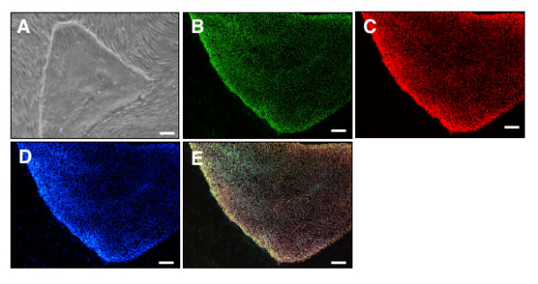
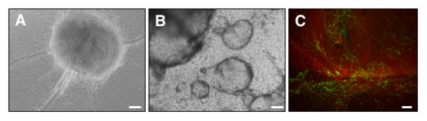
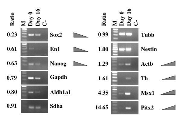
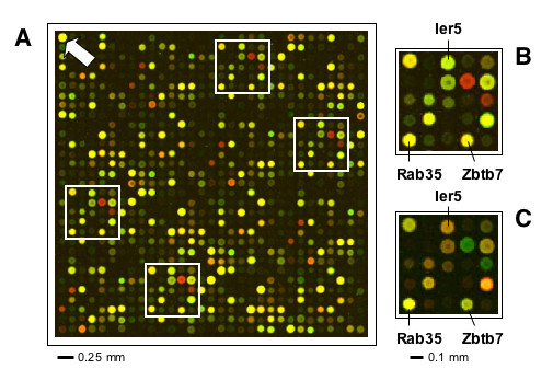
Results: We have designed a focused DNA (oligonucleotide-based) large-scale microarray platform (named "NeuroStem Chip") and used it to study gene expression patterns in hSCs as they differentiate into neurons. We have selected genes that are relevant to cells (i) being stem cells, (ii) becoming neurons, and (iii) being neurons. The NeuroStem Chip has over 1,300 pre-selected gene targets and multiple controls spotted in quadruplicates (approximately 46,000 spots total). In this study, we present the NeuroStem Chip in detail and describe the special advantages it offers to the fields of experimental neurology and stem cell biology. To illustrate the utility of NeuroStem Chip platform, we have characterized an undifferentiated population of pluripotent human embryonic stem cells (hESCs, cell line SA02). In addition, we have performed a comparative gene expression analysis of those cells versus a heterogeneous population of hESC-derived cells committed towards neuronal/dopaminergic differentiation pathway by co-culturing with PA6 stromal cells for 16 days and containing a few tyrosine hydroxylase-positive dopaminergic neurons.

Conclusion: We characterized the gene expression profiles of undifferentiated and dopaminergic lineage-committed hESC-derived cells using a highly focused custom microarray platform (NeuroStem Chip) that can become an important research tool in human stem cell biology. We propose that the areas of application for NeuroStem microarray platform could be the following: (i) characterization of the expression of established, pre-selected gene targets in hSC lines, including newly derived ones, (ii) longitudinal quality control for maintained hSC populations, (iii) following gene expression changes during differentiation under defined cell culture conditions, and (iv) confirming the success of differentiation into specific neuronal subtypes.


Figures

Figure 1
Figure 1
Human Embryonic Stem Cells (hESCs) used in a study. (A) Phase contrast image of unaltered hESC colony. Immunocytochemical analysis of (B) Ki67, (C) OCT3/4, (D)DAPI, (E) Merge. Scale bars = 100 μm.
Figure 2
Figure 2
Human Embryonic Stem Cell (hESCs)-derived cells committed toward neuronal/dopaminergic differentiation pathway by co-culturing with PA6 stromal cells for 16 days. (A, B) Phase contrast images of structures formed in hESC colonies. (C) Immunocytochemical analysis of cell composition: tyrosine hydroxylase (TH), green; human nuclei marker, red. Scale bars = 100 μm.
Figure 3
Figure 3
RT-PCR analysis of RNA samples used and validation of microarray results. Ratio, ratio of differentiated (Day 16)/undifferentiated (Day 0) hESC sample normalized spot intensity as detected by microarray analysis (average value from all spots). M, 100 bp DNA ladder; Day 0, undifferentiated hESCs; Day 16, hESCs committed to neuronal/dopaminergic differentiation pathway by co-culturing with PA6 stromal cells for 16 days; C-, No template control. Sox2, SRY-box 2; En1, Engrailed 1; Gapdh, glyceraldehydes-3-phosphate dehydrogenase; Aldh1a1, aldehyde dehydrogenase 1 family, member A1; Sdha, succinate dehydrogenase 2, flavoprotein sububit; Tubb, β-tubulin; Actb, β-actin; Th, tyrosine hydroxylase; Msx1, homolog of Drosophila muscle segment homolog 1; Pitx2, paired-like homeodomain transcription factor 2.
Figure 4
Figure 4
NeuroStem Chip layout (a fragment). (A) Representative block (31 × 31 spots), as hybridized with actual hESC (Cy3)/universal reference (Cy5) sample. Arrow indicates β-actin (Actb) spot, which serves as a control for grid orientation; white boxes (B) highlight repetitive patterns, illustrating quadriplication of individual spots. (B, C) Dye-swap (hESC (Cy5)/universal reference (Cy3)) indicates accuracy of fluorescent dye incorporation and hybridization chemistry. Ier5, Immediate early response 5; Rab35, RAS-associated protein RAB35; Zbtb7, Zink finger and BTB domain containing 7. Spot-to-spot center distance 140 μm; average spot size 90–110 μm.
Figure 5
Figure 5
Normalization and reproducibility of the NeuroStem Chip arrays. (A-B) Representative plots depict Log2(ratio) versus log10(intensities) prior to (A) and following (B) the normalization of one technical replicate (hESCs (Cy3) : universal reference (Cy5), 10:5 pmol). Green lines represent fitted values after normalization. (C) Centroid graph of a K-means classifier of 101 genes clustered as highly up-regulated in the 4 technical replicates, indicating high reproducibility. Pink line is an average of the Log2 ratio values of the 101 genes. See [Additional file 1] for a list of genes in this cluster.

References

    1. Ahn JI, Lee KH, Shin DM, Shim JW, Lee JS, Chang SY, Lee YS, Brownstein MJ, Lee SH, Lee YS. Comprehensive transcriptome analysis of differentiation of embryonic stem cells into midbrain and hindbrain neurons. Dev Biol. 2004;265:491–501. doi: 10.1016/j.ydbio.2003.09.041. - DOI - PubMed
    1. Grimm J, Mueller A, Hefti F, Rosenthal A. Molecular basis for catecholaminergic neuron diversity. Proc Natl Acad Sci USA. 2004;101:13891–13896. doi: 10.1073/pnas.0405340101. - DOI - PMC - PubMed
    1. Thuret S, Bhatt L, O'Leary DD, Simon HH. Identification and developmental analysis of genes expressed by dopaminergic neurons of the substantia nigra pars compacta. Mol Cell Neurosci. 2004;25:394–405. doi: 10.1016/j.mcn.2003.11.004. - DOI - PubMed
    1. Chung CY, Seo H, Sonntag KC, Brooks A, Lin L, Isacson O. Cell type-specific gene expression of midbrain dopaminergic neurons reveals molecules involved in their vulnerability and protection. Hum Mol Genet. 2005;14:1709–1725. doi: 10.1093/hmg/ddi178. - DOI - PMC - PubMed
    1. Greene JG, Dingledine R, Greenamyre JT. Gene expression profiling of rat midbrain dopamine neurons: implications for selective vulnerability in parkinsonism. Neurobiol Dis. 2005;18:19–31. doi: 10.1016/j.nbd.2004.10.003. - DOI - PubMed

Publication types

MeSH terms