Comparative Study
. 2007 Feb 8;8(1):10.
doi: 10.1186/1465-9921-8-10.

Upregulation of pirin expression by chronic cigarette smoking is associated with bronchial epithelial cell apoptosis

Affiliations
Comparative Study

Upregulation of pirin expression by chronic cigarette smoking is associated with bronchial epithelial cell apoptosis

Brian D Gelbman et al. Respir Res. .

Abstract

Background: Cigarette smoke disrupts the protective barrier established by the airway epithelium through direct damage to the epithelial cells, leading to cell death. Since the morphology of the airway epithelium of smokers does not typically demonstrate necrosis, the most likely mechanism for epithelial cell death in response to cigarette smoke is apoptosis. We hypothesized that cigarette smoke directly up-regulates expression of apoptotic genes, which could play a role in airway epithelial apoptosis.

Methods: Microarray analysis of airway epithelium obtained by bronchoscopy on matched cohorts of 13 phenotypically normal smokers and 9 non-smokers was used to identify specific genes modulated by smoking that were associated with apoptosis. Among the up-regulated apoptotic genes was pirin (3.1-fold, p < 0.002), an iron-binding nuclear protein and transcription cofactor. In vitro studies using human bronchial cells exposed to cigarette smoke extract (CSE) and an adenovirus vector encoding the pirin cDNA (AdPirin) were performed to test the direct effect of cigarette smoke on pirin expression and the effect of pirin expression on apoptosis.

Results: Quantitative TaqMan RT-PCR confirmed a 2-fold increase in pirin expression in the airway epithelium of smokers compared to non-smokers (p < 0.02). CSE applied to primary human bronchial epithelial cell cultures demonstrated that pirin mRNA levels increase in a time-and concentration-dependent manner (p < 0.03, all conditions compared to controls). Overexpression of pirin, using the vector AdPirin, in human bronchial epithelial cells was associated with an increase in the number of apoptotic cells assessed by both TUNEL assay (5-fold, p < 0.01) and ELISA for cytoplasmic nucleosomes (19.3-fold, p < 0.01) compared to control adenovirus vector.

Conclusion: These observations suggest that up-regulation of pirin may represent one mechanism by which cigarette smoke induces apoptosis in the airway epithelium, an observation that has implications for the pathogenesis of cigarette smoke-induced diseases.


Figures
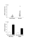
Figure 1
Figure 1
Mean RNA expression levels for pirin in smokers and non-smokers A. Microarray relative expression values (non-smokers n = 9, smokers n = 13, right and left lung samples for each individual). The relative expression level for pirin represents the normalized expression level in each array divided by the median expression level for pirin in all 44 microarrays. B. Comparison of microarray and TaqMan relative expression values (nonsmokers n = 3, smokers n = 9). Shown are the mean expression level of pirin ± standard error in airway epithelium of nonsmokers and smokers quantified by either microarray (p < 0.01) and TaqMan (p < 0.01).
Figure 2
Figure 2
Mean RNA expression levels for pirin assessed by quantitative PCR in human bronchial epithelial (HBE) cells exposed to varying concentrations of cigarette smoke extract (CSE) over time. RNA was extracted from HBE cells after exposure to 10% and 100% CSE and pirin RNA levels were quantified relative to the control gene GAPDH by real-time quantitative PCR by TaqMan. Each data point was generated from triplicate wells; and a total of three separate data points were obtained for each condition at each time point. The ordinate represents the fold change in pirin expression in samples exposed to 10% and 100% CSE over time relative to the background pirin expression level in cells cultured in the absence of CSE.
Figure 3
Figure 3
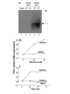
Mean RNA expression levels for pirin assessed by quantitative PCR in the human bronchial epithelial (BEAS-2B) cell line infected with AdPirin, AdNull, and negative control (naive) over varying concentrations (particle units, pu) and time. RNA was extracted from BEAS-2B cells that were infected with 103 and 104 particle units of AdPirin or AdNull. A. Northern analysis for Pirin RNA demonstrating the full length (1.1 kb) pirin mRNA directed by AdPirin 24 hr after infection. Lane 1 – control (naive); lane 2 – AdNull, 103 particle units; lane3 – AdNull, 104 particle units; lane 4 – AdPirin, 103 particle units; and lane 5 – AdPirin, 104 particle units. B. Concentration-dependent increase in pirin mRNA following infection with AdPirin. Pirin mRNA was quantified by TaqMan analysis. C. Time-dependent pirin mRNA expression over time evaluated by TaqMan.
Figure 4
Figure 4
Apoptosis in BEAS-2B bronchial epithelial cells following up-regulation of pirin levels by infection with AdPirin. BEAS-2B cells were infected with AdPirin, AdNull, or control (naive) and TdT-mediated dUTP nick end labeling (TUNEL) was used to assess apoptosis after 24 hr. A. control (naive); B. AdNull (104 particle units); and C. AdPirin (104 particle units). D. Quantitative assessment of apoptosis in BEAS-2B bronchial epithelial cells exposed to AdPirin, AdNull or control by TUNEL assay. The percentage of apoptotic cells per 10× field were manually counted in 10 fields per slide, with three replicates.
Figure 5
Figure 5
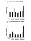
Evaluation of BEAS-2B bronchial epithelial cells exposed to varying concentrations of cigarette smoke extract, AdPirin and AdNull. A. Cytoplasmic nucleosome ELISA for apoptosis index. After cells were lysed and centrifuged to pellet nuclei, the supernatant was assessed by ELISA for the presence of cytoplasmic nucleosomes. The apoptotic index is the relative fluorescence value for given conditions, normalized to the naive control cells. Significant increases seen in apoptosis for cells exposed to 50% CSE, AdPirin (104 particle units) and AdPirin (103 particle units).B. Mean RNA expression levels for pirin assessed by quantitative TaqMan PCR in BEAS-2B cells exposed to varying concentrations of cigarette smoke extract (CSE), AdPirin and AdNull over time. The ordinate represents the fold change in pirin expression ± standard error in samples exposed to various conditions relative to the background pirin expression level in naive cells. Asterisk indicates a significant difference (p < 0.05) for conditions compared to naive control cells.

Similar articles

Cited by

References

    1. Mills PR, Davies RJ, Devalia JL. Airway epithelial cells, cytokines, and pollutants. Am J Respir Crit Care Med. 1999;160:S38–S43. - PubMed
    1. Maestrelli P, Saetta M, Mapp CE, Fabbri LM. Remodeling in response to infection and injury. Airway inflammation and hypersecretion of mucus in smoking subjects with chronic obstructive pulmonary disease. Am J Respir Crit Care Med. 2001;164:S76–S80. - PubMed
    1. Spurzem JR, Rennard SI. In: Asthma and COPD. Barns P, Drazen J, Rennard S, Thompson N, editor. Amsterdam; 2002. p. 145.
    1. Hogg JC. Pathophysiology of airflow limitation in chronic obstructive pulmonary disease. Lancet. 2004;364:709–721. doi: 10.1016/S0140-6736(04)16900-6. - DOI - PubMed
    1. van d V, Postma DS, Timens W, ten Hacken NH. Acute effects of cigarette smoke on inflammation and oxidative stress: a review. Thorax. 2004;59:713–721. doi: 10.1136/thx.2003.012468. - DOI - PMC - PubMed

Publication types

LinkOut - more resources