. 2007 Apr 15;304(2):556-66.
doi: 10.1016/j.ydbio.2007.01.010. Epub 2007 Jan 12.

Directed differentiation of embryonic stem cells into bladder tissue

Affiliations

Directed differentiation of embryonic stem cells into bladder tissue

Siam Oottamasathien et al. Dev Biol. .

Abstract

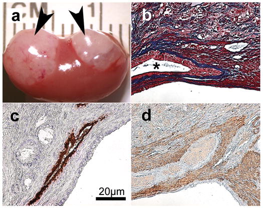
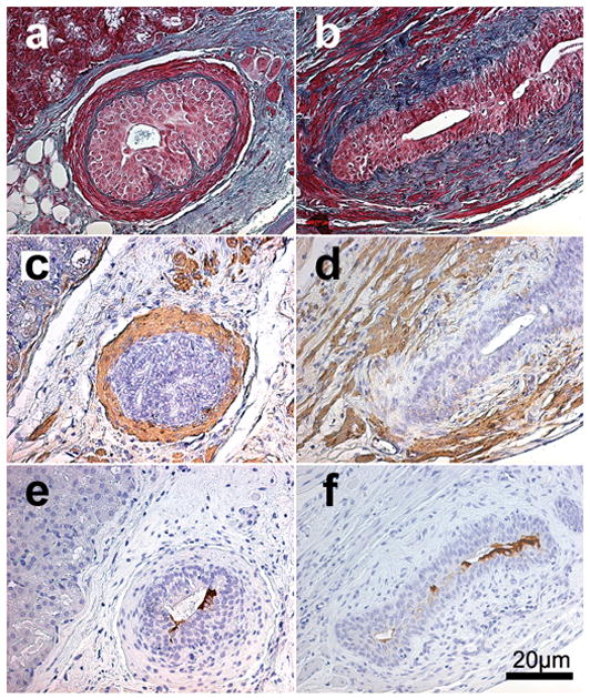
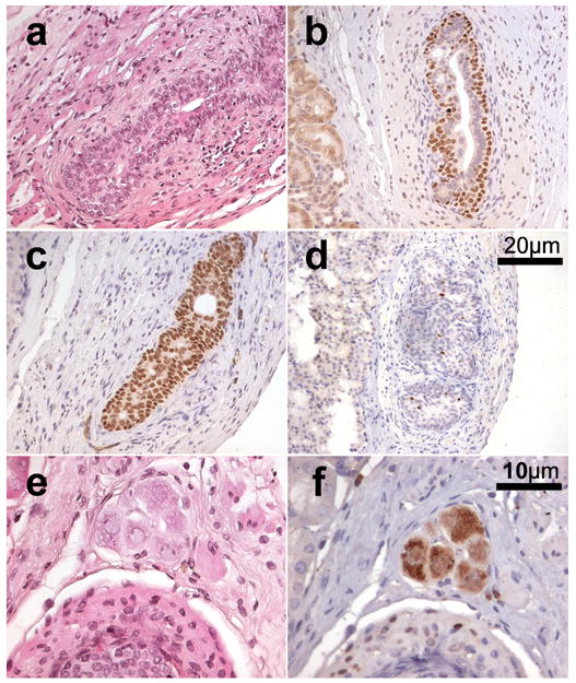
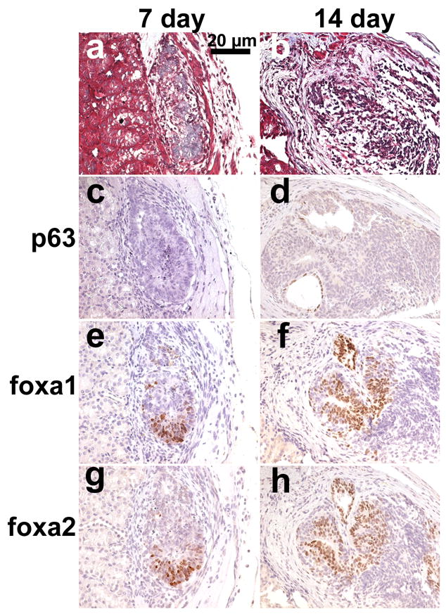
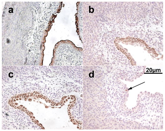
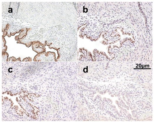
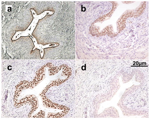
Manipulatable models of bladder development which interrogate specific pathways are badly needed. Such models will allow a systematic investigation of the multitude of pathologies which result from developmental defects of the urinary bladder. In the present communication, we describe a model in which mouse embryonic stem (ES) cells are directed to differentiate to form bladder tissue by specific interactions with fetal bladder mesenchyme. This model allows us to visualize the various stages in the differentiation of urothelium from ES cells, including the commitment to an endodermal cell lineage, with the temporal profile characterized by examining the induction of specific endodermal transcription factors (Foxa1 and Foxa2). In addition, final functional urothelial differentiation was characterized by examining uroplakin expression. It is well established that ES cells will spontaneously develop teratomas when grown within immunocompromised mouse hosts. We determined the specific mesenchymal to ES cell ratios necessary to dictate organ-specific differentiation while completely suppressing teratomatous growth. Embryonic mesenchyme is well established as an inductive tissue which dictates organ-specific programming of epithelial tissues. The present study demonstrates that embryonic bladder mesenchyme can also steer ES cells towards developing specific endodermal derived urothelium. These approaches allow us to capture specific stages of stem cell differentiation and to better define stem cell hierarchies.


Figures

Figure 1
Figure 1
Xenografts harvested at 42 days. a. Gross appearance of 1000 embryonic stem (ES) cell grafts on host kidney (arrowheads denote whitish-tan appearing grafts). b–d. Histology and immunohistochemical staining of 1000 ES cell + 1 embryonic bladder mesenchyme shell recombinant graft. b. Gomori’s trichrome, star denotes central lumen of bladder structure, urothelial cells surrounding central lumen (red) with sub-urothelial connective tissue (blue) were seen. c. Immunohistochemical detection of broad-spectrum uroplakin (brown) denoting mature urothelium. d. Immunohistochemical detection of smooth muscle α-actin (brown) denoting smooth muscle fibrils.
Figure 2
Figure 2
Xenografts of 1500 embryonic stem cells + 4 embryonic bladder mesenchymal shells/graft harvested at 42 days. a&b. Gomori’s trichrome, multilayered cuboidal urothelial cells (red) with sub-urothelial connective tissue (blue) were seen. c&d. Immunohistochemical detection of smooth muscle α-actin (brown) denoting smooth muscle fibrils. e&f. Immunohistochemical detection of broad-spectrum uroplakin (brown) denoting mature urothelium.
Figure 3
Figure 3
Xenografts of 1500 embryonic stem (ES) cells + 4 embryonic bladder mesenchymal shells/graft harvested at 42 days. a. H&E staining. b. Immunohistochemical detection of p63 (brown) illustrating complete spatial orientation of ES cell derived urothelium. c. Immunohistochemical detection of Foxa1 (brown) seen in all ES cell derived urothelium. d. Immunohistochemical detection of Foxa2 (brown) seen strongly positive in only a few urothelial cells. e&f. H&E and immunohistochemical detection of neuronal nuclear antigen A60 (NeuN) (brown) confirming the presence of neuronal cells clustering in the form of a ganglion. Scale bars of 20 μm apply to images a–d. Scale bars of 10 μm apply to images e&f.
Figure 4
Figure 4
Xenografts of 1500 embryonic stem cells + 4 embryonic bladder mesenchymal shells/graft harvested at 7 (a,c,e,g) and 14 (b,d,f,h) days. a&b. Gomori’s trichrome, cellular organization occurring in cells that look destined to become urothelium (blue). c&d. Immunohistochemical detection of p63 (brown) illustrating lack of expression within 7 day tissues and early expression observed in 14 day tissues. e&f. Immunohistochemical detection of Foxa1 (brown) denoting endodermal cells. g&h. Immunohistochemical detection of Foxa2 (brown) denoting endodermal cells.
Figure 5
Figure 5
Native embryonic day 16 mouse bladder. a. Immunohistochemical detection of broad-spectrum uroplakin (brown) denoting mature urothelium. b. Immunohistochemical detection of p63 (brown) demonstrating urothelial basilar orientation. c. Immunohistochemical detection of Foxa1 (brown) seen in all urothelial cells denoting endodermal derivation of the cells. d. Immunohistochemical detection of Foxa2 (brown), arrow denotes one positive staining cell within the section.
Figure 6
Figure 6
Native postnatal day 1 of life mouse bladder. a. Immunohistochemical detection of broad-spectrum uroplakin (brown) denoting mature urothelium. b. Immunohistochemical detection of p63 (brown) demonstrating urothelial basilar orientation. c. Immunohistochemical detection of Foxa1 (brown) seen in all urothelial cells denoting endodermal derivation of the cells. d. Complete lack of immunohistochemical detection of Foxa2.
Figure 7
Figure 7
Native adult mouse bladder. a. Immunohistochemical detection of broad-spectrum uroplakin (brown) denoting mature urothelium. b. Immunohistochemical detection of p63 (brown) demonstrating urothelial basilar orientation. c. Immunohistochemical detection of Foxa1 (brown) seen in all urothelial cells denoting endodermal derivation of the cells. d. Complete lack of immunohistochemical detection of Foxa2.

References

    1. Aboseif S, El-Sakka A, Young P, Cunha G. Mesenchymal reprogramming of adult human epithelial differentiation. Differentiation. 1999;65:113–8. - PubMed
    1. Alison MR, Poulsom R, Forbes S, Wright NA. An introduction to stem cells. J Pathol. 2002;197:419–23. - PubMed
    1. Baskin LS, Hayward SW, Young P, Cunha GR. Role of mesenchymal-epithelial interactions in normal bladder development. J Urol. 1996a;156:1820–7. - PubMed
    1. Baskin LS, Hayward SW, Young PF, Cunha GR. Ontogeny of the rat bladder: smooth muscle and epithelial differentiation. Acta Anat (Basel) 1996b;155:163–71. - PubMed
    1. Besnard V, Wert SE, Hull WM, Whitsett JA. Immunohistochemical localization of Foxa1 and Foxa2 in mouse embryos and adult tissues. Gene Expr Patterns. 2004;5:193–208. - PubMed

Publication types

Substances

LinkOut - more resources