. 2007 Mar 6;581(5):858-64.
doi: 10.1016/j.febslet.2007.01.058. Epub 2007 Feb 2.

The Krüppel-like zinc finger protein Glis2 functions as a negative modulator of the Wnt/beta-catenin signaling pathway

Affiliations

The Krüppel-like zinc finger protein Glis2 functions as a negative modulator of the Wnt/beta-catenin signaling pathway

Yong-Sik Kim et al. FEBS Lett. .

Abstract

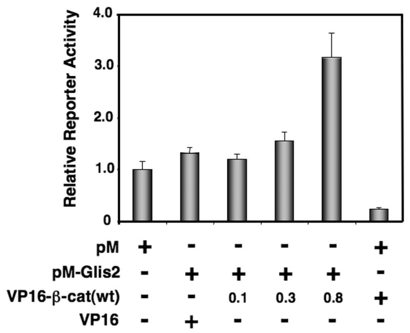
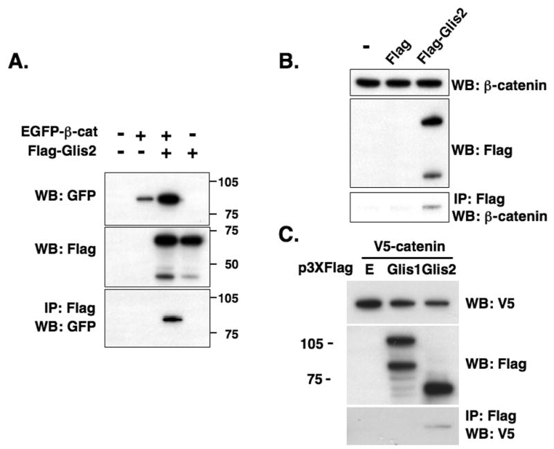
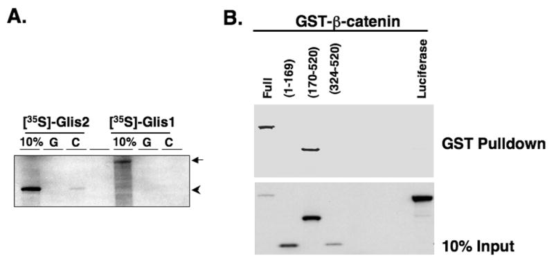
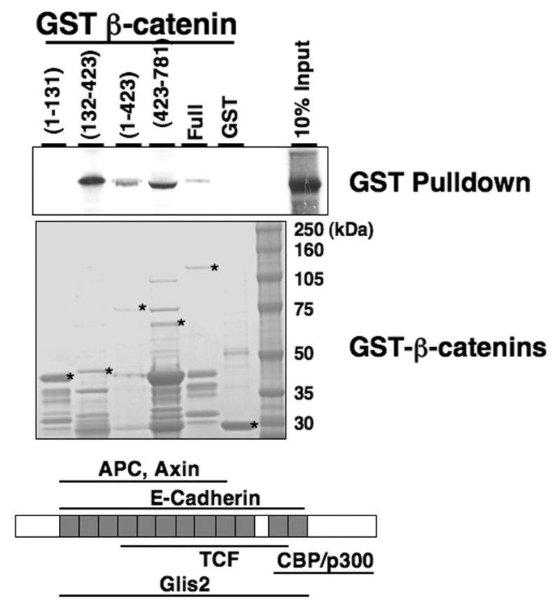
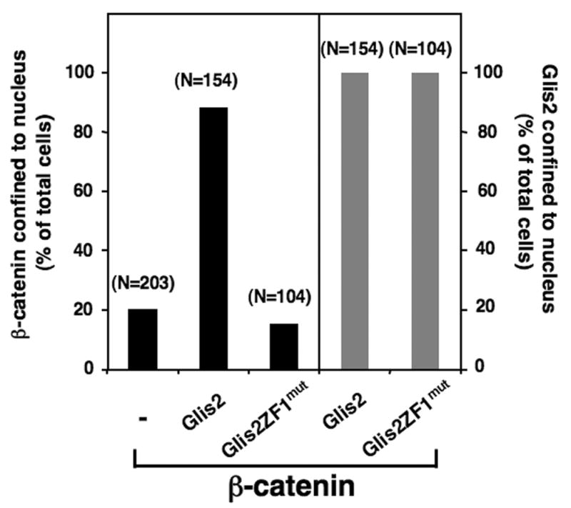
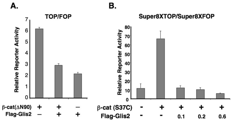
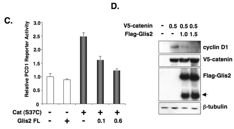
To gain insight into the mechanism by which Gli-similar 2 (Glis2) regulates transcription, we performed yeast-two hybrid cDNA library screening using Glis2 as bait. This screening identified beta-catenin as a potential Glis2-interacting protein. Mammalian two-hybrid, co-immunoprecipitation, and GST-pulldown analyses supported the interaction between Glis2 and beta-catenin. Pulldown analyses with several Glis2 deletion mutants indicated that the 1st zinc finger motif of Glis2 is critical for its interaction with beta-catenin, while the armadillo repeats of beta-catenin are important in its interaction with Glis2. Reporter analyses showed that Glis2 represses T-cell factor (TCF)-mediated transcriptional activation. In addition, Glis2 represses the expression of the TCF target gene cyclin D1. Our results indicate that Glis2 interacts with beta-catenin and suggest that Glis2 functions as a negative modulator of beta-catenin/TCF-mediated transcription.


Figures

Fig. 1
Fig. 1
(A) Identification of β-catenin as a Glis2-interacting protein by mammalian two-hybrid analysis. HCT116 cells were co-transfected with (UAS)5-LUC, pRL-SV40, pM, pVP16, pM-Glis2, and increasing amounts of pVP16-β-catenin as indicated. After 48 h, cells were harvested and assayed for reporter activities. The relative LUC activity was calculated and plotted.
Fig. 2
Fig. 2
Glis2 and β-catenin are associated with the same protein complex. (A) HCT116 cells were transfected with p3xFlag-CMV-Glis2, p3xFlag-CMV, and pEGFP-wt-β-catenin as indicated. Forty-eight h after transfection, protein lysates were prepared and one part was used in Western blot (WB) analysis with an anti-GFP (upper panel) or anti-Flag M2 antibody (middle panel). The remaining was used for immunoprecipitation (IP) using anti-Flag M2 affinity resin. Bound proteins were then examined by Western blot analysis with anti-GFP antibody (lower panel). (B) Glis2 interacts with endogenous β-catenin. HCT116 cells were transfected with p3xFlag-CMV-Glis2 or p3xFlag-CMV. Forty-eight h after transfection, cell lysates were prepared and Flag-Glis2 protein complexes immunoprecipitated with anti-Flag affinity resin. Bound proteins were examined by Western blot analysis with anti-β-catenin or anti-Flag antibodies. (C) The interaction of β-catenin is specific for Glis2. HCT116 cells were transfected with V5-β-catenin, p3xFlag-CMV, p3xFlag-CMV-Glis1, or p3xFlag-CMV-Glis2. Proteins in the cellular lysates and immunoprecipitated proteins were examined by Western blot analysis using anti-Flag M2 or anti-V5 antibodies.
Fig. 3
Fig. 3
Glis2 interacts directly with β-catenin. (A) Interaction of GST and GST-β-catenin fusion proteins with [35S]-methionine-labeled Glis2 or Glis1 was examined as described in Materials and Methods. First lane, 10% input; G, GST; C; GST-β-catenin. Arrow indicated Glis1 and arrow head indicated Glis2. (B) Glis2 interacts with β-catenin through its ZFD. GST-pulldown analysis with [35S]-methionine-labeled Glis2, Glis2(1–169), Glis2(170–520), Glis2(324–520) or luciferase which served as negative control. Bound radiolabeled proteins were separated by PAGE and visualized by autoradiography. (C, D) Co-immunoprecipitation assay using Glis2 mutants. HCT116 cells were transfected with V5-β-catenin, p3xFlag-CMV empty vector, p3xFlag-CMV-Glis2, p3xFlag-CMV-Glis2(1–382), p3xFlag-CMV-Glis2(1–200), p3xFlag-CMV-Glis2(1–143), p3xFlag-CMV-Glis2(170–377), or p3xFlag-CMV-Glis2ZF1mut containing the C175A mutation in ZF1 (ZF1mut). Proteins in the cellular extracts and immunoprecipitated proteins were examined by Western blot analysis using anti-Flag M2 or anti-V5 antibodies.
Fig. 3
Fig. 3
Glis2 interacts directly with β-catenin. (A) Interaction of GST and GST-β-catenin fusion proteins with [35S]-methionine-labeled Glis2 or Glis1 was examined as described in Materials and Methods. First lane, 10% input; G, GST; C; GST-β-catenin. Arrow indicated Glis1 and arrow head indicated Glis2. (B) Glis2 interacts with β-catenin through its ZFD. GST-pulldown analysis with [35S]-methionine-labeled Glis2, Glis2(1–169), Glis2(170–520), Glis2(324–520) or luciferase which served as negative control. Bound radiolabeled proteins were separated by PAGE and visualized by autoradiography. (C, D) Co-immunoprecipitation assay using Glis2 mutants. HCT116 cells were transfected with V5-β-catenin, p3xFlag-CMV empty vector, p3xFlag-CMV-Glis2, p3xFlag-CMV-Glis2(1–382), p3xFlag-CMV-Glis2(1–200), p3xFlag-CMV-Glis2(1–143), p3xFlag-CMV-Glis2(170–377), or p3xFlag-CMV-Glis2ZF1mut containing the C175A mutation in ZF1 (ZF1mut). Proteins in the cellular extracts and immunoprecipitated proteins were examined by Western blot analysis using anti-Flag M2 or anti-V5 antibodies.
Fig. 4
Fig. 4
β-catenin interacts with Glis2 through its specific regions. GST and several GST-β-catenin fusion proteins were bound to glutathione-Sepharose 4B beads and then incubated with [35S]-methionine-labeled Glis2. Bound radiolabeled proteins were visualized by autoradiography. The input of the various GST-β-catenin proteins is shown in the lower panel. Asterisks indicate the respective GST-β-catenin proteins.
Fig. 5
Fig. 5
Glis2 enhances nuclear localization of β-catenin. NEH293 cells were transiently transfected with p3xFlag-CMV-Glis2, p3xFlag-CMV-Glis2ZF1mut, and/or pEGFP-β-catenin. The percentage of cells in which the localization of Glis2 or β-catenin was restricted to the nucleus, was calculated and plotted. The number of cells counted is indicated above each bar.
Fig. 6
Fig. 6
Glis2 represses TCF-mediated transcriptional activity and the induction of cyclin D1 by β-catenin. (A) HCT116 cells were cotransfected with TOPFlash and FOPFlash reporters, pCMVbeta, β-catenin(ΔN90), and p3xFlag-CMV-Glis2 as indicated. Forty-eight h after transfection, cells were assayed for reporter activities as described in Materials and Methods. (B) HCT116 cells were cotransfected with Super8XTOPFlash and Super8XFOPFlash reporters, pRL-SV40, pCMV-V5-β-catenin (β-cat(S37C)) and different concentrations of p3xFlag-CMV-Glis2 expression vector as indicated and then processed as described under A. (C) HCT116 cells were cotransfected with the cyclin D1 reporter PCD1, pRL-SV40, the constitutively-active pCMV-V5-β-catenin (S37C) expression vector, and p3xFlag-CMV-Glis2 as indicated and then processed as described under A. (D) HCT116 cells were transfected with pCMV-V5-β-catenin and p3xFlag-CMV-Glis2 (1.0 and 1.5 μg) as indicated. Thirty-six h later, cellular proteins were analyzed by Western blot analysis using antibodies against V5, Flag, β-tubulin, and cyclin D1. The lower molecular weight band (arrow head) in the Flag-Glis2 Western blot represents a proteolytically processed form of Glis2.
Fig. 6
Fig. 6
Glis2 represses TCF-mediated transcriptional activity and the induction of cyclin D1 by β-catenin. (A) HCT116 cells were cotransfected with TOPFlash and FOPFlash reporters, pCMVbeta, β-catenin(ΔN90), and p3xFlag-CMV-Glis2 as indicated. Forty-eight h after transfection, cells were assayed for reporter activities as described in Materials and Methods. (B) HCT116 cells were cotransfected with Super8XTOPFlash and Super8XFOPFlash reporters, pRL-SV40, pCMV-V5-β-catenin (β-cat(S37C)) and different concentrations of p3xFlag-CMV-Glis2 expression vector as indicated and then processed as described under A. (C) HCT116 cells were cotransfected with the cyclin D1 reporter PCD1, pRL-SV40, the constitutively-active pCMV-V5-β-catenin (S37C) expression vector, and p3xFlag-CMV-Glis2 as indicated and then processed as described under A. (D) HCT116 cells were transfected with pCMV-V5-β-catenin and p3xFlag-CMV-Glis2 (1.0 and 1.5 μg) as indicated. Thirty-six h later, cellular proteins were analyzed by Western blot analysis using antibodies against V5, Flag, β-tubulin, and cyclin D1. The lower molecular weight band (arrow head) in the Flag-Glis2 Western blot represents a proteolytically processed form of Glis2.

References

    1. Kim YS, Lewandoski M, Perantoni AO, Kurebayashi S, Nakanishi G, Jetten AM. Identification of Glis1, a novel Gli-related, Kruppel-like zinc finger protein containing transactivation and repressor functions. J. Biol. Chem. 2002;277:30901–30913. - PubMed
    1. Kim YS, Nakanishi G, Lewandoski M, Jetten AM. GLIS3, a novel member of the GLIS subfamily of Kruppel-like zinc finger proteins with repressor and activation functions. Nucleic Acids Res. 2003;31:5513–5525. - PMC - PubMed
    1. Lamar E, Kintner C, Goulding M. Identification of NKL, a novel Gli-Kruppel zinc-finger protein that promotes neuronal differentiation. Development. 2001;128:1335–1346. - PubMed
    1. Zhang F, Nakanishi G, Kurebayashi S, Yoshino K, Perantoni A, Kim YS, Jetten AM. Characterization of Glis2, a novel gene encoding a Gli-related, Kruppel-like transcription factor with transactivation and repressor functions. Roles in kidney development and neurogenesis. J. Biol. Chem. 2002;277:10139–10149. - PubMed
    1. Kasper M, Regl G, Frischauf AM, Aberger F. GLI transcription factors: mediators of oncogenic Hedgehog signalling. Eur J Cancer. 2006;42:437–445. - PubMed

Publication types

MeSH terms