. 2007 Apr;89(4):521-31.
doi: 10.1016/j.ygeno.2006.12.004. Epub 2007 Feb 6.

Obscurin-like 1, OBSL1, is a novel cytoskeletal protein related to obscurin

Affiliations

Obscurin-like 1, OBSL1, is a novel cytoskeletal protein related to obscurin

Sarah B Geisler et al. Genomics. 2007 Apr.

Abstract

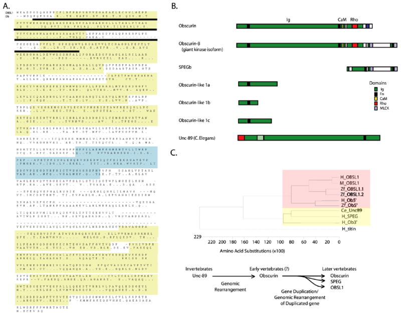
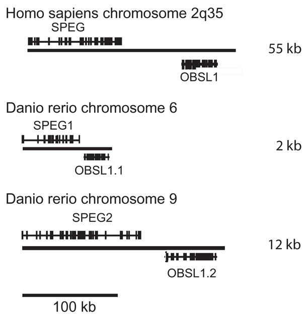
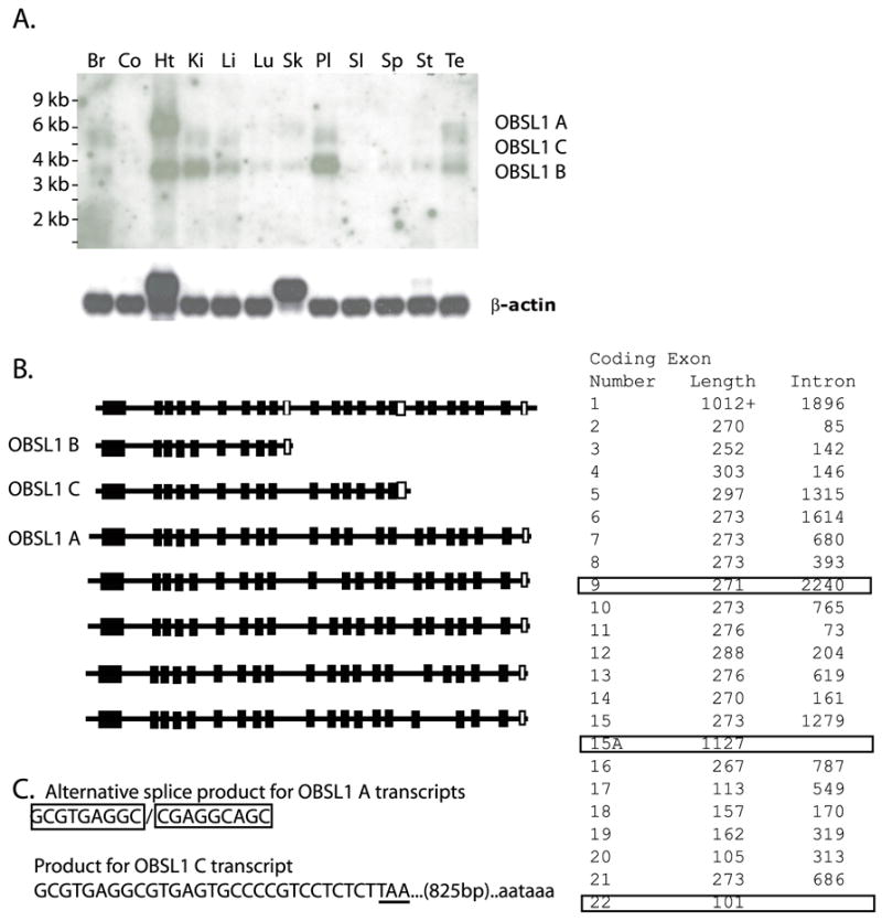
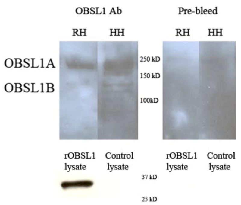
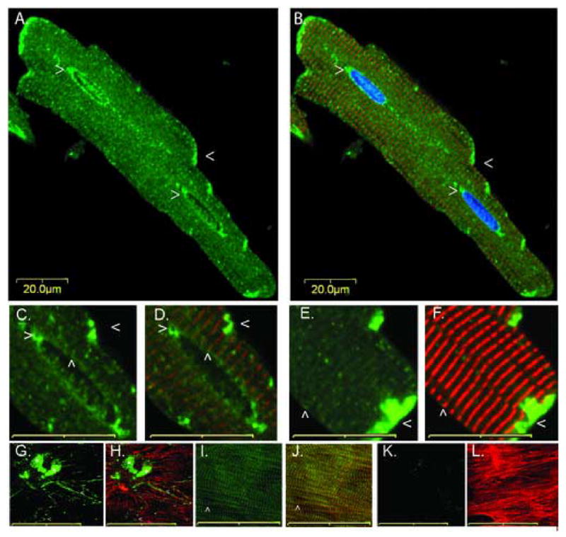
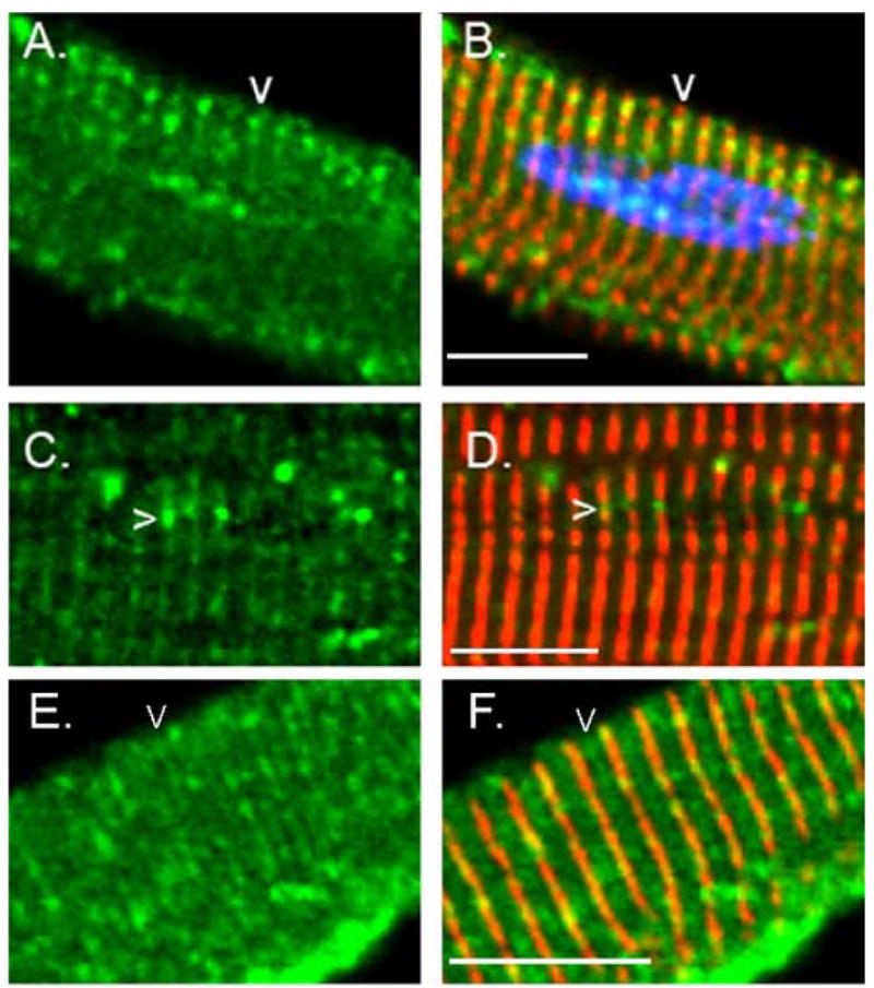
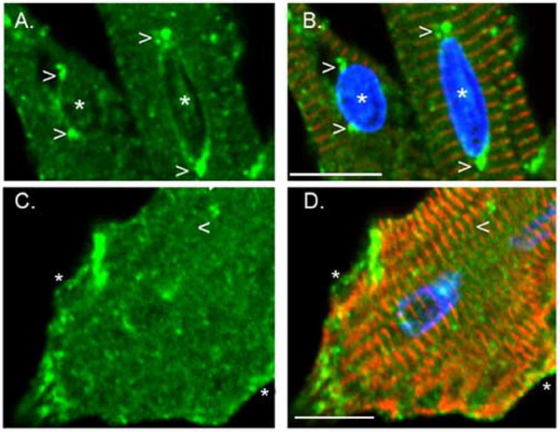
Cytoskeletal adaptor proteins serve vital functions in linking the internal cytoskeleton of cells to the cell membrane, particularly at sites of cell-cell and cell-matrix interactions. The importance of these adaptors to the structural integrity of the cell is evident from the number of clinical disease states attributable to defects in these networks. In the heart, defects in the cytoskeletal support system that surrounds and supports the myofibril result in dilated cardiomyopathy and congestive heart failure. In this study, we report the cloning and characterization of a novel cytoskeletal adaptor, obscurin-like 1 (OBSL1), which is closely related to obscurin, a giant structural protein required for sarcomere assembly. Multiple isoforms arise from alternative splicing, ranging in predicted molecular mass from 130 to 230 kDa. OBSL1 is located on human chromosome 2q35 within 100 kb of SPEG, another gene related to obscurin. It is expressed in a broad range of tissues and localizes to the intercalated discs, to the perinuclear region, and overlying the Z lines and M bands of adult rat cardiac myocytes. Further characterization of this novel cytoskeletal linker will have important implications for understanding the physical interactions that stabilize and support cell-matrix, cell-cell, and intracellular cytoskeletal connections.


Figures

Fig. 1
Fig. 1
The Unc-89/obscurin family of genes. (A) Sequence alignment of the human OBSL1 B isoform (top row) and the amino terminal end of obscurin (amino terminal, bottom row) demonstrating the arrangement of their immuoglobulin- (Ig) and fibronectin-like (Fn3) domains. Ig domains are indicated in yellow and the Fn3 domain in blue. Amino acids that are common to both sequences are represented by dots (.), while gaps in the alignment are represented by dashes (-). Alignment demonstrates 31% amino acid identity and 46% similarity between OBSL1 and the corresponding region of obscurin. The sequence used to generate the antibody is underlined. (B) Schematic representation of the Unc-89/obscurin gene family. Represented motifs include the Ig (green), Fn3 (black), CaM-binding (yellow), RhoGEF (blue) and MLCK-like (red) domains. Obscurin isoforms A and B have been previously described [9,24]. (C) Phylogenetic tree produced by Clustal W alignment of the Fn3 domains from the human (H), mouse (M), and zebrafish (Zf) OBSL1, obscurin [amino (Ob5) and carboxy (Ob3) terminal Fn3 domains], C. elegans Unc-89 (Ce_Unc89), SPEG, and titin genes. Zebrafish has two OBSL1 orthologues, 1.1 and 1.2 (see text for details). Note that the OBSL1 Fn3 domain is most closely related to the corresponding amino terminal domain of obscurin (boxed in red). The obscurin carboxy terminal, Unc-89, and SPEG Fn3 domains are highly homologous to one another (boxed in yellow) but more distantly related to the OBSL1 Fn3 domain. Below the tree is the proposed evolutionary relationship of the Unc-89/obscurin gene family.
Fig. 2
Fig. 2
OBSL1 and SPEG genes are physically linked in both the zebrafish and human genomes. Transcripts corresponding to the zebrafish (Danio rerio) and human OBSL1 and SPEG genes were identified by BLAST sequence homology search. Mapping information was derived from the Ensembl database (www.ensembl.org) and has not been independently verified. The approximate distances between the polyadenylation signals for the SPEG and OBSL1 genes are noted at the right side of the figure. Note that in both the zebrafish and human genomes, the SPEG and OBSL1 genes are closely physically linked but transcribed in opposite directions. The scale bar represents 100 kb of sequence and estimated distances are approximate. For clarity, transcripts other than those of OBSL1 and SPEG have not been included in the figure.
Fig. 3
Fig. 3
OBSL1 mRNA expression. (A) A northern blot of adult human tissue mRNA was performed using a cDNA probe from the 5′ end of the OBSL1 that detects the OBSL1 A, B, and C transcripts. The OBSL1 A transcript (5.9 kb) is expressed predominantly in the heart (Ht) with some expression in skeletal muscle (Sk) and the brain (Br) Multiple splice variants are expressed in all the tissues examined including the placenta (Pl) and testes (Te). OBSL1 B (3.3 kb) is expressed in multiple tissues, including all of the tissues mentioned above as well as kidney (Ki), liver (Li), lung (Lu), small intestine (SI), stomach (St), and spleen (Sp). Co lane is colon tissue. The blot was hybridized with a β-actin probe to assess for differences in RNA loading. (B) Schematic representation of some of the splicing variations involving the OBSL1 transcripts that have been detected by EST sequencing and cDNA amplification. Coding exons are represented by filled boxes and 3′ untranslated regions by empty boxes. Based on the transcript sizes from northern analysis, additional splice variants are suspected. (C) Alternative splice site at the end of coding exon 15 (5′ non-coding exons have not yet been characterized). A conserved splice donor site within this exon can be utilized to generate longer transcripts corresponding to OBSL1 A (top sequence). If this splice donor is not utilized then a conserved translation stop codon (TAA) is encountered followed by a conserved polyadenlyation (AATAAA) signal (bottom sequence). (Sequence elements were conserved in human, dog, chimpanzee, rat and mouse genomes as per the alignment provided by the UCSC Genome Bioinformatics server at the University of California, Santa Cruz). To the right of panels B and C is a table summarizing the sizes of each exon and the length of intron sequence between that exon and the following one. The three exons containing translation stop codons are highlighted with boxes.
Fig. 4
Fig. 4
Western analysis of OBSL1. Adult rat and human heart lysates were probed with an antibody recognizing the Ig-Fn3 domains of OBSL1. A prominent band was noted at 230 kD, the expected size for the OBSL1 A isoform, and a band, that was far more prominent in the human heart than the rat heart lysate, was noted at 130 kD, the predicted size of the OBSL1 B isoform. In the human heart lysate, additional bands were noted between 170 and 200 kD and one at 120 kD. The 170–200 kD bands likely correspond to the OBSL1 C isoform or splice variants of OBSL1 A while the smaller band may represent either another isoform or a breakdown product of one of the larger isoforms. A bacterial lysate expressing a portion of OBSL1 common to all known isoforms was used as a positive control (rOBSL1) and a bacterial lysate using the same expression vector was included as a negative control (Control Lysate). On the right panel, the prebleed control serum was used on the same samples under identical reaction conditions.
Fig. 5
Fig. 5
Cellular localization of OBSL1 in remodeling adult cardiac myocytes. Freshly isolated adult rat cardiac myocytes were immunostained for OBSL1 (A–H: green), and α-actinin (B,D,F,H,J,L: red). Nuclei were counterstained with DAPI (B: blue). OBSL1 prominently localizes to intercalated discs (A–F: <) and the perinuclear region (A–D: >). There is also localization overlying the Z bands (C–F: ^) as noted by colocalization of OBSL1 and α-actinin. (C,D) 2x magnification of a nucleus in Fig. 5A,B showing a dotted pattern of circumferential perinuclear OBSL1 localization overlying the Z bands (C,D: ^) and accumulation at the polar ends (C,D: >) of the elongated nucleus. (G–L) Adult rat cardiac myocytes at 14 days post-plating. Note that later in the remodeling process that OBSL1 localizes to new cell-cell (G,H: <) and cell-matrix (G,H: *) contacts but has not yet regained a striated pattern overlying the Z lines and M bands. This is in marked contrast to obscurin, which at this timepoint displays a prominent striated pattern (I,J: green, ^) suggesting that there is no cross-reactivity of the OBSL1 antibody with obscurin. Also, note that there is no immunostaining with the pre-bleed control serum ((K,L: green) under identical reaction conditions. Scale bars are 20 μM (A–F) and 50 μM (G–L).
Fig. 6
Fig. 6
Accumulation of OBSL1 overlying the Z line and M band. Freshly isolated adult rat cardiac myocytes were immunostained for OBSL1 (green) and α-actinin (red) and nuclei were counterstained with DAPI (B: blue). Note that OBSL1 is distributed along the Z band with accumulation between (A–D: arrowheads) adjacent myofibrils. Adjacent but not fused myofibrils are defined by a discontinuous or interrupted pattern of α-actinin immunostaining. Some immunolocalization of OBSL1 was also noted overlying the M band (E–F arrowheads) but this was usually far less prominent than was noted over the Z line (see A–D for comparison). Scale bar is 20μm.
Fig. 7
Fig. 7
Redistribution of OBSL1 during early cardiac myocyte remodeling. Primary adult rat cardiac myocytes were allowed to remodel in culture for 3 days in the presence of serum. During this time, the adult cardiac myocytes attach to the substrate, begin to disassemble their contractile structures and assume a more rounded appearance forming new focal contacts with the substrate. After 3 days in culture, the cells were fixed and immunostained for OBSL1 (green) and α-actinin (red). Nuclei were counterstained with DAPI (blue). (A, B) Nuclei (*) from adjacent cells demonstrating polar (cell on the right) or eccentric (cell on the left) accumulations of OBSL1. Polar accumulations were noted to be much more common in freshly isolated, elongated cells with elongated nuclei. As the cells and their nuclei remodeled to be more spherical in shape, the perinuclear distribution was more diffuse, usually without significant areas of accumulation. (C, D) In this remodeling cardiac myocyte, note that the cellular localization of OBSL1 has become more granular or reticular (>) despite persistence of the Z band architecture as noted by the striated pattern of α-actinin staining (<). Also, note that the perinuclear distribution of OBSL1 remains intact and that OBSL1 has begun to distribute to new focal contacts that have developed between the cell and the substrate (*). Scale bar is 20 μm.

References

    1. Kostin S, Hein S, Arnon E, Scholz D, Schaper J. The cytoskeleton and related proteins in the human failing heart. Heart Fail Rev. 2000;5:271–80. - PubMed
    1. Maeda M, Holder E, Lowes B, Valent S, Bies RD. Dilated cardiomyopathy associated with deficiency of the cytoskeletal protein metavinculin. Circulation. 1997;95:17–20. - PubMed
    1. Sepp R, Severs NJ, Gourdie RG. Altered patterns of cardiac intercellular junction distribution in hypertrophic cardiomyopathy. Heart. 1996;76:412–7. - PMC - PubMed
    1. Su X, Sekiguchi M, Endo M. An ultrastructural study of cardiac myocytes in postmyocardial infarction ventricular aneurysm representative of chronic ischemic myocardium using semiquantitative and quantitative assessment. Cardiovasc Pathol. 2000;9:1–8. - PubMed
    1. Rohr S. Role of gap junctions in the propagation of the cardiac action potential. Cardiovasc Res. 2004;62:309–22. - PubMed

Publication types