Validity of large-deformation high dimensional brain mapping of the basal ganglia in adults with Tourette syndrome
- PMID: 17289354
- PMCID: PMC2859464
- DOI: 10.1016/j.pscychresns.2006.08.006
Validity of large-deformation high dimensional brain mapping of the basal ganglia in adults with Tourette syndrome
Abstract
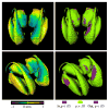
The basal ganglia and thalamus may play a critical role for behavioral inhibition mediated by prefrontal, parietal, temporal, and cingulate cortices. The cortico-basal ganglia-thalamo-cortical loop with projections from frontal cortex to striatum, then to globus pallidus or to substantia nigra pars reticulata, to thalamus and back to cortex, provides the anatomical substrate for this function. In-vivo neuroimaging studies have reported reduced volumes in the thalamus and basal ganglia in individuals with Tourette Syndrome (TS) when compared with healthy controls. However, patterns of neuroanatomical shape that may be associated with these volume differences have not yet been consistently characterized. Tools are being developed at a rapid pace within the emerging field of computational anatomy that allow for the precise analysis of neuroanatomical shape derived from magnetic resonance (MR) images, and give us the ability to characterize subtle abnormalities of brain structures that were previously undetectable. In this study, T1-weighted MR scans were collected in 15 neuroleptic-naïve adults with TS or chronic motor tics and 15 healthy, tic-free adult subjects matched for age, gender and handedness. We demonstrated the validity and reliability of large-deformation high dimensional brain mapping (HDBM-LD) as a tool to characterize the basal ganglia (caudate, globus pallidus and putamen) and thalamus. We found no significant volume or shape differences in any of the structures in this small sample of subjects.
Figures


Similar articles
-
Altered structural connectivity of cortico-striato-pallido-thalamic networks in Gilles de la Tourette syndrome.Brain. 2015 Feb;138(Pt 2):472-82. doi: 10.1093/brain/awu311. Epub 2014 Nov 11. Brain. 2015. PMID: 25392196 Free PMC article.
-
Basal ganglia shape abnormalities in the unaffected siblings of schizophrenia patients.Biol Psychiatry. 2008 Jul 15;64(2):111-20. doi: 10.1016/j.biopsych.2008.01.004. Epub 2008 Mar 4. Biol Psychiatry. 2008. PMID: 18295189 Free PMC article.
-
Structural analysis of the basal ganglia in schizophrenia.Schizophr Res. 2007 Jan;89(1-3):59-71. doi: 10.1016/j.schres.2006.08.031. Epub 2006 Oct 30. Schizophr Res. 2007. PMID: 17071057 Free PMC article.
-
Neuroimaging assessment of basal ganglia volumes in Tourette Syndrome: a systematic review and meta-analysis.Cogn Neuropsychiatry. 2024 Jul-Sep;29(4-5):256-267. doi: 10.1080/13546805.2024.2439800. Epub 2024 Dec 13. Cogn Neuropsychiatry. 2024. PMID: 39671252
-
Putting the Pieces Together in Gilles de la Tourette Syndrome: Exploring the Link Between Clinical Observations and the Biological Basis of Dysfunction.Brain Topogr. 2017 Jan;30(1):3-29. doi: 10.1007/s10548-016-0525-z. Epub 2016 Oct 25. Brain Topogr. 2017. PMID: 27783238 Free PMC article. Review.
Cited by
-
Levodopa effects on [ (11)C]raclopride binding in the resting human brain.F1000Res. 2015 Jan 23;4:23. doi: 10.12688/f1000research.5672.1. eCollection 2015. F1000Res. 2015. PMID: 26180632 Free PMC article.
-
Northwestern University schizophrenia data sharing for SchizConnect: A longitudinal dataset for large-scale integration.Neuroimage. 2016 Jan 1;124(Pt B):1196-1201. doi: 10.1016/j.neuroimage.2015.06.030. Epub 2015 Jun 16. Neuroimage. 2016. PMID: 26087378 Free PMC article.
-
The New Tics study: A Novel Approach to Pathophysiology and Cause of Tic Disorders.J Psychiatr Brain Sci. 2020;5:e200012. doi: 10.20900/jpbs.20200012. Epub 2020 May 27. J Psychiatr Brain Sci. 2020. PMID: 32587895 Free PMC article.
-
Computational analysis of LDDMM for brain mapping.Front Neurosci. 2013 Aug 27;7:151. doi: 10.3389/fnins.2013.00151. eCollection 2013. Front Neurosci. 2013. PMID: 23986653 Free PMC article.
-
Tics are caused by alterations in prefrontal areas, thalamus and putamen, while changes in the cingulate gyrus reflect secondary compensatory mechanisms.BMC Neurosci. 2014 Jan 7;15:6. doi: 10.1186/1471-2202-15-6. BMC Neurosci. 2014. PMID: 24397347 Free PMC article.
References
-
- American Psychiatric Association. Diagnostic and Statistical Manual of Mental Disorders. 4. APA; Washington, DC: 2000.
-
- Ashburner J, Csernansky JG, Davatzikos C, Fox NC, Frisoni GB, Thompson PM. Computer-assisted imaging to assess brain structure in healthy and diseased brains. Lancet Neurology. 2003;2:79–88. - PubMed
-
- Berthier ML, Bayes A, Tolosa ES. Magnetic resonance imaging in patients with concurrent Tourette’s disorder and Asperger’s syndrome. Journal of the American Academy of Child Adolescent Psychiatry. 1993;32:633–639. - PubMed
-
- Black KJ, Webb H. Tourette syndrome and other tic disorders. eMedicine Journal. 2005. http://www.emedicine.com/neuro/topic664.htm.
-
- Castellanos FX, Giedd JN, Hamburger SD, Marsh WL, Rapoport JL. Brain morphometry in Tourette’s syndrome: the influence of comorbid attention-deficit/hyperactivity disorder. Neurology. 1996;47:1581–1583. - PubMed
Publication types
MeSH terms
Grants and funding
LinkOut - more resources
Full Text Sources
Medical
Research Materials
Miscellaneous

