. 2007 Feb 9;128(3):477-90.
doi: 10.1016/j.cell.2006.12.040.

Kinetochore orientation during meiosis is controlled by Aurora B and the monopolin complex

Affiliations

Kinetochore orientation during meiosis is controlled by Aurora B and the monopolin complex

Fernando Monje-Casas et al. Cell. .

Abstract

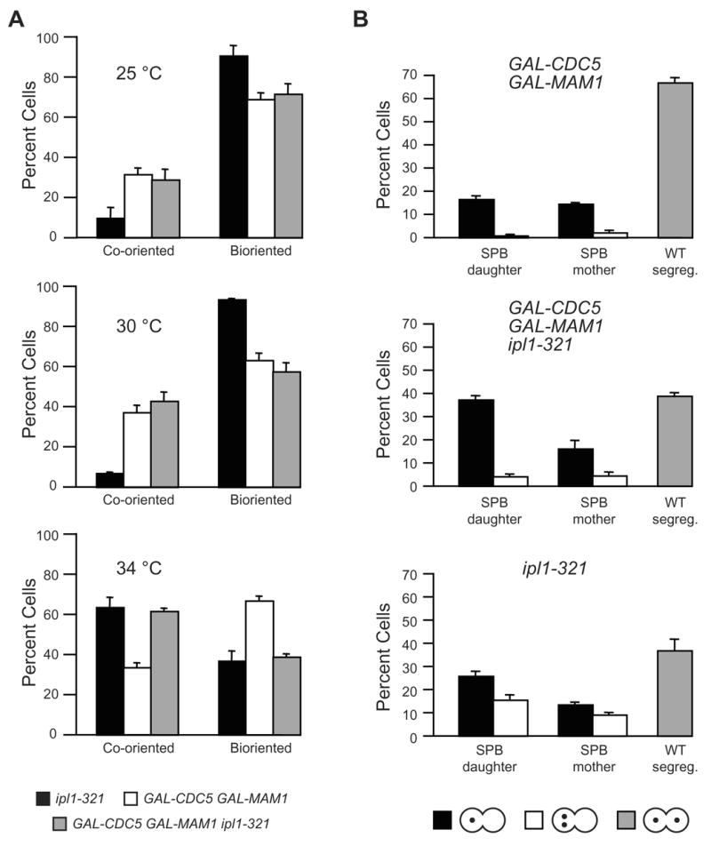
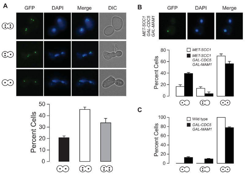
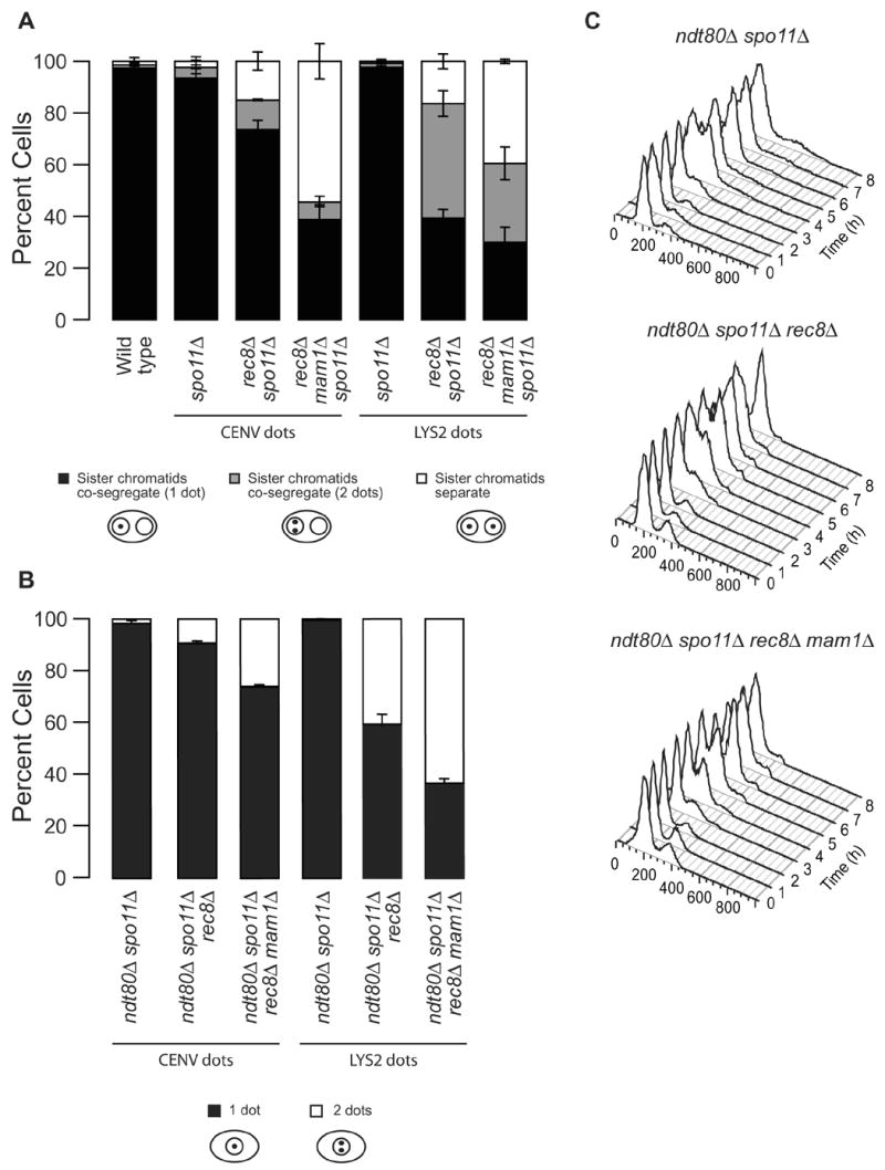
Kinetochores of sister chromatids attach to microtubules emanating from the same pole (coorientation) during meiosis I and microtubules emanating from opposite poles (biorientation) during meiosis II. We find that the Aurora B kinase Ipl1 regulates kinetochore-microtubule attachment during both meiotic divisions and that a complex known as the monopolin complex ensures that the protein kinase coorients sister chromatids during meiosis I. Furthermore, the defining of conditions sufficient to induce sister kinetochore coorientation during mitosis provides insight into monopolin complex function. The monopolin complex joins sister kinetochores independently of cohesins, the proteins that hold sister chromatids together. We propose that this function of the monopolin complex helps Aurora B coorient sister chromatids during meiosis I.


Figures

Figure 1
Figure 1. Ipl1 regulates meiosis I chromosome segregation
(A) pSCC1-3HA-IPL1 cells (A10423) were induced to sporulate to examine 3HA-Ipl1 levels at the indicated times. vATPase was used as a loading control. (B) Wild type (A5811) and pSCC1-3HA-IPL1 (A10423) cells were induced to sporulate to determine DNA content by FACS. (C – E) Wild type (A5715) and pSCC1-3HA-IPL1 (A14502) cells, both carrying homozygous CENV GFP dots were induced to sporulate to determine spindle morphology (C), nuclear morphology (D) and CENV GFP dot segregation (E). (F) Strains described in (C) were resuspended in sporulation (SPO) medium containing 120 μg/ml benomyl (benomyl) or DMSO (1%; mock) 4 hours after induction of sporulation. After 30 minutes cells were washed and resuspended in SPO medium. Samples were taken between 3–6 hours thereafter and CENV GFP dot segregation was determined. Note that we only determined the presence but not the number of GFP dots per nucleus.
Figure 2
Figure 2. IPL1 controls multiple meiosis II events
(A) Wild type (A5811) and pSCC1-3HA-IPL1 (A10423) cells carrying heterozygous CENV GFP dots were induced to sporulate to determine GFP dot segregation at the indicated times. (B) spo11Δ(A9498) and spo11Δ pSSC1-3HA-IPL1 (A10425) cells carrying heterozygous CENV GFP dots were induced to sporulate to determine GFP dot segregation at the indicated times. (C) Wild type (A10483), Ipl1-depleted (A15201) or Sgo1-depleted (A15056) cells carrying a NDC10-6HA and a REC8-13MYC fusion were induced to sporulate. Chromosome spreads were prepared at 5, 6 and 8 hours after sporulation induction and Rec8 localization was analyzed in binucleate cells (n=50). (D) Wild type (A10461), spo13Δ (A10755) and pSCC1-3HA-IPL1 (A15169) cells carrying a NDC10-6HA and a SGO1-9MYC fusion were induced to sporulate to examine Sgo1 localization as described in (C). Note that we only determined the presence but not the number of GFP dots per nucleus.
Figure 3
Figure 3. The Ipl1-depletion phenotype is epistatic to that caused by the inactivation of MAM1 or SPO13
Wild type (A5811), pSCC1-3HA-IPL1 (A10423), mam1Δ spo11Δ (A8128) and mam1Δ spo11Δ pSCC1-3HA-IPL1 (A15164) cells in (A) and wild type (A5811), pSCC1-3HA-IPL1 (A10423), spo13Δ spo11Δ (A7170) and spo13Δ spo11Δ pSCC1-3HA-IPL1 (A11432) cells in (B), all carrying heterozygous CENV GFP dots, were induced to sporulate to determine GFP dot segregation at the indicated times.
Figure 4
Figure 4. An active monopolin complex is sufficient to promote sister kinetochore co-orientation
(A) Wild type (A5244), GAL-MAM1 (A12315), GAL-CDC5 (A12325), GAL-CDC5 GAL-MAM1 (A12312), lrs4Δ (A15911), GAL-CDC5 GAL-MAM1 lrs4Δ (A15910) and GAL-CDC5 GAL-MAM1 lrs4Δ csm1Δ (A16882) cells, all carrying CENIV GFP dots, were arrested in G1 using 5 μg/ml α factor, and treated with galactose for 1 hour prior to release. When arrest was complete, cells were released into medium lacking pheromone and containing 2% galactose. Samples were taken to determine GFP dot segregation (data represent the average of 3 experiments; statistically significant changes relative to wild type (p ≤ 0.001) are indicated by ***). (B) Wild type (A15127), GAL-CDC5 (A15926), and GAL-CDC5 GAL-MAM1 (A15925) cells carrying LRS4–6HA and NDC80-GFP were grown as described in (A) to determine the localization of Lrs4-HA on chromosome spreads. Lrs4-6HA is shown in red, Ndc80-GFP in green and DNA in blue. (C, D) Wild type (A15912, squares) and GAL-CDC5 GAL-MAM1 (A15915, circles) cells, all carrying PDS1-3HA fusions, were grown as in (A) except α-factor was re-added (5 μg/ml) 90 minutes after release from the G1 arrest. Samples were taken to determine the percentage of metaphase (closed symbols; C) and anaphase (open circles; C) spindles and Pds1-3HA protein levels (D). Pgk1 was used as a loading control.
Figure 5
Figure 5. Effects of overproducing Cdc5 and Mam1 on ipl1-321 mutants
GAL-CDC5 GAL-MAM1 (A12312), ipl1-321 (A16485), and GAL-CDC5 GAL-MAM1 ipl1-321 (A15931) cells, all carrying CENIV GFP dots, were arrested in G1 as described in Figure 4A, followed by release into medium lacking pheromone and containing 2% galactose at 25ºC, 30ºC or 34ºC. (A) The percentage of co-segregating and correctly segregating (bi-oriented) sister chromatids was determined in anaphase cells (data represent the average of 3 experiments). (B) The percentage of the following three classes of anaphase cells was determined at 34°C: (i) co-segregating sister chromatids that segregated into the bud (SPB daughter), (ii) co-segregating sister chromatids that segregated into the mother (SPB mother) and (iii) correctly segregating sister chromatids (WT segreg.). Within classes (i) and (ii) the following distinctions were made: co-segregating sister chromatids tightly paired (black bars); co-segregating sister chromatids not paired (white bars).
Figure 6
Figure 6. The monopolin complex fuses sister kinetochores together
(A) Haploid wild-type cells carrying GFP dots on both chromosome IV and V (A15978) were arrested in G1 using 5μg/ml α factor. Galactose was added 1 hour prior to release. When arrest was complete cells were released into medium containing galactose but lacking pheromone. The presence of one or two GFP dots in each nuclear lobe was determined in anaphase cells. (B) Haploid MET-SCC1 (A16486) and GAL-CDC5 GAL-MAM1 MET-SCC1 (A16023) cells carrying CENV GFP dots were arrested in G1 in media lacking methionine using 5 μg/ml α factor, preinduced with 2% galactose and 8 mM methionine for 1 h, and released into YEP medium lacking pheromone and containing 2% galactose and 8 mM methionine at 25ºC. Cells were analyzed as in (A). (C) Wild type (A5237) and GAL-CDC5 GAL-MAM1 (A16883) cells carrying TELV GFP dots, were grown as described in Figure 4A to determine GFP dot segregation. In this set of strains only 25% of cells co-segregated sister chromatids. The reasons for the lower levels of co-segregation in this strain are unclear.
Figure 7
Figure 7. The co-segregation of sister chromatids observed in REC8Δ cells depends in part on MAM1
(A) Wild type (A5811), spo11 (A9498), rec8Δ spo11 Δ (A16020) and rec8 Δ mam1 Δ spo11 Δ (A16342) cells carrying heterozygous CENV GFP dots, and spo11 Δ (A16725) rec8 Δ spo11Δ (A16838) and rec8Δ mam1Δ spo11Δ (A16839) all carrying heterozygous LYS2 GFP dots, were induced to sporulate to examine the distribution of GFP dots in binucleate cells 6 hours after induction. Strains A16838 and A16839 were analyzed after 8 hours. A later time point was chosen for the LYS2 GFP dot strains to allow for a more complete segregation of chromosome arms away from the midzone. (n=100 cells for A16838 and n=150 cells for A16839, n=200 cells for all other strains). (B, C) spo11Δ ndt80 Δ (A16840) rec8Δ spo11Δ ndt80Δ (A16841) and rec8 Δ mam1Δ spo11Δ ndt80Δ (A16842) cells all carrying heterozygous CENV GFP dotsΔ and spo11Δ ndt80Δ (A16835), rec8Δ spo11Δ ndt80Δ (A16836) and rec8Δ mam1Δ spo11Δ ndt80Δ (A16837) cells all carrying heterozygous LYS2 GFP dots, were induced to sporulate to examine DNA content (C; CENV dot strains are shown) and the association of GFP dots (B; 6h time point was analyzed).

Similar articles

Cited by

References

    1. Bergerat A, de Massy B, Gadelle D, Varoutas PC, Nicolas A, Forterre P. An atypical topoisomerase II from Archaea with implications for meiotic recombination. Nature. 1997;386:414–417. - PubMed
    1. Biggins S, Severin FF, Bhalla N, Sassoon I, Hyman AA, Murray AW. The conserved protein kinase Ipl1 regulates microtubule binding to kinetochores in budding yeast. Genes Dev. 1999;13:532–544. - PMC - PubMed
    1. Bishop JD, Han Z, Schumacher JM. The Caenorhabditis elegans Aurora B kinase AIR-2 phosphorylates and is required for the localization of a BimC kinesin to meiotic and mitotic spindles. Mol Biol Cell. 2005;16:742–756. - PMC - PubMed
    1. Cheeseman IM, Anderson S, Jwa M, Green EM, Kang J, Yates JR, 3rd, Chan CS, Drubin DG, Barnes G. Phospho-regulation of kinetochore-microtubule attachments by the Aurora kinase Ipl1p. Cell. 2002;111:163–172. - PubMed
    1. Clyne RK, Katis VL, Jessop L, Benjamin KR, Herskowitz I, Lichten M, Nasmyth K. Polo-like kinase Cdc5 promotes chiasmata formation and cosegregation of sister centromeres at meiosis I. Nat Cell Biol. 2003;5:480–485. - PubMed

Publication types

MeSH terms

Substances