. 2007 Feb 9;128(3):533-46.
doi: 10.1016/j.cell.2007.01.019.

Death domain assembly mechanism revealed by crystal structure of the oligomeric PIDDosome core complex

Affiliations

Death domain assembly mechanism revealed by crystal structure of the oligomeric PIDDosome core complex

Hyun Ho Park et al. Cell. .

Abstract

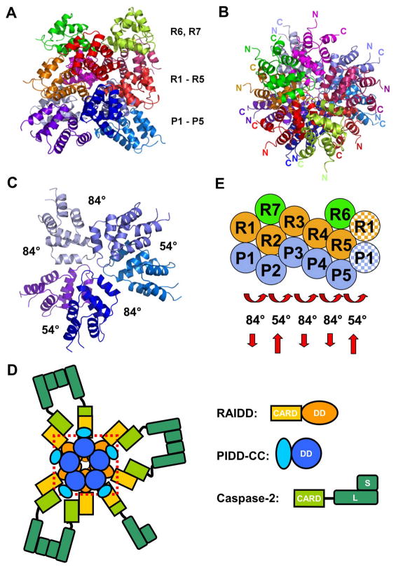
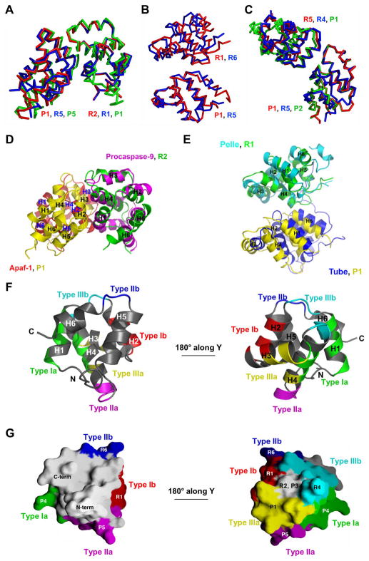
Proteins of the death domain (DD) superfamily mediate assembly of oligomeric signaling complexes for the activation of caspases and kinases via unknown mechanisms. Here we report the crystal structure of the PIDD DD and RAIDD DD complex, which forms the core of the caspase-2-activating complex PIDDosome. Although RAIDD DD and PIDD DD are monomers, they assemble into a complex that comprises seven RAIDD DDs and five PIDD DDs. Despite the use of an asymmetric assembly mechanism, all DDs in the complex are in quasi-equivalent environments. The structure provided eight unique asymmetric interfaces, which can be classified into three types. These three types of interactions together cover a majority of the DD surface. Mutagenesis on almost all interfaces leads to disruption of the assembly, resulting in defective caspase-2 activation. The three types of interactions may represent most, if not all, modes of interactions in the DD superfamily for assembling complexes of different stoichiometry.


Figures

Figure 1
Figure 1
Characterization of the PIDD DD: RAIDD DD complex. A. Domain organizations of the PIDDosome components, PIDD, RAIDD and caspase-2. The cleavage fragments of PIDD are shown. B. Gel filtration profiles of PIDD DD alone (cyan), RAIDD DD alone (blue) and the complex (red). C. Determination of the molar mass of the complex by multi-angle light scattering. D. Representative class averages obtained with negatively stained sample. Each class contains 50–170 particles. Scale bar is 10 nm.
Figure 2
Figure 2
Overview of the PIDD DD: RAIDD DD complex. A. Side view of the complex. The top layer contains 2 RAIDD DD molecules (green and yellow). The middle layer contains 5 RAIDD DD molecules (red, purple, orange, magenta and pink). The bottom layer contains 5 PIDD DD molecules (different shades of blue). B. Top view of the complex. C. Top view of the 5 PIDD DDs at the bottom layer, showing the rotational relationships. D. A model of the PIDDosome for caspase-2 activation, showing the PIDD DD: RAIDD DD complex inside a dotted red box and the rest of the domains and molecules. Caspase-2 molecules are schematically dimerized to illustrate proximity induced dimerization in the PIDDosome. E. A schematic planar diagram for the construction of the complex, showing the successive screw rotations of a hypothetical PR sub-complex. The shaded R1 and P1 molecules at the right indicate that the rotations have brought the PR sub-complex back to the beginning and completed the ring.
Figure 3
Figure 3
Quasi-equivalent environment. A. Superposition of the 5 PIDD DD: RAIDD DD PR sub-complexes. B. Superposition of RAIDD DD (purple) and PIDD DD (green). C. Quasi-equivalence of the contacts on the plane of the staggered hexagonal construction. R5 and its neighboring DDs (gray) are superimposed with P1 and its neighboring DDs (different colors). D. Structure-based sequence alignment between RAIDD DD and PIDD DD. Residues of RAIDD DD and PIDD DD involved in the 8 different interfaces, which are classified into 3 types of interactions, are highlighted in yellow and marked. P: PIDD. R: RAIDD. Type Ia, IIa and IIIa residues are marked in red and type Ib, IIb and IIIb residues are marked in blue.
Figure 4
Figure 4
The 3 types of interactions and their subtypes, a total of 8 interactions. A. A schematic diagram for the locations of the 3 types of contacts in the PIDD DD: RAIDD DD complex. B. The 3 different subtypes of the type I interaction. C. The 2 different subtypes of the type II interaction. D. The 3 different subtypes of the type III interaction. Important residues and hydrogen bonding interactions are labeled. E. A hypothetical Fas DD: FADD DD complex constructed from the same 3 types of interactions. The same view is used as in A and the Fas DD: FADD DD complex may be considered as a portion of the PIDD DD: RAIDD DD complex composed of R7, R2, P2, R3, P3 and R4.
Figure 5
Figure 5
Conservation, plasticity and coverage of the type I, II and III interactions. A. Comparison of the R:P (red), R:R (blue) and P:P (green) subtypes of the type I interaction. One molecule in each subtype is superimposed. B. Comparison of the R:P (red) and R:R (blue) subtypes in the type II interaction. One molecule in each subtype is superimposed. C. Comparison of the R:P (red), R:R (blue) and P:P (green) subtypes in the type III interaction. One molecule in each subtype is superimposed. D. Comparison of the type I interaction (R2:P1) with the procaspase-9 CARD: Apaf-1 CARD interaction. R2 is superimposed with procaspase-9 CARD. E. Comparison of the type II interaction (R1:P1) with the Pelle DD: Tube DD interaction. R1 is superimposed with Pelle DD. F. The 6 types of regions of R5 in its interaction with neighboring DDs in the complex. Two views of R5 are shown. Green and red: type Ia and Ib regions. Magenta and blue: type IIa and IIb regions. Yellow and cyan: type IIIa and IIIb regions. G. Surface representation of R5, showing the same 6 surfaces of contacts. Same color coding is used as in F. The small gray area of surface at the 180° rotated view of R5 that does not contact any of the 6 immediate neighboring molecules interact with R2 and P3 in the three-dimensional assembly.
Figure 6
Figure 6
Mutational analysis of the PIDD DD: RAIDD DD interaction. A. Structure-based mutations and their effects on assembly of the PIDD DD: RAIDD DD complex in vitro. ** and * show mutations that completely and partially disrupted complex formation, respectively. B. HEK293T cells were transiently transfected with expression vectors encoding wild type or mutant Flag-PIDD and wild type VSV-RAIDD. PIDD or RAIDD was immunoprecipitated from the lysates with anti-Flag or anti-VSV antibodies respectively, and co-immunoprecipitated proteins were revealed by Western blotting (WB). * corresponds to the p20 subunit of caspase-2, which is due the presence of overexpressed RAIDD (PIDD independent). C. Instead of PIDD mutants, the activity of RAIDD mutants was analyzed. D. as B, but PIDD proteins were expressed in the absence of overexpressed RAIDD.

References

    1. Baliga BC, Read SH, Kumar S. The biochemical mechanism of caspase-2 activation. Cell Death Differ. 2004;11:1234–1241. - PubMed
    1. Bergeron L, Perez GI, Macdonald G, Shi L, Sun Y, Jurisicova A, Varmuza S, Latham KE, Flaws JA, Salter JC, et al. Defects in regulation of apoptosis in caspase-2-deficient mice. Genes Dev. 1998;12:1304–1314. - PMC - PubMed
    1. Brunger AT, Adams PD, Clore GM, DeLano WL, Gros P, Grosse-Kunstleve RW, Jiang JS, Kuszewski J, Nilges M, Pannu NS, et al. Crystallography & NMR system: A new software suite for macromolecular structure determination. Acta Crystallogr. 1998;D54:905–921. - PubMed
    1. Duan H, Dixit VM. RAIDD is a new ‘death’ adaptor molecule. Nature. 1997;385:86–89. - PubMed
    1. Frank J, Radermacher M, Penczek P, Zhu J, Li Y, Ladjadj M, Leith A. SPIDER and WEB: processing and visualization of images in 3D electron microscopy and related fields. J Struct Biol. 1996;116:190–199. - PubMed

MeSH terms

Substances

Associated data