. 2007;35(5):1578-88.
doi: 10.1093/nar/gkm011. Epub 2007 Feb 8.

Ribosomal protein L20 controls expression of the Bacillus subtilis infC operon via a transcription attenuation mechanism

Affiliations

Ribosomal protein L20 controls expression of the Bacillus subtilis infC operon via a transcription attenuation mechanism

Nasslie Choonee et al. Nucleic Acids Res. 2007.

Abstract

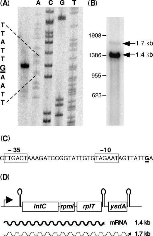
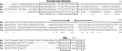
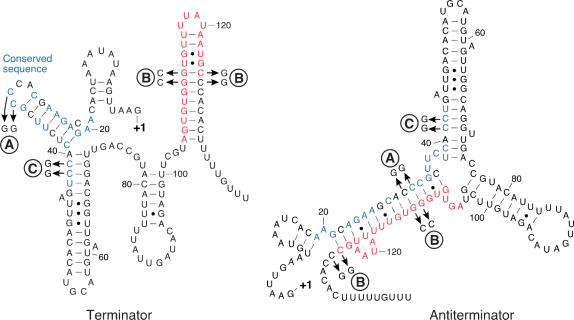
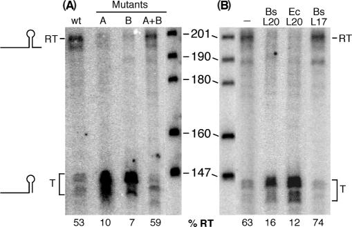
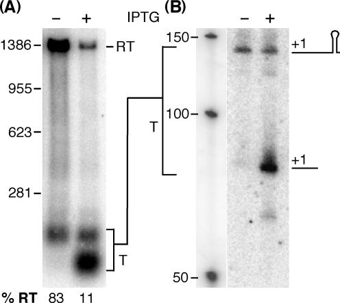
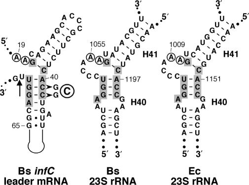
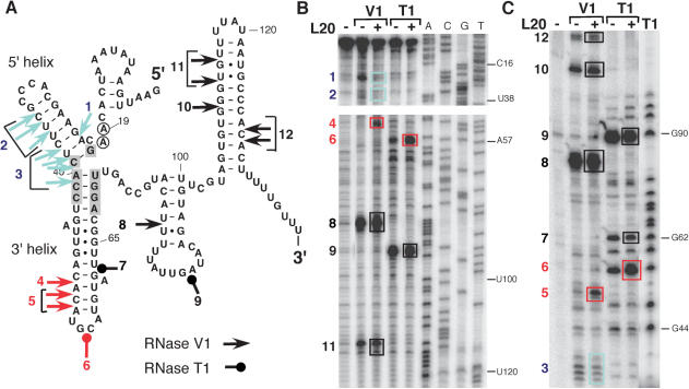
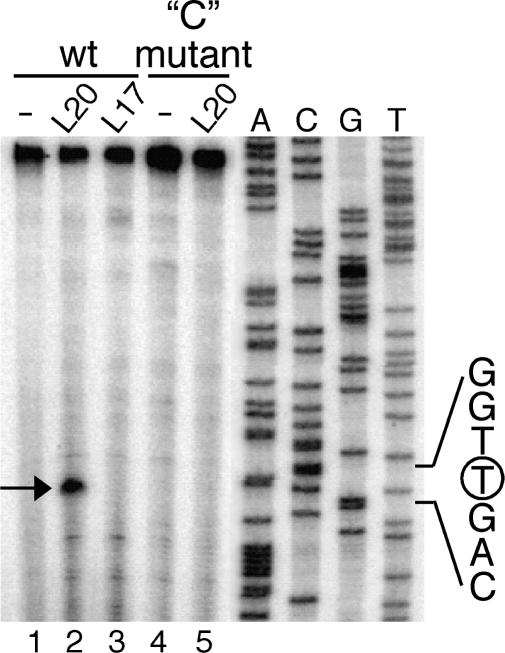
In contrast to Escherichia coli no molecular mechanism controlling the biosynthesis of ribosomal proteins has been elucidated in Gram-positive organisms. Here we show that the expression of the Bacillus subtilis infC-rpmI-rplT operon encoding translation factor IF3 and the ribosomal proteins L35 and L20 is autoregulated by a complex transcription attenuation mechanism. It implicates a 200-bp leader region upstream of infC which contains two conserved regulatory elements, one of which can act as a transcription terminator. Using in vitro and in vivo approaches we show that expression of the operon is regulated at the level of transcription elongation by a change in the structure of the leader mRNA which depends upon the presence of ribosomal protein L20. L20 binds to a phylogenetically conserved domain and provokes premature transcription termination at the leader terminator. Footprint and toeprint experiments support a regulatory model involving molecular mimicry between the L20-binding sites on 23S rRNA and the mRNA. Our data suggest that Nomura's model of ribosomal protein biosynthesis based on autogenous control and molecular mimicry is also valid in Gram-positive organisms.


Figures

Figure 1.
Figure 1.
Transcription profile of the B. subtilis infC operon. (A) Determination of the transcription initiation start point by extension of primer HP667; the +1 guanosine residue is underlined, the sequence ladder was generated with oligonucleotide HP667. (B) Northern analysis of infC operon transcripts with a probe spanning the four genes. (C) −35 and −10 promoter sequences upstream of the transcription start point which is underlined. (D) Schematic view of the infC operon transcripts.
Figure 2.
Figure 2.
Alignment of the infC leader sequences from four Gram-positive organisms. A 5′ conserved domain and the Shine–Dalgarno sequences are boxed. Inverted arrows depict factor-independent transcription terminators. Potential infC translation initiation codons (which are generally not ATG/GTG) are underlined and identical residues are marked by an asterisk. Bs = Bacillus subtilis, Ba = Bacillus anthracis, Sa = Staphylococcus aureus, Ca = Clostridium acetobutylicum. The numbering corresponds to the B. subtilis sequence.
Figure 3.
Figure 3.
Putative alternative RNA foldings within the B. subtilis infC leader. Identical nucleotides of the 5′ conserved domain (boxed in Figure 2) are in blue and residues of the terminator structure involved in the formation of the antiterminator are in red. ‘A’ and ‘B’ indicate double and quadruple point mutations, respectively, as shown in the figure. They have been introduced to analyze the folding pattern of the leader mRNA (see text). ‘C’ depicts a double mutation used for the toeprint assay (see text below).
Figure 4.
Figure 4.
Effect of infC leader mutations and the presence of L20 on the transcription profile in vitro. Single round in vitro transcription assays were performed on PCR templates comprising the promoter and 5′ noncoding region of infC using B. subtilis RNA polymerase. RT and T on the left side indicate read-through or prematurely terminated infC leader transcripts. The size of the marker fragments in bases is indicated. Numbers at the bottom indicate the percentage of read-through transcripts (%RT = (RT/(T + RT) × 100). (A) Templates were wild type (wt) or contained the A, B or A + B mutations depicted in Figure 3. (B) Where indicated proteins were added in 50-fold molar excess over the template during the elongation phase of the single-round transcription assay. The B. subtilis L20 (Bs L20) and L17 (Bs L17) r-proteins were native, E. coli L20 (Ec L20) only contained the C-terminal half of the protein.
Figure 5.
Figure 5.
Effect of L20 overproduction on the transcription profile of the infC operon. Northern analysis of total RNA of a B. subtilis wild-type strain harboring plasmid pHML17 carrying an IPTG inducible copy of the rplT (L20) gene was performed using two different probes. (A) RNA was separated on a 0.8% agarose gel and probed with an infC specific probe including the entire leader sequence (see Materials and methodssection). (B) RNA separated on a 8% polyacrylamide gel was probed with oligonucleotide HP1080 complementary to positions +8 to +34 of the infC leader. Where indicated IPTG (1 mM) was added to mid-log cultures for 20 min prior to isolation of the RNA. RT = read-through transcript, T = terminated transcript. The positions of the molecular size markers are indicated. Percentages of read-through transcripts are indicated below the gel.
Figure 6.
Figure 6.
Secondary structure diagrams of the L20-binding sites on B. subtilis and E. coli 23S rRNA and a region of the B. subtilis infC leader. A segment of the infC leader (nts 19–73) comprising the conserved 5′ domain (Figure 2) is drawn to highlight the structural resemblance between the mRNA fold and the L20-binding sites at the junction of helices 40 and 41 (H40/41) on 23S rRNA from B. subtilis and E. coli. Numbering corresponds to the infC mRNA and the respective mature 23S rRNA molecules. The arrow indicates the position of the reverse transcription arrest observed in the presence of L20 (see text and Figure 7). The two A residues conserved in eubacterial 23S rRNA are encircled, nucleotides at the interhelical junction conserved between the structures are shown in grey boxes.
Figure 7.
Figure 7.
RNase probing of the infC leader mRNA structure in the absence or presence of L20 protein. An in vitro infC leader transcript (nts 1–168) was subjected to cleavage by RNases V1 or T1. Where indicated purified B. subtilis L20 protein was added prior to RNase cleavage. (A) Cleavages by RNase V1 and RNase T1 are shown on the infC leader mRNA structure. Colors indicate the change in cleavage efficiency observed in the presence of L20: black (no change), red (increase), blue (decrease). The numbers next to the symbols locate the corresponding cleavages on the gels (boxes). Shaded and encircled nucleotides correspond to positions conserved at the L20-binding site on 23S rRNA as shown in Figure 6. (B) Cleavages on an unlabeled transcript were detected by primer extension with labeled oligonucleotide HP697 (positions 168–141 on infC leader). (C) Cleavages on a 5′ labeled transcript were analyzed directly.
Figure 8.
Figure 8.
Toeprint caused by L20 binding to the B. subtilis infC leader mRNA. Oligonucleotide HP697 was extended on a wild type and ‘C’ mutant (CC41,42 → GG, see Figure 5) in vitro transcript of the infC leader region. Where indicated the transcript was incubated with the B. subtilis r-proteins L20 or L17 prior to the extension reaction. The reverse transcriptase stop caused by L20 bound to the transcript is indicated by an arrow; it corresponds to a primer extension arrest at uridine residue 72 (see Figure 6). The sequence ladder was generated with the same primer HP697, the encircled nucleotide indicates the arrest position on the cDNA.
Figure 9.
Figure 9.
Regulatory model for the control of the B. subtilis infC operon. (A) L20 is primarily recruited by 23S rRNA to integrate the ribosome (in grey). When in excess, free L20 will bind at the operator in the infC leader which is similar to its binding site on 23S rRNA and inhibit formation of the antiterminator structure causing premature transcription termination. (B) In the absence of free L20 (i.e. rapid growth) a part of the operator (blue line) will hybridize to bases included in the terminator (red line) to form an antiterminator structure and provoke transcriptional read-through.

References

    1. Zengel JM, Lindahl L. Diverse mechanisms for regulating ribosomal protein synthesis in Escherichia coli. Prog. Nucleic Acid Res. Mol. Biol. 1994;47:331–370. - PubMed
    1. Mattheakis LC, Nomura M. Feedback regulation of the spc operon in Escherichia coli: translational coupling and mRNA processing. J. Bacteriol. 1988;170:4484–4492. - PMC - PubMed
    1. Fallon AM, Jinks CS, Strycharz GD, Nomura M. Regulation of ribosomal protein synthesis in Escherichia coli by selective mRNA inactivation. Proc. Natl. Acad. Sci. U.S.A. 1979;76:3411–3415. - PMC - PubMed
    1. Nomura M, Yates JL, Dean D, Post LE. Feedback regulation of ribosomal protein gene expression in Escherichia coli: structural homology of ribosomal RNA and ribosomal protein MRNA. Proc. Natl. Acad. Sci. U.S.A. 1980;77:7084–7088. - PMC - PubMed
    1. Merianos HJ, Wang J, Moore PB. The structure of a ribosomal protein S8/spc operon mRNA complex. RNA. 2004;10:954–964. - PMC - PubMed

Publication types

MeSH terms