. 2007 Feb 20;104(8):2991-6.
doi: 10.1073/pnas.0611170104. Epub 2007 Feb 8.

Molecular determinants for the interaction between AMPA receptors and the clathrin adaptor complex AP-2

Affiliations

Molecular determinants for the interaction between AMPA receptors and the clathrin adaptor complex AP-2

Kathrin Kastning et al. Proc Natl Acad Sci U S A. .

Abstract

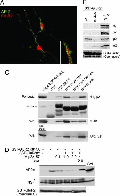
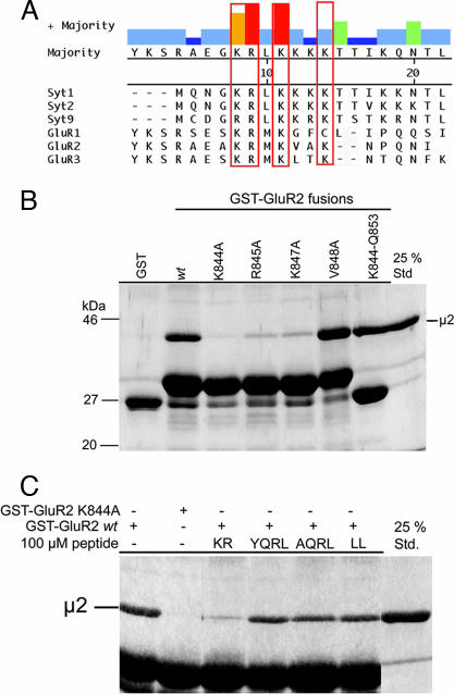
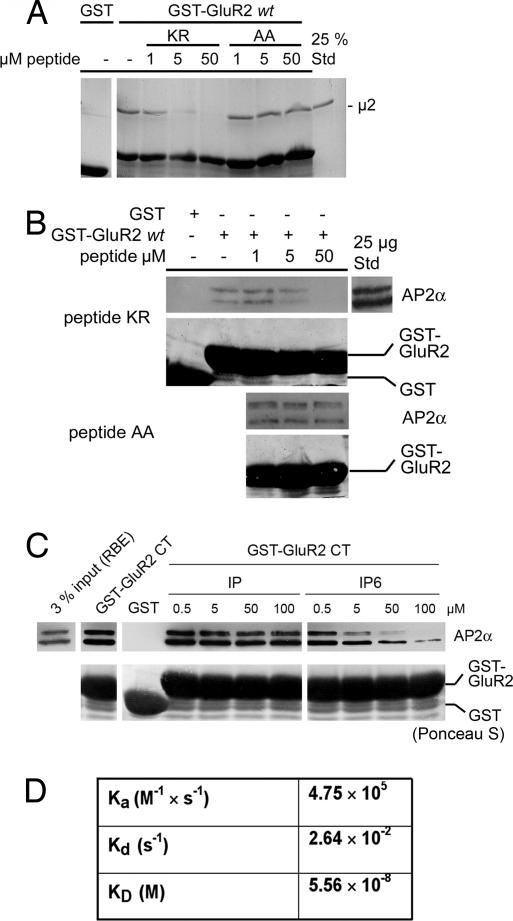
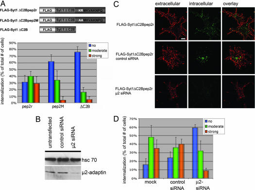
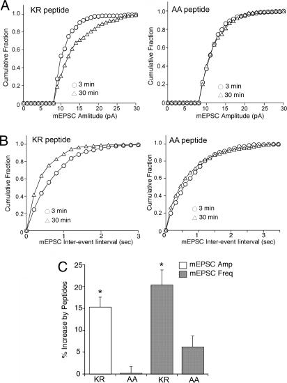
alpha-Amino-3-hydroxy-5-methylisoxazole-4-propionic acid (AMPA)-type glutamate receptors undergo constitutive and ligand-induced internalization that requires dynamin and the clathrin adaptor complex AP-2. We report here that an atypical basic motif within the cytoplasmic tails of AMPA-type glutamate receptors directly associates with mu2-adaptin by a mechanism similar to the recognition of the presynaptic vesicle protein synaptotagmin 1 by AP-2. A synaptotagmin 1-derived AP-2 binding peptide competes the interaction of the AMPA receptor subunit GluR2 with AP-2mu and increases the number of surface active glutamate receptors in living neurons. Moreover, fusion of the GluR2-derived tail peptide with a synaptotagmin 1 truncation mutant restores clathrin/AP-2-dependent internalization of the chimeric reporter protein. These data suggest that common mechanisms regulate AP-2-dependent internalization of pre- and postsynaptic membrane proteins.


Conflict of interest statement

The authors declare no conflict of interest.

Figures

Fig. 1.
Fig. 1.
AMPA receptor CTs associate directly with AP2μ. (A) Localization of AP-2α and GluR2 in K+-glutamate-stimulated hippocampal neurons (100 μM for 10 min at 37°C). GluR2 colocalizes partially with AP-2 within dendrites (yellow in the overlay). (Inset) Magnification (×5) of selected area. (Scale bar: 10 μm.) (B) GluR2 CT directly binds to AP-2μ. 35S-labeled αχ, β2, μ2, or σ2-adaptins translated in vitro were incubated with GST, GST-GluR2 CT, or an AP-2 binding-defective mutant (K844A) (100 μg). Bound material was analyzed by SDS/PAGE and autoradiography. 25% Std., 25% of the total amount of radiolabeled protein added to the assay. (C) Binding of purified His6-tagged μ2 (157–435; top blot) or AP-2 (bottom blot) to GluR1–3 CTs. Immobilized GST fusion proteins (30 μg) were incubated with recombinant His6-tagged μ2 (157–435) or Triton X-100-extracted rat brain lysates and washed extensively, and complexes were resolved by SDS/PAGE and staining with Ponceau S or analyzed by immunoblotting. RBE input was 20 μg of rat brain lysate. (D) Recombinant μ2 (157–435) competes the association of native AP-2 with GST-GluR2. Affinity purification of AP-2 using GST-GluR2 CT (20 μg) was performed in the presence of increasing concentrations of μ2 (157–435) or BSA. Samples were analyzed by immunoblotting for AP-2 or NSF. ∗, Cross-reactive band decorated with anti-NSF antibodies; Std, Triton X-100-extracted rat brain lysate (50 μg total protein).
Fig. 2.
Fig. 2.
Binding of μ2-adaptin to the GluR2 CT involves basic residues and can be competed by a synaptotagmin 1-derived AP-2 binding peptide. (A) Multiple sequence alignment of the AP-2μ binding motifs from synaptotagmin 1 (residues 317–335), synaptotagmin 2 (318–336), synaptotagmin 9 (397–415) (C2B domains from rat), GluR1 (residues 830–850), GluR2 (residues 837–856), and GluR3 (residues 842–861) CTs (rat) using the Clustal W algorithm. Conserved basic residues are boxed (red). (B) GluR2 CT or point mutants thereof were used for pull-downs with His6-tagged μ2 (157–435) (see legend to Fig. 1C) and analyzed by Coomassie blue staining. K844-Q853 is a fusion protein between GST and amino acids 844–853 of GluR2. (C) Direct binding of His6-tagged μ2 (157–435) to GST-GluR2 can be competed by a synaptotagmin 1 C2B domain-derived AP-2 binding peptide, but not by tyrosine (YQRL) or dileucine (LL) sorting motif peptides (100 μM each). A nonfunctional tyrosine motif peptide (AQRL) was used as a control. Samples were analyzed as described (see legend to Fig. 1C).
Fig. 3.
Fig. 3.
Direct high-affinity binding of μ2-adaptin to the GluR2 CT. (A and B) The inhibitory effect of the synaptotagmin 1-derived peptide on the association of μ2 (157–435; A) or AP-2 (B) with GST-GluR2 requires basic residues. Affinity purification was performed in the presence of increasing concentrations of wild-type (KR) or mutant (AA) synaptotagmin 1 C2B domain peptides (see legend to Fig. 2). Coomassie blue (A) and Ponceau S (B) staining were used to verify equal loading of GST or GST-GluR2 fusion proteins. (C) Affinity purification from rat brain extracts was done in the presence of increasing concentrations of inositol(1)-monophosphate (IP) or inositol(1,2,3,4,5,6)-hexakisphosphate. Immunoblotted material was stained with Ponceau S or antibodies against AP-2αA/C. (D) Surface plasmon resonance analysis of the binding of purified μ2 (157–435) to immobilized GluR2-derived AP-2 binding peptide (pep2R). Rate constants and KD were derived from sensograms shown in SI Fig. 8.
Fig. 4.
Fig. 4.
The GluR2-derived AP-2μ binding peptide targets a synaptotagmin 1 ΔC2B chimeric reporter protein for internalization. (A) Synaptotagmin 1 ΔC2B chimeric proteins (Upper) were expressed in Cos7 and assayed for internalization (compare SI Fig. 9B). Cells were classified visually into three phenotypic categories. Histograms (Lower) display the averaged percentage values collected from four independent experiments (n = 4; 20 cells per experiment). Error bars correspond to the SEM. Statistical significance was analyzed by the Pearson χ2 test (P < 0.01). (B) Immunoblot analysis of Cos7 cell extracts (10 μg total protein) transfected with siRNAs against a control protein (γ-BAR) or AP-2μ (μ2 siRNA) using antibodies against hsc70 or μ2-adaptin (72 h after transfection). (C) Internalization of synaptotagmin 1 ΔC2B-pep2r chimera in Cos7 cells transfected with siRNAs against a control protein or AP-2μ. Surface-exposed chimeric protein was labeled with Alexa Fluor 594 (red) under nonpermeabilizing conditions. Endocytosed anti-FLAG primary antibody was detected after Triton X-100 permeabilization of the cells followed by decoration with Alexa Fluor 488-conjugated secondary antibodies (green). (Scale bar: 15 μm.) (D) Semiquantitative analysis (see A) of FLAG-Syt1 ΔC2Bpep2r internalization in Cos7 cells depleted of AP-2μ. Histograms display the averaged percentage values collected from four independent experiments (n = 4; 20 cells per experiment). Error bars correspond to the SEM. The decrease of FLAG-Syt1 ΔC2Bpep2r endocytosis from 39 ± 11% in control transfected cells to 8 ± 3% in μ2-adaptin depleted cells is statistically significant (P < 0.01).
Fig. 5.
Fig. 5.
The Syt-1 AP-2 binding peptide increases mEPSC amplitude and frequency. (A and B) Cumulative plots of the distribution of mEPSC amplitude (A) and frequency (B; shown as interevent interval) from the Syt-1 KR peptide or mutant AA control peptide-injected neurons. (C) Bar plot summary showing the different effects of Syt-1 KR and mutant AA peptides on mEPSC amplitude and frequency. (Scale bar: 50 pA and 2 sec.)

References

    1. Fukata Y, Tzingounis AV, Trinidad JC, Fukata M, Burlingame AL, Nicoll RA, Bredt DS. J Cell Biol. 2005;169:399–404. - PMC - PubMed
    1. Bredt DS, Nicoll RA. Neuron. 2003;40:361–379. - PubMed
    1. Malinow R, Malenka RC. Annu Rev Neurosci. 2002;25:103–126. - PubMed
    1. Perez-Otano I, Ehlers MD. Trends Neurosci. 2005;28:229–238. - PubMed
    1. Sheng M, Kim MJ. Science. 2002;298:776–780. - PubMed

Publication types

MeSH terms

LinkOut - more resources