. 2007 May;148(5):2148-56.
doi: 10.1210/en.2006-1491. Epub 2007 Feb 8.

Role of microsomal retinol/sterol dehydrogenase-like short-chain dehydrogenases/reductases in the oxidation and epimerization of 3alpha-hydroxysteroids in human tissues

Affiliations

Role of microsomal retinol/sterol dehydrogenase-like short-chain dehydrogenases/reductases in the oxidation and epimerization of 3alpha-hydroxysteroids in human tissues

Olga V Belyaeva et al. Endocrinology. 2007 May.

Abstract

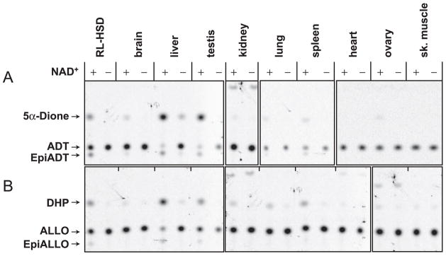
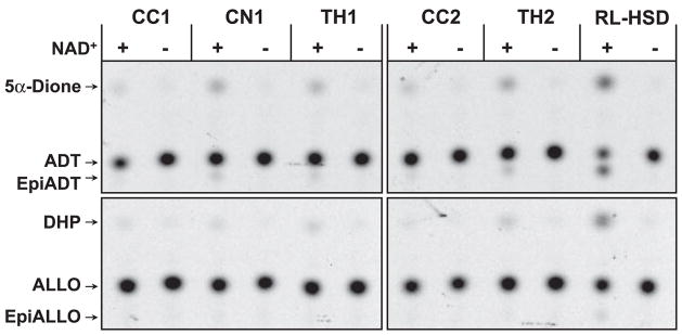
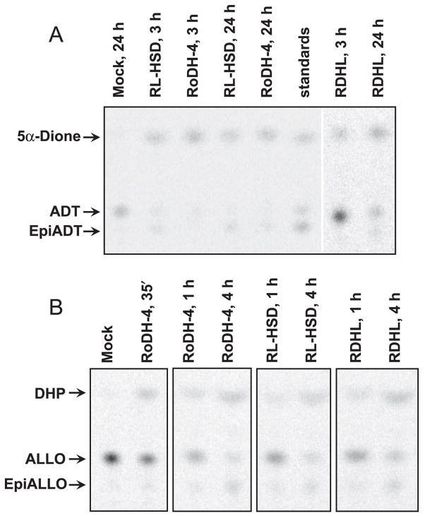
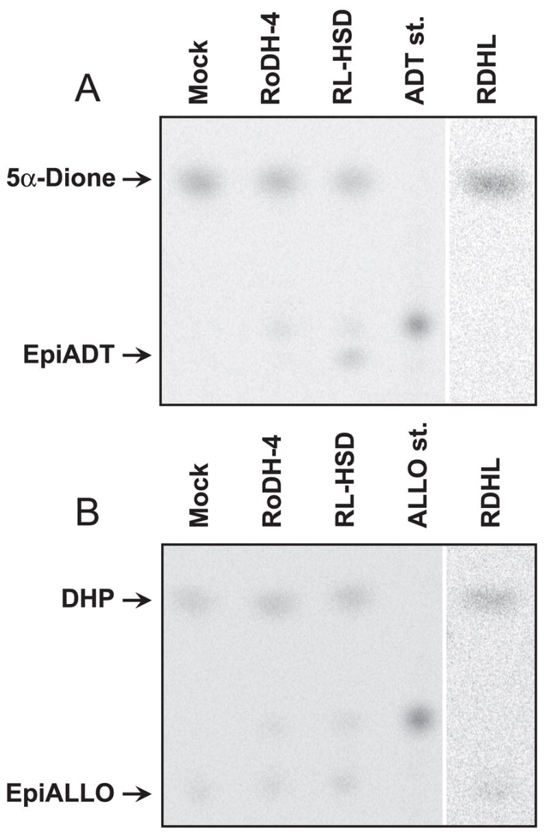
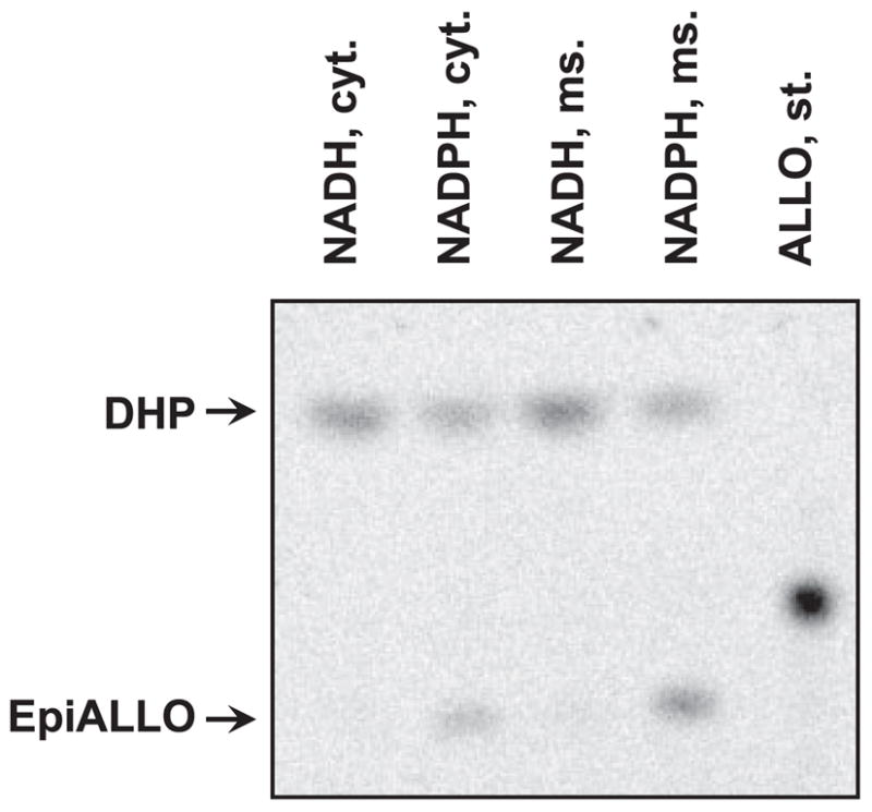
Allopregnanolone (ALLO) and androsterone (ADT) are naturally occurring 3alpha-hydroxysteroids that act as positive allosteric regulators of gamma-aminobutyric acid type A receptors. In addition, ADT activates nuclear farnesoid X receptor and ALLO activates pregnane X receptor. At least with respect to gamma-aminobutyric acid type A receptors, the biological activity of ALLO and ADT depends on the 3alpha-hydroxyl group and is lost upon its conversion to either 3-ketosteroid or 3beta-hydroxyl epimer. Such strict structure-activity relationships suggest that the oxidation or epimerization of 3alpha-hydroxysteroids may serve as physiologically relevant mechanisms for the control of the local concentrations of bioactive 3alpha-hydroxysteroids. The exact enzymes responsible for the oxidation and epimerization of 3alpha-hydroxysteroids in vivo have not yet been identified, but our previous studies showed that microsomal nicotinamide adenine dinucleotide-dependent short-chain dehydrogenases/reductases (SDRs) with dual retinol/sterol dehydrogenase substrate specificity (RoDH-like group of SDRs) can oxidize and epimerize 3alpha-hydroxysteroids in vitro. Here, we present the first evidence that microsomal nicotinamide adenine dinucleotide-dependent 3alpha-hydroxysteroid dehydrogenase/epimerase activities are widely distributed in human tissues with the highest activity levels found in liver and testis and lower levels in lung, spleen, brain, kidney, and ovary. We demonstrate that RoDH-like SDRs contribute to the oxidation and epimerization of ALLO and ADT in living cells, and show that RoDH enzymes are expressed in tissues that have microsomal 3alpha-hydroxysteroid dehydrogenase/epimerase activities. Together, these results provide further support for the role of RoDH-like SDRs in human metabolism of 3alpha-hydroxysteroids and offer a new insight into the enzymology of ALLO and ADT inactivation.


Figures

Fig. 1
Fig. 1
Distribution of the NAD+-dependent microsomal 3α-HSD/3(αβ)-HSE activity in human tissues. The light membrane fractions were incubated in the presence of 1 mM NAD+ and 1 μM ADT (A) or 1 μM ALLO (B) for 1 h at 37 C. Ten micrograms of liver and testis membranes were used in the reaction with ADT and 2.5 micrograms were used in the reaction with ALLO. For all other tissues, the reactions contained 62 μg of the membrane protein. Control samples contained the same amount of protein but lacked the cofactor. A sample with recombinant RL-HSD was included as a positive control for the 3α-HSD activity. It should be noted that different postmortem collection times could differentially influence enzyme activities in various tissues, although the activities of recombinant RoDH-like SDRs in microsomal preparations are rather stable.
Fig. 2
Fig. 2
The NAD+-dependent microsomal 3α-HSD/3(αβ)-HSE activity of human brain. The light membrane fractions from corpus callosum (CC), caudate nucleus (CN), and thalamus from two different donors (TH1 and TH2) were incubated with 1 μM ADT or 1 μM ALLO in the presence or absence of 1 mM NAD+ at 37 C for 2 h. The amount of protein used in each reaction was as follows: 140 μg CC1, 340 μg CN1, 160 μg TH1, 190 μg CC2, and 90 μg TH2. Caudate nucleus from donor 2 is shown in Fig. 1.
Fig. 3
Fig. 3
Radiochromatogram of the products of RL-HSD, RoDH-4, and RDHL enzymatic activities in living cells. HEK293 cells stably transfected with SDR cDNAs (RoDH-4, RL-HSD, RDHL) or with empty vector (Mock) were incubated with either 1 μM ADT (A) or 1 μM ALLO (B) for indicated times. Reaction products were extracted and analyzed by TLC.
Fig. 4
Fig. 4
Metabolism of DHP and 5α-Dione in cells stably transfected with RL-HSD, RoDH-4, and RDHL. The cells were incubated with 0.58 μM 5α-Dione for 1 h (A) or with 0.72 μM DHP for 3.5 h (B). The products were extracted and analyzed by radiochromatography. ALLO st., ALLO standard; ADT st., ADT standard.
Fig. 5
Fig. 5
Subcellular localization and cofactor preference of the endogenous 3β-HSOR activity of HEK293 cells. Mock-transfected HEK293 cells were fractionated to obtain the cytosolic and microsomal fractions. Equal portions (1/10) of each fraction were used in activity assays. These corresponded to 135 μg of cytosolic and 30 μg of microsomal protein per reaction. Samples were incubated with 0.55 μM DHP and 1 mM NADH or reduced nicotinamide adenine dinucleotide phosphate (NADPH) for 4 h at 37 C. Cyt., Cytosol; ms., microsomes; st., standard.
Fig. 6
Fig. 6
Characterization of antibodies and Western blot analysis of liver and testis. A, Antibodies against the N-terminal fragment of RL-HSD at a 1:3000 dilution. B, Antibodies against the C-terminal fragment of RoDH-4 at a 1:5000 dilution. C, Antibodies against the peptide specific for RL-HSD. The amount of protein was as follows: liver, 5 μg; testis, 16 μg; RoDH-4 microsomes, 1 μg; RL-HSD microsomes, 0.5 μg; RDHL microsomes, 9 μg; 11-cis-RDH (11cRDH) microsomes, 4.5 μg.
Fig. 7
Fig. 7
Immunolocalization of RoDH-4 in human brain. A, Cerebellum, × 100 magnification; inset at the bottom of the figure represent another field at a higher magnification (× 400), showing neuron-specific immunostaining in more detail. B, Preimmune serum, × 400. C, Cerebral cortex, × 400. D, Diencephalon, × 400. Note immunopositive staining in neurons (marked by arrows).

Similar articles

Cited by

References

    1. Paul SM, Purdy RH. Neuroactive steroids. FASEB J. 1992;6:2311–2322. - PubMed
    1. Majewska MD, Harrison NL, Schwartz RD, Barker JL, Paul SM. Steroid hormone metabolites are barbiturate-like modulators of the GABA receptor. Science. 1986;232:1004–1007. - PubMed
    1. Lambert JJ, Belelli D, Hill-Venning C, Peters JA. Neurosteroids and GABAA receptor function. Trends Pharmacol Sci. 1995;16:295–303. - PubMed
    1. Baulieu EE, Schumacher M. Progesterone as a neuroactive neurosteroid, with special reference to the effect of progesterone on myelination. Hum Reprod. 2000;15:1–13. - PubMed
    1. Schumacher M, Weill-Engerer S, Liere P, Robert F, Franklin RJ, Garcia-Segura LM, Lambert JJ, Mayo W, Melcangi RC, Parducz A, Suter U, Carelli C, Baulieu EE, Akwa Y. Steroid hormones and neurosteroids in normal and pathological aging of the nervous system. Prog Neurobiol. 2003;71:3–29. - PubMed

Publication types

MeSH terms