Comparative Study
. 2007 Feb 1;21(3):316-31.
doi: 10.1101/gad.398207.

Defective N-sulfation of heparan sulfate proteoglycans limits PDGF-BB binding and pericyte recruitment in vascular development

Affiliations
Comparative Study

Defective N-sulfation of heparan sulfate proteoglycans limits PDGF-BB binding and pericyte recruitment in vascular development

Alexandra Abramsson et al. Genes Dev. .

Abstract

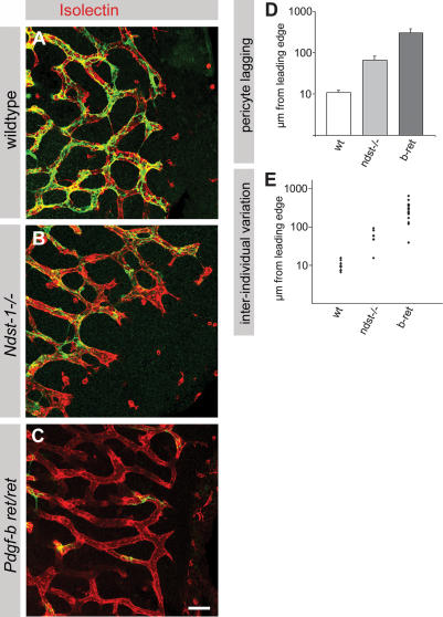
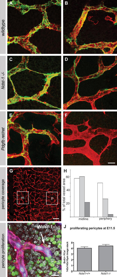
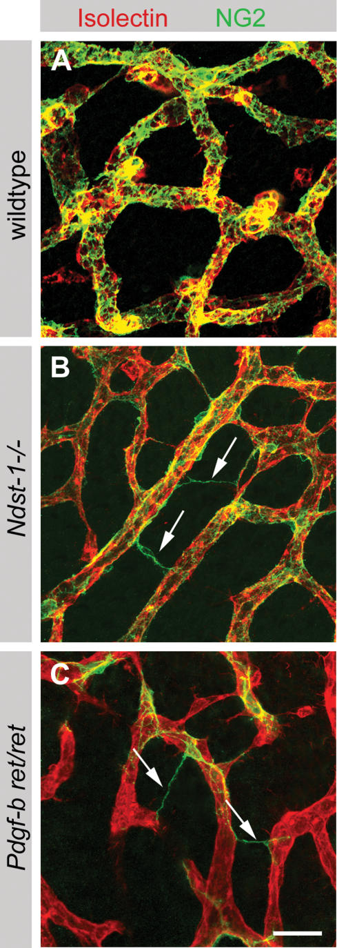
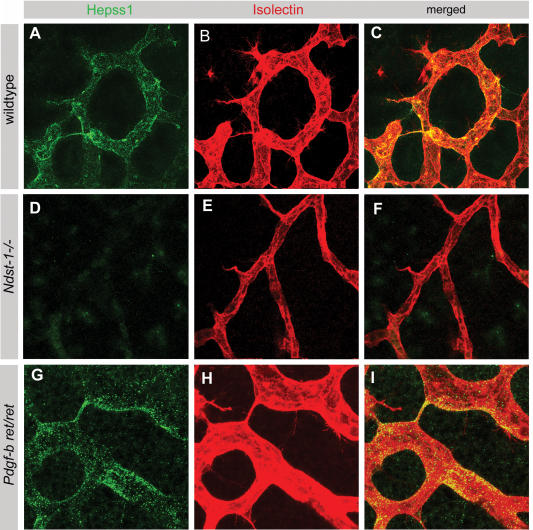
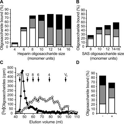
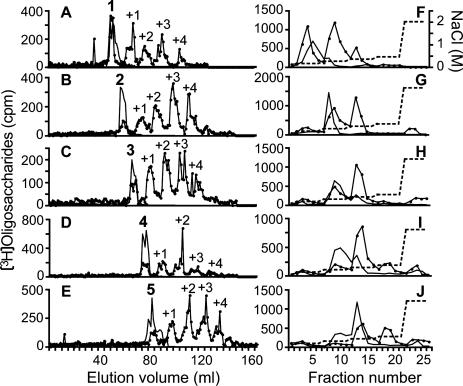
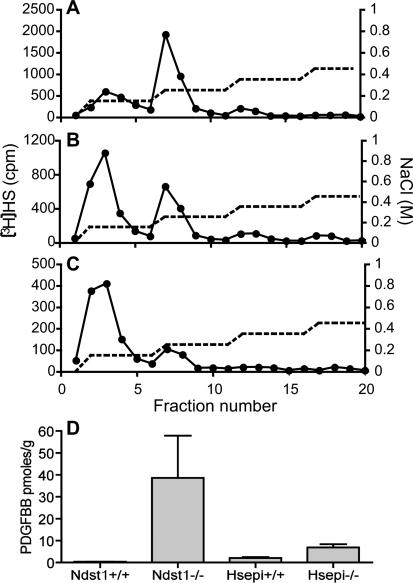
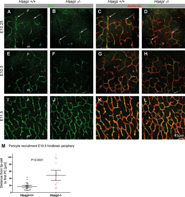
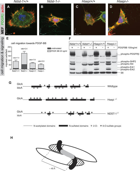
During vascular development, endothelial platelet-derived growth factor B (PDGF-B) is critical for pericyte recruitment. Deletion of the conserved C-terminal heparin-binding motif impairs PDGF-BB retention and pericyte recruitment in vivo, suggesting a potential role for heparan sulfate (HS) in PDGF-BB function during vascular development. We studied the participation of HS chains in pericyte recruitment using two mouse models with altered HS biosynthesis. Reduction of N-sulfation due to deficiency in N-deacetylase/N-sulfotransferase-1 attenuated PDGF-BB binding in vitro, and led to pericyte detachment and delayed pericyte migration in vivo. Reduced N-sulfation also impaired PDGF-BB signaling and directed cell migration, but not proliferation. In contrast, HS from glucuronyl C5-epimerase mutants, which is extensively N- and 6-O-sulfated, but lacks 2-O-sulfated L-iduronic acid residues, retained PDGF-BB in vitro, and pericyte recruitment in vivo was only transiently delayed. These observations were supported by in vitro characterization of the structural features in HS important for PDGF-BB binding. We conclude that pericyte recruitment requires HS with sufficiently extended and appropriately spaced N-sulfated domains to retain PDGF-BB and activate PDGF receptor beta (PDGFRbeta) signaling, whereas the detailed sequence of monosaccharide and sulfate residues does not appear to be important for this interaction.


Figures

Figure 1.
Figure 1.
Delayed PC coverage of the vasculature lacking NDST-1 or the PDGF-B retention motif. Staining of hindbrain endothelium and associated PCs from wild-type (A), Ndst-1−/− (B), and Pdgf-bret/ret (C) embryos with isolectin (red) and NG2 (green). Wild-type PCs cover the endothelium up to the sprouting front. In contrast, large parts of sprouting vessels remain uncovered in the Ndst-1−/− and Pdgf-bret/ret embryos. Bar, 50 μm. (D) Quantification of the total length from the sprouting endothelial tip to the first PC. (E) Illustration of interindividual variation.
Figure 2.
Figure 2.
Reduced PC coverage of endothelium in Ndst-1−/− and Pdgf-bret/ret hindbrain. Comparison of PC coverage at the midline (A,C,E) and periphery (B,D,F) of hindbrains of wild-type (A,B), Ndst-1−/− (C,D), and Pdgf-bret/ret (E,F) embryos stained with isolectin (red) and NG2 (green). (G) Illustration of the hindbrain areas representing midline (m) and periphery (p). Bars: AE, 20 μm; G; 100 μm. (H) Relative proportion of endothelial staining overlapping with PC staining in percent of total vessel area (see also Materials and Methods; Abramsson et al. 2003) at the midline and periphery of wild-type (white bars), Ndst-1−/− (light-gray bars), and Pdgf-bret/ret (dark-gray bars). (I,J) PC proliferation is unaffected in Ndst-1−/− hindbrains. (I) Illustration of Ndst-1−/− hindbrain sample labeled for PDGFRβ (red), isolectin (blue-purple), BrdU (after 2-h pulse; green), and DAPI (gray-white). The arrow points to proliferating PC with PDGFRβ labeling on the cell surface. Bar, 10 μm. (J) Quantification of BrdU-positive PC (see Materials and Methods for details; see Supplementary Fig. 1 for illustration of confocal stacks analyzed for quantification).
Figure 3.
Figure 3.
Defective PC association to endothelium in Ndst-1−/− and Pdgf-bret/ret embryos. (A) High-resolution images of wild-type hindbrain stained with isolectin (red) and NG2 (green) revealed a tight and circumferential cover of PCs. In hindbrains of Ndst-1−/− (B) and Pdgf-bret/ret (C) mutants, pericytic processes were frequently found to stretch out into the surrounding tissue or to make contact with neighboring vessels (arrows in B,C). Bar, 40 μm.
Figure 4.
Figure 4.
HS localizes to vascular structures in developing hindbrain. HS distribution in hindbrain was visualized using the HepSS-1 antibody (green), while isolectin (red) was used as an endothelial marker. In wild-type (AC) and Pdgf-bret/ret (GI) embryos, HS overlapped with the vasculature. (DF) The HS staining was absent or highly reduced in hindbrains of Ndst-1−/− mutants. Bar, 40 μm.
Figure 5.
Figure 5.
Binding of heparin and HS oligosaccharides to PDGF-BB. (A) The size of heparin oligosaccharides able to bind to PDGF-BB. Size-defined 3H-end-labeled fragments were allowed to bind to the PDGF-BB affinity column and were then eluted as described in Materials and Methods. The amounts of radioactivity in fractions eluted with 0.15 M (white bars), 0.2 M (gray bars), and 0.4 M (black bars) NaCl were determined and plotted as the percent of total radioactivity applied. Not shown is material unretarded upon application in 50 mM Tris-HCl (pH 7.4). (B) The same type of affinity separation was performed with SAS oligosaccharides isolated by partial cleavage of HS chains. (C) Affinity-purified SAS fragments (∼8–18-mer) eluted with 0.4 M NaCl from PDGF-BB were separated on a sizing column before (filled symbols) and after (open symbols) cleavage at chemically N-deacetylated glucosamine units as described in Materials and Methods. Size-defined, standard heparin oligosaccharides run on the same column are indicated with arrows. (D) The same SAS fragments were also reapplied to PDGF-affinity chromatography before (indicated with −) and after (marked with +) selective cleavage at N-deacetylated glucosamine units. The amounts of radioactivity in each effluent fraction were determined and plotted as the percent of total radioactivity as described in A.
Figure 6.
Figure 6.
Assessment of O-sulfation required for optimal binding. An oligosaccharide library was made from 3H-radiolabeled, 6-O-desulfated heparin dodecasaccharides that were separated according to their number of 2-O-sulfate groups (one to five 2-O-sulfate groups as indicated with bold numbers above black lines in AE) and then separately subjected to enzymatic 6-O-sulfation as described in Materials and Methods. The modified structures (containing additional 6-O-sulfate groups as indicated, +1 to +4) were reapplied to the anion-exchange column (filled symbols in AE) as described. The nonmodified structures (black lines) and the pools of biosynthetically modified dodecasaccharides (filled symbols) were subjected to affinity chromatography using a stepwise salt gradient as shown in FJ.
Figure 7.
Figure 7.
PDGF-BB binding of HS isolated from genetically modified mouse embryos. N-[3H]acetyl-labeled HS from wild-type (A), Hsepi−/− (B), and Ndst-1−/− (C) embryos was affinity-separated on immobilized PDGF-BB, using a stepwise salt gradient as indicated. (D) Three proximity ligation assay measuring PDGF-BB in medium of transfected MEFs isolated from Ndst-1+/+, Ndst-1−/−, Hsepi+/+, and Hsepi−/− cells.
Figure 8.
Figure 8.
Transiently delayed PC recruitment in Hsepi-deficient mice. (AD) At E10.25, PCs (arrows) stained for NG2 (green) were already abundant on hindbrain vessels (isolectin staining, red) in wild-type embryos (A,C) both near the midline (m) and near the avascular periphery (p). (B,D) In Hsepi-null embryos, PCs (arrows) were only sparse on vessels closest to the midline and absent in the periphery. (EH) At E10.5, PC recruitment was still slightly delayed in Hsepi-null embryos compared with wild-type littermates (quantified in M). (IL) By E11.5, PC recruitment was equally advanced in wild-type and Hsepi-null hindbrains (cf. I and J, and K and L). (M) Quantification of the distance from the growing vascular front in the periphery to the first pericytic process at this stage indicated significantly delayed longitudinal recruitment in Hsepi-null embryos (wild type, 16.8 ± 9.3 μm; Hsepi −/−, 48.7 ± 33.4 μm; n = 63 and 74, respectively; four hindbrains each; p < 0.001).
Figure 9.
Figure 9.
Ndst-1−/− MEF cells are deficient in PDGF-BB signaling and chemotaxis. (AD) Representative images of wild-type and HS mutant MEF cells expressing PDGRβ (green; counterlabeling for actin, red, and nuclei, DAPI, blue). (E) Quantification of a modified Boyden chamber chemotaxis assay using 20 ng/mL PDGF-BB as chemoattractant (24 h) in the bottom chamber. Control medium without PDGF-BB was used to assess baseline migration (unstimulated, dark gray). Note that although all cells migrated toward PDGF-BB (light-gray bars), the ratio of unstimulated/stimulated migration was reduced from ∼7 in wild-type to 2.4 in Ndst-1−/− cells. (F) Wild-type and Hsepi−/− MEF cells stimulated for 10 min with 100 ng/mL PDGF-BB show similar phosphorylation of PDGFRβ, SHP-2, Akt, Erk1, and Erk2. However, Ndst-1−/− cells show reduced phosphorylation of PDGFRβ, SHP-1, Erk1, and Erk2, and very little if any additional phosphorylation of Akt. Ribosomal protein S6 served as loading control. (G,H) Schematic model of mutated HS structures (G) and of HS–PDGF-BB interaction (H). (G) Structures of wild-type, Hsepi−/−, and Ndst-1−/− HS with schematic indication of NS domains (black bars) and O-sulfation (circles). Notably, the Hsepi−/− HS is devoid of IdoA units and 2-O-sulfate groups, whereas the Ndst-1−/− HS contains fewer N-sulfate groups and thus IdoA units and 2-O-sulfate groups. (H) Illustration of a PDGF homodimer in interaction with an HS chain. A HS ≥12-mer SAS domain joins the two PDGF monomers through interactions between its N-sulfated regions (black, separated by N-acetylated structure, gray) and the retention motifs (striped) on the protein.

Similar articles

Cited by

References

    1. Abramsson A., Lindblom P., Betsholtz C., Lindblom P., Betsholtz C., Betsholtz C. Endothelial and nonendothelial sources of PDGF-B regulate pericyte recruitment and influence vascular pattern formation in tumors. J. Clin. Invest. 2003;112:1142–1151. - PMC - PubMed
    1. Beauvais D.M., Rapraeger A.C., Rapraeger A.C. Syndecans in tumor cell adhesion and signaling. Reprod. Biol. Endocrinol. 2004;2:3. - PMC - PubMed
    1. Betsholtz C. Insight into the physiological functions of PDGF through genetic studies in mice. Cytokine Growth Factor Rev. 2004;15:215–228. - PubMed
    1. Bjarnegard M., Enge M., Norlin J., Gustafsdottir S., Fredriksson S., Abramsson A., Takemoto M., Gustafsson E., Fassler R., Betsholtz C., Enge M., Norlin J., Gustafsdottir S., Fredriksson S., Abramsson A., Takemoto M., Gustafsson E., Fassler R., Betsholtz C., Norlin J., Gustafsdottir S., Fredriksson S., Abramsson A., Takemoto M., Gustafsson E., Fassler R., Betsholtz C., Gustafsdottir S., Fredriksson S., Abramsson A., Takemoto M., Gustafsson E., Fassler R., Betsholtz C., Fredriksson S., Abramsson A., Takemoto M., Gustafsson E., Fassler R., Betsholtz C., Abramsson A., Takemoto M., Gustafsson E., Fassler R., Betsholtz C., Takemoto M., Gustafsson E., Fassler R., Betsholtz C., Gustafsson E., Fassler R., Betsholtz C., Fassler R., Betsholtz C., Betsholtz C. Endothelium-specific ablation of PDGFB leads to pericyte loss and glomerular, cardiac and placental abnormalities. Development. 2004;131:1847–1857. - PubMed
    1. Couchman J.R., Woods A., Woods A. Syndecan-4 and integrins: Combinatorial signaling in cell adhesion. J. Cell Sci. 1999;112:3415–3420. - PubMed

Publication types

MeSH terms