. 2007 Feb 1;21(3):347-59.
doi: 10.1101/gad.407107.

Cdk phosphorylation of the Ste11 transcription factor constrains differentiation-specific transcription to G1

Affiliations

Cdk phosphorylation of the Ste11 transcription factor constrains differentiation-specific transcription to G1

Søren Kjaerulff et al. Genes Dev. .

Abstract

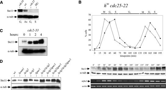
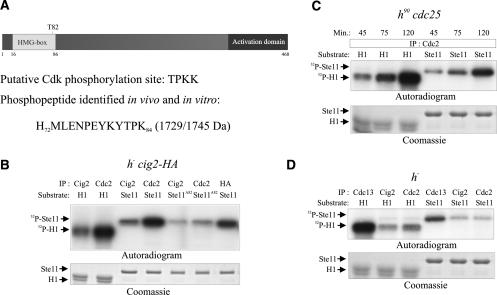
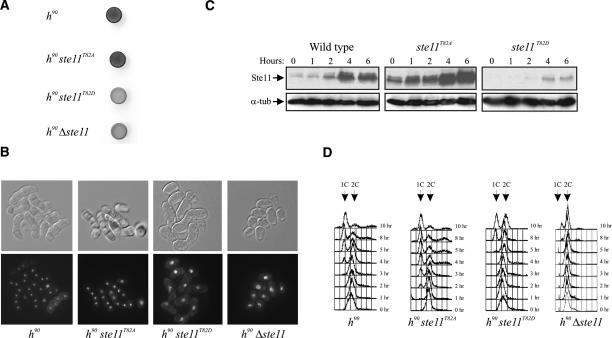
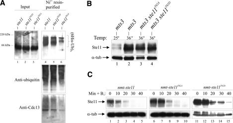
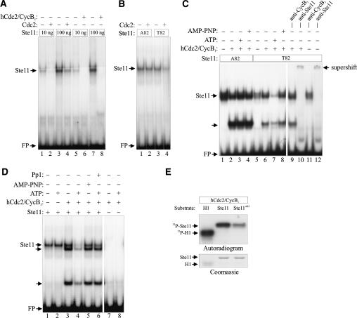
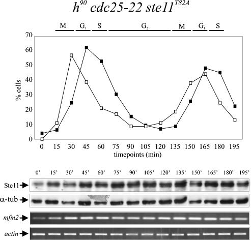
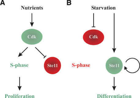
Eukaryotic cells normally differentiate from G(1); here we investigate the mechanism preventing expression of differentiation-specific genes outside G(1). In fission yeast, induction of the transcription factor Ste11 triggers sexual differentiation. We find that Ste11 is only active in G(1) when Cdk activity is low. In the remaining part of the cell cycle, Ste11 becomes Cdk-phosphorylated at Thr 82 (T82), which inhibits its DNA-binding activity. Since the ste11 gene is autoregulated and the Ste11 protein is highly unstable, this Cdk switch rapidly extinguishes Ste11 activity when cells enter S phase. When we mutated T82 to aspartic acid, mimicking constant phosphorylation, cells no longer underwent differentiation. Conversely, changing T82 to alanine rendered Ste11-controlled transcription constitutive through the cell cycle, and allowed mating from S phase with increased frequency. Thus, Cdk phosphorylation mediates periodic expression of Ste11 and its target genes, and we suggest this to be part of the mechanism restricting differentiation to G(1).


Figures

Figure 1.
Figure 1.
Periodicity of Ste11 protein levels and activity during the cell cycle. (A) Western analysis of Ste11 protein from asynchronously (AS) growing cells and cells arrested in G1, S, and G2. cdc10-V50 and cdc25-22 strains were arrested in G1 and G2, respectively, by raising the temperature to 36°C for 4 h. Wild-type cells were arrested in S phase by treatment with 20 mM hydroxy-urea (HU) for 4 h. (α-tub) Tubulin, which here serves as an internal control. (B) An exponentially growing cdc25-22 strain was shifted from 25°C to 35.5°C, and after 3.5 h of incubation the culture was released at the permissive temperature. After the release, the levels of Ste11 protein and mfm2 transcript were followed by Western analysis (middle panel) and RT–PCR (bottom panel), respectively. (Top panel) The synchrony of the culture was followed by determining the percentage of cells in anaphase (open squares) and cells with a septum (closed squares). (C) Ste11 accumulates when Cdc2 is inactivated. Western analysis of Ste11 protein from a cdc2-33 strain incubated at 36°C for 0, 1, 2, and 4 h. (D) Ste11 levels are up-regulated in cig2 mutants. Western analysis of Ste11 protein from exponentially growing cultures of wild-type cells and rum1, cig2, cig1, puc1, cig1 cig2, cig1 puc1, cig2 puc1, and cig1 cig2 puc1 mutants.
Figure 2.
Figure 2.
Ste11 is phosphorylated by Cdc2 in vitro and in vivo. (A) Schematic representation of Ste11 showing T82 that occurs in a Cdk phosphorylation consensus sequence, S/T-P-X-K/R. The 1729-kDa phosphopeptide identified from in vitro and in vivo phosphorylated Ste11 by mass spectrometry is shown below (for details, see Supplemental Material). (B–D) In vitro kinase assays. (B) h cig2-HA cells were grown to mid-exponential phase in minimal medium. Protein extracts were prepared and immunoprecipitated with anti-Cdc2, anti-Cig2, and anti-HA antibodies. Protein kinase activities were measured using histone H1 (H1), wild-type Ste111–113 (Ste11), and Ste11T82A1–113 (Ste11T82A) as substrates. Kinase assays were carried out for 15 min at 30°C, and the samples were separated by SDS-PAGE, followed by autoradiography (top panel) or Coomassie staining (bottom panel). (C) Samples from time points 45, 75, and 120 min from the synchronized cdc25 cells (see Fig. 1B) were analyzed for Cdc2 activity. Protein extracts were immunoprecipitated with anti-Cdc2 antibodies and kinase activities were measured as described above using histone H1 and wild-type Ste111–113 as substrates. (D) h cells were grown to mid-exponential phase in minimal medium. Protein extracts were immunoprecipitated with anti-Cdc13, anti-Cdc2, and anti-Cig2 antibodies. Kinase activities were measured as described above using histone H1 and wild-type Ste111–113 as substrates.
Figure 3.
Figure 3.
Characterization of ste11T82 mutants in fission yeast. (A,B) ste11T82D mutants are semisterile. (A) Iodine staining of cells plated on sporulation medium. Homothallic wild-type, ste11T82A, ste11T82D, and Δste11 strains were plated on MSA and incubated for 48 h prior to iodine staining. The same cells were fixed and micrographed. (B, bottom panel) Cells stained with DAPI. DIC images of the same cells are shown above. (C) T82 mutations affect the level of Ste11 protein in vegetatively growing cells and in nitrogen-starved cells. Western analysis of Ste11 protein from wild-type, ste11T82A, and ste11T82D strains nitrogen-starved for 0, 1, 2, 4, and 6 h. (D) The same strains together with a Δste11 strain were nitrogen-starved for 0, 1, 2, 3, 4, 5, 8, and 10 h, and the DNA content of the cells was analyzed by flow cytometry.
Figure 4.
Figure 4.
Ste11 is poly\ubiquitinated independently of T82 phosphorylation. (A) mts3-1, mts3-1 ste11T82A, and mts3-1 ste11T82D strains were grown at 25°C and shifted to 36°C for 4 h. The level of Ste11 was determined by Western analysis. (B) His6-ubiquitin was expressed from the nmt1 promoter in the same cells for 18 h at 25°C followed by 4 h at 36°C. (Left panel) Cell extracts were prepared from these cultures and analyzed for the presence of Ste11 by Western blotting (input) and subjected to Ni2+-NTA chromatography. Purified ubiquitin conjugates were analyzed by Western blotting using anti-Ste11 (top panel), anti-ubiquitin (middle panel), and anti-Cdc13 (bottom panel) antibodies. Western blot using anti-ubiquitin here serves as a loading control. (C) The half-life of wild-type Ste11, Ste11T82A, and Ste11T82D is approximately the same. Δste11 cells expressing wild-type Ste11 (lanes 15), Ste11T82A (lanes 610), and Ste11T82D (lanes 1115) from the nmt1 promoter were grown in the absence of thiamine. At time point 0, thiamine (6 μM) and cycloheximide (100 μg/mL) were added, and samples were taken every 10 min to determine Ste11 protein levels by Western blotting.
Figure 5.
Figure 5.
Cdc2 inhibits the DNA-binding activity of bacterially produced Ste11. (A) Purified GST-Ste11 (10–100 ng) was either directly assayed by EMSA for DNA-binding activity or was first incubated with either fission yeast Cdc2 immune complexes or with 2.5 ng (50 U) of recombinant human Cdc2–Cyclin B1 complex (New England Biolabs) in the presence of ATP. (B) Ten nanograms of GST-Ste11 and GST-Ste11T82A were either directly assayed by EMSA for DNA-binding activity or were first incubated with ATP and Cdc2 immunoprecipitated from fission yeast cells. (C) Twenty-five nanograms of GST-Ste11 and GST-Ste11T82A were either directly assayed by EMSA for DNA-binding activity or were first incubated with 0.5 ng of human Cdc2–Cyclin B1 complex in the presence or the absence of ATP or AMP-PNP. In addition, lanes 9 + 11 and lanes 10 + 12 contained anti-Cyclin B1 and anti-Ste11 antibodies, respectively. (D) In a similar assay, the effect of adding Protein Phosphatase 1 (Pp1) to Cdc2–Cyclin B1-phosphorylated GST-Ste11 was examined (cf. lanes 4 and 6). In addition the DNA-binding activity of Cdc2–Cyclin B1 was assayed in the absence of GST-Ste11 (lanes 7 + 8). (E) In vitro kinase assay. Protein kinase activities of recombinant human Cdc2–Cyclin B1 were measured using histone H1 (H1), wild-type Ste111–113 (Ste11), and Ste11T82A1–113 (Ste11T82A) as substrates. Kinase assays were carried out for 15 min at 30°C, and the samples were separated by SDS-PAGE, followed by autoradiography and Coomassie staining.
Figure 6.
Figure 6.
In ste11T82A mutants, the periodicity in expression of Ste11 and mfm2 is lost. An exponentially growing cdc25-22 ste11T82A strain was shifted from 25°C to 35.5°C, and after 3.5 h of incubation, the culture was released at the permissive temperature. After the release, the levels of Ste11 protein and mfm2 transcript were followed by Western analysis (middle panel) and RT–PCR (bottom panel), respectively. (Top panel) The synchrony of the culture was followed by determining the percentage of cells in anaphase (open squares) and cells with a septum (closed squares).
Figure 7.
Figure 7.
Model for activation of differentiation by a Cdk-sensitive autocatalytic switch. (A) When supply of nutrients is sufficient to support vegetative growth, Cdk activation in late G1 will trigger S phase and inactivate Ste11. (B) Following nutritional starvation, Cdk activity will be down-regulated, causing G1 arrest. This also activates the Ste11 positive feedback loop that drives cells into differentiation.

Similar articles

Cited by

References

    1. Benito J., Martin-Castellanos C., Moreno S., Martin-Castellanos C., Moreno S., Moreno S. Regulation of the G1 phase of the cell cycle by periodic stabilization and degradation of the p25rum1 CDK inhibitor. EMBO J. 1998;17:482–497. - PMC - PubMed
    1. Booher R.N., Alfa C.E., Hyams J.S., Beach D.H., Alfa C.E., Hyams J.S., Beach D.H., Hyams J.S., Beach D.H., Beach D.H. The fission yeast cdc2/cdc13/suc1 protein kinase: Regulation of catalytic activity and nuclear localization. Cell. 1989;58:485–497. - PubMed
    1. Colomina N., Gari E., Gallego C., Herrero E., Aldea M., Gari E., Gallego C., Herrero E., Aldea M., Gallego C., Herrero E., Aldea M., Herrero E., Aldea M., Aldea M. G1 cyclins block the Ime1 pathway to make mitosis and meiosis incompatible in budding yeast. EMBO J. 1999;18:320–329. - PMC - PubMed
    1. Connolly T., Beach D., Beach D. Interaction between the Cig1 and Cig2 B-type cyclins in the fission yeast cell cycle. Mol. Cell. Biol. 1994;14:768–776. - PMC - PubMed
    1. Costanzo M., Nishikawa J.L., Tang X., Millman J.S., Schub O., Breitkreuz K., Dewar D., Rupes I., Andrews B., Tyers M., Nishikawa J.L., Tang X., Millman J.S., Schub O., Breitkreuz K., Dewar D., Rupes I., Andrews B., Tyers M., Tang X., Millman J.S., Schub O., Breitkreuz K., Dewar D., Rupes I., Andrews B., Tyers M., Millman J.S., Schub O., Breitkreuz K., Dewar D., Rupes I., Andrews B., Tyers M., Schub O., Breitkreuz K., Dewar D., Rupes I., Andrews B., Tyers M., Breitkreuz K., Dewar D., Rupes I., Andrews B., Tyers M., Dewar D., Rupes I., Andrews B., Tyers M., Rupes I., Andrews B., Tyers M., Andrews B., Tyers M., Tyers M. CDK activity antagonizes Whi5, an inhibitor of G1/S transcription in yeast. Cell. 2004;117:899–913. - PubMed

Publication types

MeSH terms

LinkOut - more resources