

Global and regional effects of type 2 diabetes on brain tissue volumes and cerebral vasoreactivity
- PMID: 17290035
- PMCID: PMC2031924
- DOI: 10.2337/dc06-2052
Global and regional effects of type 2 diabetes on brain tissue volumes and cerebral vasoreactivity
Abstract
Objective: The aim of this study was to evaluate the regional effects of type 2 diabetes and associated conditions on cerebral tissue volumes and cerebral blood flow (CBF) regulation.
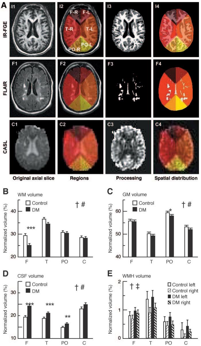
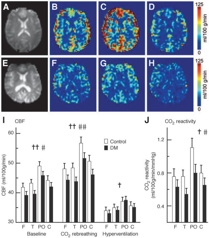
Research design and methods: CBF was examined in 26 diabetic (aged 61.6 +/- 6.6 years) and 25 control (aged 60.4 +/- 8.6 years) subjects using continuous arterial spin labeling (CASL) imaging during baseline, hyperventilation, and CO2 rebreathing. Regional gray and white matter, cerebrospinal fluid (CSF), and white matter hyperintensity (WMH) volumes were measured on a T1-weighted inversion recovery fast-gradient echo and a fluid attenuation inversion recovery magnetic resonance imaging at 3 Tesla.
Results: The diabetic group had smaller global white (P = 0.006) and gray (P = 0.001) matter and larger CSF (36.3%, P < 0.0001) volumes than the control group. Regional differences were observed for white matter (-13.1%, P = 0.0008) and CSF (36.3%, P < 0.0001) in the frontal region, for CSF (20.9%, P = 0.0002) in the temporal region, and for gray matter (-3.0%, P = 0.04) and CSF (17.6%, P = 0.01) in the parieto-occipital region. Baseline regional CBF (P = 0.006) and CO2 reactivity (P = 0.005) were reduced in the diabetic group. Hypoperfusion in the frontal region was associated with gray matter atrophy (P < 0.0001). Higher A1C was associated with lower CBF (P < 0.0001) and greater CSF (P = 0.002) within the temporal region.
Conclusions: Type 2 diabetes is associated with cortical and subcortical atrophy involving several brain regions and with diminished regional cerebral perfusion and vasoreactivity. Uncontrolled diabetes may further contribute to hypoperfusion and atrophy. Diabetic metabolic disturbance and blood flow dysregulation that affects preferentially frontal and temporal regions may have implications for cognition and balance in elderly subjects with diabetes.
Figures
References
-
- U.S. Department of Health and Human Services, Centers for Disease Control and Prevention . National Diabetes Fact Sheet: General Information and National Estimates on Diabetes in the United States, 2003. Revised ed. Centers for Disease Control and Prevention; Atlanta, GA: 2004.
-
- Trauernicht AK, Sun H, Patel KP, Mayhan WG. Enalapril prevents impaired nitric oxide synthase-dependent dilatation of cerebral arterioles in diabetic rats. Stroke. 2003;34:2698–2703. - PubMed
-
- Makimattila S, Malmberg-Ceder K, Hakkinen AM, Vuori K, Salonen O, Summanen P, Yki-Jarvinen H, Kaste M, Heikkinen S, Lundbom N, Roine RO. Brain metabolic alterations in patients with type 1 diabetes-hyperglycemia-induced injury. J Cereb Blood Flow Metab. 2004;24:1393–1399. - PubMed
-
- Vazquez LA, Amado JA, Garcia-Unzueta MT, Quirce R, Jimenez-Bonilla JF, Pazos F, Pesquera C, Carril JM. Decreased plasma endothelin-1 levels in asymptomatic type I diabetic patients with regional cerebral hypoperfusion assessed by Spect. J Diabetes Complications. 1999;13:325–331. - PubMed
-
- Jimenez-Bonilla JF, Carril JM, Quirce R, Gomez-Barquin R, Amado JA, Gutierrez-Mendiguchia C. Assessment of cerebral blood flow in diabetic patients with no clinical history of neurological disease. Nucl Med Commun. 1996;17:790–794. - PubMed
Publication types
MeSH terms
Substances
Grants and funding
LinkOut - more resources
Full Text Sources
Medical
