. 2007 May 1;175(9):911-8.
doi: 10.1164/rccm.200605-695OC. Epub 2007 Feb 8.

CD48 is critically involved in allergic eosinophilic airway inflammation

Affiliations

CD48 is critically involved in allergic eosinophilic airway inflammation

Ariel Munitz et al. Am J Respir Crit Care Med. .

Abstract

Rationale: Despite ongoing research, the molecular mechanisms controlling asthma are still elusive. CD48 is a glycosylphosphatidylinositol-anchored protein involved in lymphocyte adhesion, activation, and costimulation. Although CD48 is widely expressed on hematopoietic cells and commonly studied in the context of natural killer and cytotoxic T cell functions, its role in helper T cell type 2 settings has not been examined.

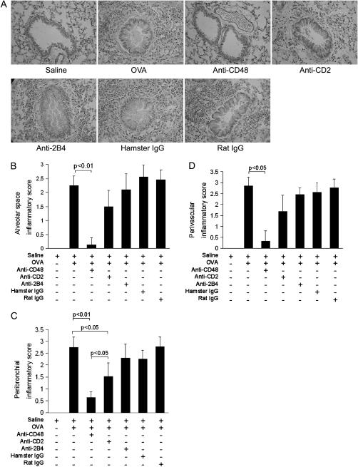
Objectives: To evaluate the expression and function of CD48, CD2, and 2B4 in a murine model of allergic eosinophilic airway inflammation.

Methods: Allergic eosinophilic airway inflammation was induced by ovalbumin (OVA)-alum sensitization and intranasal inoculation of OVA or, alternatively, by repeated intranasal inoculation of Aspergillus fumigatus antigen in wild-type, STAT (signal transducer and activator of transcription)-6-deficient, and IL-4/IL-13-deficient BALB/c mice. Gene profiling of whole lungs was performed, followed by Northern blot and flow cytometric analysis. Anti-CD48, -CD2, and -2B4 antibodies were administered before OVA challenge and cytokine expression and histology were assessed.

Measurements and main results: Microarray data analysis demonstrated upregulation of CD48 in the lungs of OVA-challenged mice. Allergen-induced CD48 expression was independent of STAT-6, IL-13, and IL-4. Neutralization of CD48 in allergen-challenged mice abrogated bronchoalveolar lavage fluid and lung inflammation. Neutralization of CD2 inhibited the inflammatory response to a lesser extent and neutralization of 2B4 had no effect.

Conclusions: Our results suggest that CD48 is critically involved in allergic eosinophilic airway inflammation. As such, CD48 may provide a new potential target for the suppression of asthma.


Figures

<b>Figure 1.</b>
Figure 1.
DNA microarray analysis identifies CD48 as an allergen-induced gene in allergic eosinophilic airway inflammation. Expression of CD48 in ovalbumin (OVA)–challenged mice (A) and Aspergillus fumigatus (Asp)–challenged mice (B) as measured by gene chip analysis is shown. The average difference for the hybridization signal after saline (shaded columns) and allergen (solid columns) challenge is depicted (n = 3 mice for Aspergillus control group, n = 4 mice for OVA control group, and n = 4 mice for OVA and Aspergillus experimental groups). *p < 0.05; **p < 0.01; ns = not significant. (C) The induction of CD48, 2B4, and CD2 in allergen-challenged mice as measured by Northern blot analysis. Total RNA was electrophoresed, transferred, and hybridized with a radiolabeled sequence-confirmed CD48 cDNA probe. The location of 18S RNA is shown. Each lane represents an extract from one separate mouse. EtBr = ethidium bromide.
<b>Figure 2.</b>
Figure 2.
CD48 expression is independent of signal transducer and activator of transcription (STAT)-6, IL-4, and IL-13. RNA was extracted from the lungs of wild-type (WT) mice (AD, left), STAT-6–deficient mice (A and B, right), and IL-13– and IL-4/IL-13–deficient mice (C and D, middle and right, respectively). Mice that express tetracycline-inducible IL-13 were fed doxycycline (Dox)–containing food for the indicated time periods (E). For all Northern blot assays, total RNA was electrophoresed, transferred, and hybridized with a radiolabeled sequence-confirmed CD48 cDNA probe. The location of 18S RNA is shown. Each lane represents an extract from one separate mouse. Asp = Aspergillus; OVA = ovalbumin.
<b>Figure 3.</b>
Figure 3.
Cellular source of CD48, CD2, and 2B4 in the lungs of mice sensitized with ovalbumin (OVA)–alum and challenged with OVA. Eighteen hours after the last allergen challenge the lungs were harvested and expression of CD48, CD2, and 2B4 on various cell types was analyzed. Data are represented as the change in mean fluorescence intensity (ΔMFI) ± SD (n = 4 mice). Eos = eosinophils; Neut = neutrophils; Lymph = CD4+ lymphocytes; Mac = macrophages; NK = NK cells; NKT = NKT cells. Values in parentheses indicate the percentage of the indicated cell type in bronchoalveolar lavage fluid (BALF).
<b>Figure 4.</b>
Figure 4.
Neutralization of CD48 attenuates eosinophilic inflammation and helper T cell type 2 and proinflammatory cytokine expression in bronchoalveolar lavage fluid (BALF). Mice sensitized with ovalbumin (OVA)–alum were treated with anti-CD48, anti-CD2, or anti-2B4 monoclonal antibodies (mAbs) or control antibodies (rat IgG or hamster IgG) on Day 23 and on Days 24 and 27, 1 hour before allergen challenge (250 μg/mouse). Twenty-four hours after the last allergen challenge bronchoalveolar lavage was performed and BALF cells were stained for differential cell identification. CCR3+/VLA-4+/CD3/SSChigh (high side scatter) cells were gated and considered eosinophils (A). IL-4, IL-5, IL-13, tumor necrosis factor (TNF)-α, and eotaxin-1 (BF, respectively) in BALF were detected by ELISA according to the manufacturers' instructions. Data are presented as means ± SD of n = 3 experiments (4–6 mice per group per experiment). OVA = ovalbumin.
<b>Figure 5.</b>
Figure 5.
Neutralization of CD48 attenuates lung inflammation. Mice were sensitized, challenged, and treated as described in text. Lung tissue was fixed, paraffin embedded, and stained with hematoxylin and eosin (H&E) for assessment of inflammation. Representative photomicrographs (original magnification, ×40) of airway inflammation in the various treatment groups (A). Quantitative analysis of alveolar space (B), lung perivascular (C), and peribronchial (D) inflammation is presented. Data represent means ± SD of n = 3 experiments (4–6 mice per group per experiment). OVA = ovalbumin.
<b>Figure 6.</b>
Figure 6.
Neutralization of CD48 attenuates goblet cell hyperplasia, mucus production, and smooth muscle thickening in lungs. Mice were sensitized, challenged, and treated as described in text. Lung tissue was fixed, paraffin embedded, and stained with periodic acid–Schiff reagent (PAS) for assessment of goblet cell hyperplasia and mucus production. (A) Representative photomicrographs (original magnification, ×40) of airway PAS staining in the various treatment groups. (B) Quantitative analysis of PAS+ cells in the bronchial epithelium. Shaded circles represent the mean peribronchial smooth muscle thickness in pixels of three midsized bronchioles per mouse. (C) Analysis of peribronchial smooth muscle thickening. Data represent means ± SD of n = 3 experiments (4–6 mice per group per experiment). OVA = ovalbumin.

Similar articles

Cited by

References

    1. Busse WW, Lemanske RF Jr. Asthma. N Engl J Med 2001;344:350–362. - PubMed
    1. Bochner BS, Busse WW. Allergy and asthma. J Allergy Clin Immunol 2005;115:953–959. - PubMed
    1. Busse WW, Rosenwasser LJ. Mechanisms of asthma. J Allergy Clin Immunol 2003;111:S799–S804. - PubMed
    1. Broide DH, Firestein GS. Endobronchial allergen challenge in asthma: demonstration of cellular source of granulocyte macrophage colony-stimulating factor by in situ hybridization. J Clin Invest 1991;88:1048–1053. - PMC - PubMed
    1. Barnes PJ. New drugs for asthma. Nat Rev Drug Discov 2004;3:831–844. - PubMed

Publication types

MeSH terms