Comparative Study
. 2007 Feb 21;26(4):1068-80.
doi: 10.1038/sj.emboj.7601571. Epub 2007 Feb 8.

Death effector domain DEDa, a self-cleaved product of caspase-8/Mch5, translocates to the nucleus by binding to ERK1/2 and upregulates procaspase-8 expression via a p53-dependent mechanism

Affiliations
Comparative Study

Death effector domain DEDa, a self-cleaved product of caspase-8/Mch5, translocates to the nucleus by binding to ERK1/2 and upregulates procaspase-8 expression via a p53-dependent mechanism

Zhan Yao et al. EMBO J. .

Abstract

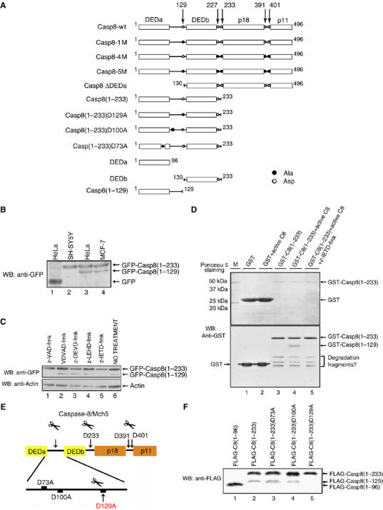
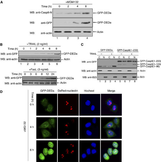
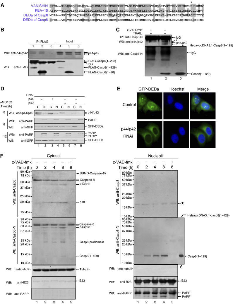
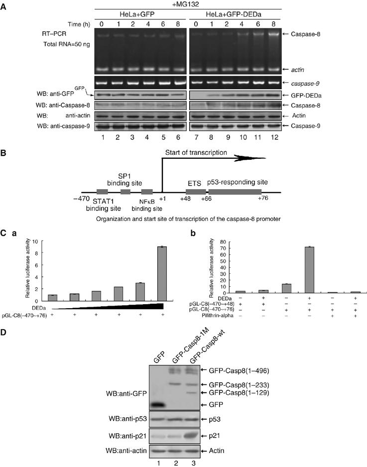
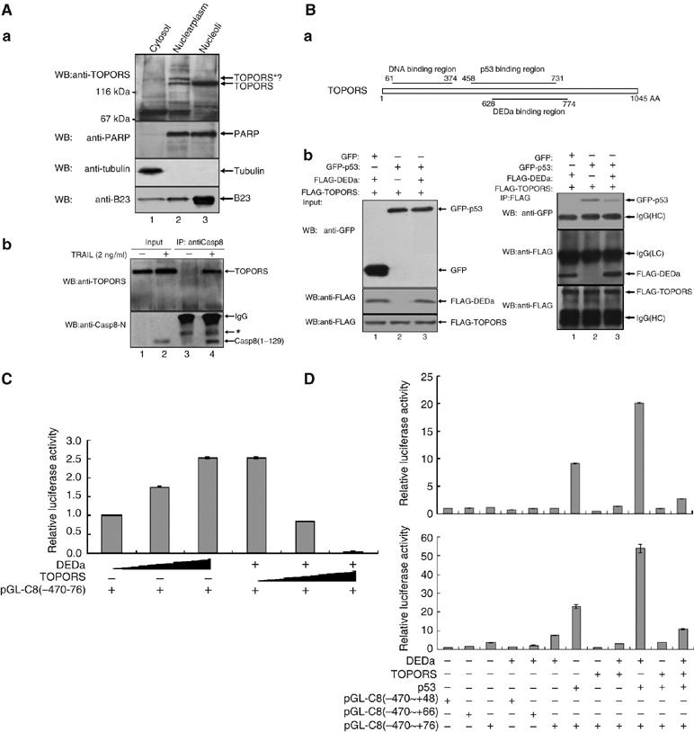
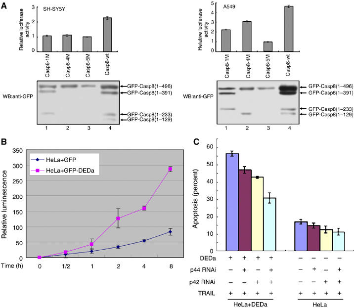
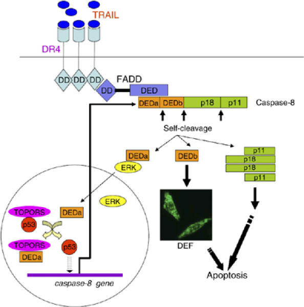
Activation of the apical caspase-8 is crucial to the extrinsic apoptotic pathway. Although the death effector domain (DED) of caspase-8 has been reported to be involved in death-inducing signaling complex formation, the detailed mechanism of how DED functions in regulating apoptosis remains largely unknown. Here, we demonstrate that the prodomain of the caspase-8/Mch5 can be further cleaved between two tandemly repeated DEDs (DEDa-DEDb) at the amino-acid residue Asp129 by caspase-8 itself. The DEDa fragment generated from the endogenous caspase-8 was detected in isolated nucleoli upon treatment with TRAIL (tumor necrosis factor-related apoptosis-inducing ligand). Cleaved DEDa appears to translocate into the nucleus by association with extracellular signal-regulated protein kinases-1/2 (ERK1/2). Elimination of ERK1/2 expression by RNA interference resulted in a significant attenuation of nuclear entry of DEDa and reduced caspase-8-dependent apoptosis. In the nucleus, DEDa interacts with TOPORS, a p53 and topoisomerase I binding protein, and possibly displaces p53 from TOPORS, allowing p53 to stimulate caspase-8 gene expression. In summary, we postulate a positive feedback loop involving DEDa, which enables the continual replenishment of procaspase-8 during apoptosis.


Figures

Figure 1
Figure 1
Processing of procaspase-8/Mch5 involves its self-induced cleavage at Asp129 in its prodomain. (A) Schematic representations of procaspase-8/Mch5 and its various mutants. Casp8, caspase-8; DED, death effecter domain; p18 and p11, the large and small subunits of mature caspase-8. The open circle represents the autocatalytic cleavage sites (aspartic acid) and the solid circle represents the mutations (alanine). The GFP or FLAG tag was fused to the N terminus of each indicated fragment in the plasmids pEGFP-C1 and p3XFLAG, respectively. (B) HeLa, SH-SY5Y and MCF-7 cell lines were transfected with pEGFP-Casp8(1–233), followed by immunoblotting with anti-GFP antibody. The empty vector pEGFP-C1 was used as a negative control. (C) HeLa cells were transfected with pEGFP-Casp8(1–233) in the presence of different caspase inhibitors as indicated. Cell lysates from the different treated cell groups were analyzed by Western blot using anti-GFP antibody. -Actin was used as the loading control. (D) Glutathione–agarose bead-tagged GST (lanes 1 and 2) or GST-C8(1–233) (lanes 3–5) were incubated with and without rh-caspase-8 (recombinant human caspase-8) (1 U) for 2 h at 37°C, and the resultant cleavage products were analyzed by immunoblotting with an anti-GST antibody (lower panel). The bacteria expressed GST and GST-C8(1–233) were visualized via Ponceau S staining (upper panel). The caspase-8-specific inhibitor z-IETD-fmk was preincubated with the active caspase-8 as indicated (lane 5). (E) A schematic illustration showing the auto-catalytic cleavage sites in procaspase-8/Mch5. The potential cleavage sites (D73, D100 and D129) between DEDa and DEDb were mutated as indicated. The arrowheads point to the actual cleavage sites. (F) HeLa cells were transfected with the indicated plasmids. Potential cleavage at the mutated sites was analyzed by Western blotting.
Figure 2
Figure 2
DEDa is stabilized and translocates from the cytoplasm into the nucleus upon apoptotic induction. (A) HeLa cell line stably expressing GFP-tagged DEDa was treated with MG132 (20 μM) over the indicated time periods. The expression level of GFP-DEDa was monitored by both anti-caspase-8 antibody (upper panel) and anti-GFP antibody (middle panel). β-Actin was used to verify equal loading (bottom panel). (B) HeLa cells stably expressing GFP-tagged DEDa were treated with TRAIL (upper panel) or FasL (lower panel) for the indicated time periods. Western blot was performed using anti-GFP antibody and anti-actin antibody. (C) HeLa cells were transiently transfected with pEGFP-DEDa (2 μg) or pEGFP-Casp8(1–233) (2 μg). Cells were treated with or without TRAIL (10 ng/ml) for 8 h and cytosolic and nuclear fractions from transfected cells were prepared. Subcellular localization of transfected GFP-DEDa (aa 1–96) and the cleaved DEDa (aa 1–129) was examined. The specificity of cytoplsmic or nuclear subcellular fractionation was confirmed by the detection of β-actin (a cytoplasm-specific protein) and B23 (nucleolus marker protein) respectively. (D) HeLa cells stably expressing GFP-DEDa (green fluorescence) were transfected with pDsRed1-C1/nucleolin (red fluorescence). Twenty-four hours post-transfection, cells were treated with MG132 (25 μM) for 0, 4 and 6 h, as indicated. In addition, the transfected cells were stained with Hoechst 33342 (10 μg/ml) for 15 min to mark the nuclei (blue fluorescence) before being subjected to fluorescence microscopy.
Figure 3
Figure 3
ERK1/2 associates with DEDa and is involved in DEDa nuclear translocation. (A) Alignment of amino-acid sequences among DEDa, DEDb and the DED regions of vanishin and PEA-15. Gray shading indicates identical residues. ‘*' indicates the conserved amino acid critical for ERK binding in PEA-15 and vanishin. (B) HeLa cells were transfected with p3XFLAG-Casp8(1–233) (lower panel, lane 4), p3XFLAG-DEDa (lower panel, lane 5) or the empty vector (lower panel, lane 6). Equal amounts of the cell extracts were then immunoprecipitated with an anti-FLAG antibody and analyzed by immunoblotting with an anti-p44/42 antibody to detect co-immunoprecipitated endogenous p42/p44 (top panel, lanes 1–3). The precipitates were also probed with an anti-FLAG antibody (bottom panel). (C) HeLa cells were treated with TRAIL for 12 h in the presence (lane 1) or absence of z-VAD-fmk (lane 2). The nucleoli were purified and the interaction of endogenous DEDa and p44/p42 was analyzed by co-IP using anti-caspase-8 (rabbit polyclonal antibody, BD Pharmingen), followed by Western blotting using rabbit anti-p44/42 antibody. The nuclear fraction of HeLa cells transfected with pcDNA3.1-Casp8(1–129) (untagged C8 1–129) was loaded as a migration control (lane 3). (D) HeLa cells stably expressing GFP-DEDa were transfected with or without siRNAs specific to p44 and p42, or both, as indicated. Seventy-two hours after two consecutive siRNA transfections with a 24-h interval, MG132 was added to allow the accumulation of DEDa for indicated periods of time (0, 6 and 12 h). Cytoplasmic and nuclear fractionations were prepared and further analyzed by immunoblotting using the indicated antibodies. Nuclear protein PARP was used as the nuclear marker and as the loading control as well. ‘*' denotes the cleaved fragments of PARP. (E) HeLa cells stably expressing GFP-DEDa were treated with or without p44/p42 siRNA as described above. MG132 (25 μM) was added for 8 h. Cells were stained with Hoechst 33342 to visualize the nuclei. Knockdown of endogenous ERK greatly diminished the nuclear localization of GFP-DEDa (visualized by green fluorescence). (F) HeLa cells (1 × 10⁸) were treated with TRAIL (2 ng/ml, R&D) in the absence or presence of z-VAD-fmk (lane 5) for the indicated time points and then fractionated into cytosolic and nucleolar fractions. Each fraction was probed with antibodies against p18 (anti-Casp8) or N-terminal (anti-Casp8-N) of caspase-8. A specific protein of about 15 kDa was detected by the anti-N-terminal caspase-8 antibody in the nucleolar fraction of HeLa cells treated with TRAIL. Untagged C8 1–129-transfected cell lysate was loaded as a migration control. B23, tubulin and PARP were used as markers.
Figure 4
Figure 4
DEDa upregulates caspase-8 in a p53-dependent manner. (A) HeLa cells stably transfected with GFP-DEDa were treated with MG132 to allow the accumulation of DEDa during the indicated time periods (0–8 h). Total RNA was isolated at each time point and analyzed by semiquantitative RT–PCR using primers specific for caspase-8 and caspase-9. The protein levels of GFP-DEDa, endogenous caspase-8 and caspase-9 were also determined by immunoblotting using the respective antibodies, as indicated. HeLa cells stably expressing GFP alone were examined as a negative control. (B) Schematic representation of the caspase-8 promoter. The transcriptional start site is indicated as +1. The sequence is numbered with respect to the start site. DNA binding sites of distinct transcription factors and the p53-responding element are denoted by gray boxes. (C) (a) A549 cells were cotransfected with a fixed amount of the luciferase reporter construct pGL3-C8(−470∼+76) (1 μg) and increasing amounts of p3XFLAG-DEDa (0, 25, 50, 100, 200 and 500 ng). The total DNA concentration in each transfection was kept constant by adjusting it with an empty vector. (b) A549 cells were cotransfected with either pGL3-C8(−470∼+48) (1 μg) or pGL3-C8(−470∼+76) (1 μg) in combination with p3XFLAG-DEDa or p3XFLAG vector (1 μg). Pifithrin-alpha (20 μM) was added (lanes 5 and 6) to inhibit p53-dependent gene transcription. For both (a) and (b), Renilla luciferase.plasmid pRL-CMV (3 ng) was introduced into all the transfected cells as an internal control. Luciferase activity was measured and plotted after normalizing with respect to Renilla luciferase activity. Data shown in both (a) and (b) are representative of three independent experiments. Vertical error bars are the average s.d.s of three independent values. (D) SH-SY5Y cells were transfected with an equal amount (1 μg) of pEGFP, pEGFP-Casp8-wt or pEGFP-Casp8-1M separately. The protein levels of endogenous p53 and its transcription target p21 were analyzed using Western blot.
Figure 5
Figure 5
DEDa interacts with TOPORS and displays p53 from the TOPORS/p53 complex. (A) (a) Cellular distribution of endogenous TOPORS was examined in cytosolic, nuclearplasmic and nucleolar fractions by immunoblotting (upper panel). PARP, tubulin and B23 were used to verify the purity of the fractions. (b) The nucleolar fractions isolated from HeLa cells treated with or without TRAIL were immunoprecipitated with anti-Casp8-N antibody. Co-precipitated TOPORS and DEDa were analyzed by immunoblotting using the indicated antibodies. (B) (a) A schematic illustration of the regions of TOPORS required for association with p53 and DEDa. Numbers indicate amino acid residues. (b) H1299 (p53−/−) cells were cotransfected with pCATCH-TOPORS (2 μg) and either pEGFP, pEGFP-p53 or pEGFP-p53 plus p3XFLAG-DEDa. Ten percent of the total cell lysate was subjected to immunoblotting to confirm an equal expression of transfected proteins (left panel). The remaining cell extracts from each transfectant were immunoprecipitated with an anti-FLAG antibody and subsequently immunoblotted with anti-GFP and anti-FLAG antibodies (right panel). (C) A549 cells were cotransfected with pGL3-C8(−470∼+76) (1 μg) and either with an increasing amount of p3XFLAG-DEDa (0–0.5 μg) (lanes 1–3) or a fixed amount of p3XFLAG-DEDa (0.5 μg) coupled with an increasing amount of pCATCH-TOPORS (0–0.5 μg) (lanes 4–6). Luciferase assay was performed identically as described above. (D) p53-null H1299 (upper panel) and p53 wild-type A549 cells (lower panel) were respectively transfected with either pGL3-C8(−470∼+48), pGL3-C8(−470∼+66) or pGL3-C8(−470∼+76) in various combinations with p3XFLAG-DEDa, pCATCH-TOPORS or p3XFLAG-p53 as indicated. Luciferase assay was performed as described above.
Figure 6
Figure 6
DEDa leads to increased amount of active caspase-8 and sensitizes HeLa cells to TRAIL-induced apoptosis. (A) SH-SY5Y and A549 cells were separately cotransfected with pGL3-C8(−470∼+76) (1 μg) and an equal amount (0.5 μg) of either pEGFPC1-Casp8-1M, pEGFPC1-Casp8-4M, pEGFPC1-Casp8-5M or pEGFPC1-Casp8-wt. Transfection efficiency was standardized against Renilla luciferase activity. Data are shown as fold induction of the luciferase activity versus control (luciferase activity obtained in cells cotransfected with pGL3-C8(−470∼+76) and pEGFPC1-Casp8-5M is defined as one-fold shown in lane 3). Immunoblotting was performed to ensure the expression and processing of these ectopic proteins using an anti-GFP antibody. (B) HeLa cells stably transfected with pEGFP-DEDa or pEGFP were treated with TRAIL (10 ng/ml, Sigma) for the indicated time periods. Activity of caspase-8 was measured using the Caspase-Glo™ 8 Assay kit (Promega) following the manufacturer's protocol. The data represent mean±s.d. of triplicate samples. (C) HeLa cell lines stably expressing GFP and GFP-DEDa were separately transfected with or without siRNAs specific for p44, p42 or both, as described in Figure 3D. Forty-eight hours after the second transfection with siRNA, cells were treated with TRAIL (10 ng/ml, Sigma). Apoptotic values were calculated as the percentage of apoptotic cells (condensed nuclei stained by Hoechst 33342) relative to the total number of cells in each random field (>200 cells) and represent the average of three independent experiments ±s.d.
Figure 7
Figure 7
A proposed model depicting a novel positive feedback circuit as an integral part of caspase-8-mediated apoptosis (see Discussion).

Similar articles

Cited by

References

    1. Andersen JS, Lyon CE, Fox AH, Leung AK, Lam YW, Steen H, Mann M, Lamond AI (2002) Directed proteomic analysis of the human nucleolus. Curr Biol 12: 1–11 - PubMed
    1. Banelli B, Casciano I, Croce M, Di Vinci A, Gelvi I, Pagnan G, Brignole C, Allemanni G, Ferrini S, Ponzoni M, Romani M (2002) Expression and methylation of CASP8 in neuroblastoma: identification of a promoter region. Nat Med 8: 1333–1335 - PubMed
    1. Barnhart BC, Lee JC, Alappat EC, Peter ME (2003) The death effector domain protein family. Oncogene 22: 8634–8644 - PubMed
    1. Boatright KM, Renatus M, Scott FL, Sperandio S, Shin H, Pedersen IM, Ricci JE, Edris WA, Sutherlin DP, Green DR, Salvesen GS (2003) A unified model for apical caspase activation. Mol Cell 11: 529–541 - PubMed
    1. Boldin MP, Goncharov TM, Goltsev YV, Wallach D (1996) Involvement of MACH, a novel MORT1/FADD-interacting protease, in Fas/APO-1- and TNF receptor-induced cell death. Cell 85: 803–815 - PubMed

Publication types

MeSH terms