Comparative Study
. 2007 Feb 21;26(4):1140-9.
doi: 10.1038/sj.emboj.7601557. Epub 2007 Feb 8.

Subcellular localization of Grb2 by the adaptor protein Dok-3 restricts the intensity of Ca2+ signaling in B cells

Affiliations
Comparative Study

Subcellular localization of Grb2 by the adaptor protein Dok-3 restricts the intensity of Ca2+ signaling in B cells

Björn Stork et al. EMBO J. .

Abstract

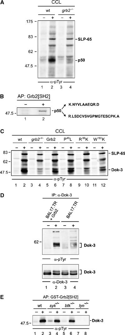
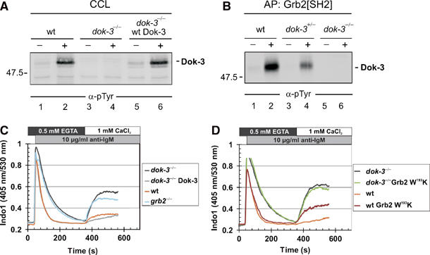
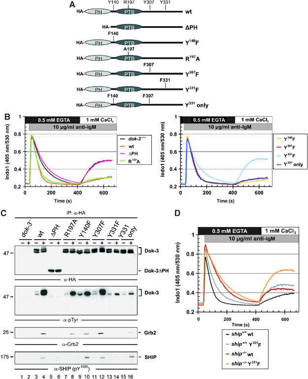
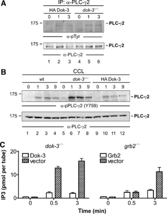
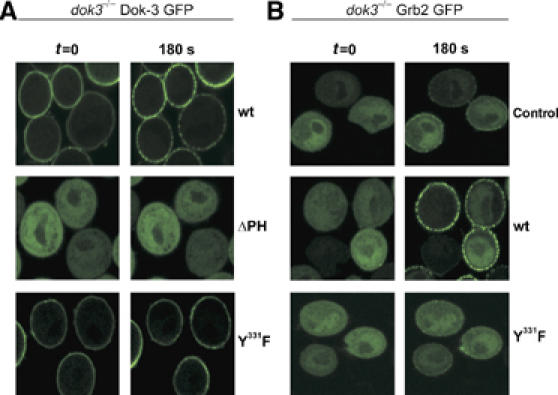
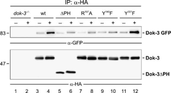
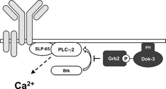
Spatial and temporal modulation of intracellular Ca2+ fluxes controls the cellular response of B lymphocytes to antigen stimulation. Herein, we identify the hematopoietic adaptor protein Dok-3 (downstream of kinase-3) as a key component of negative feedback regulation in Ca2+ signaling from the B-cell antigen receptor. Dok-3 localizes at the inner leaflet of the plasma membrane and is a major substrate for activated Src family kinase Lyn. Phosphorylated Dok-3 inhibits antigen receptor-induced Ca2+ elevation by recruiting cytosolic Grb2, which acts at this location as a negative regulator of Bruton's tyrosine kinase. This leads to diminished activation of phospholipase C-gamma2 and reduced production of soluble inositol trisphosphate. Hence, the Dok-3/Grb2 module is a membrane-associated signaling organizer, which orchestrates the interaction efficiency of Ca2+-mobilizing enzymes.


Figures

Figure 1
Figure 1
Grb2 controls Lyn-mediated phosphorylation of the adaptor protein Dok-3. (A) Wild-type (wt) and Grb2-deficient (grb2−/−) DT40 cells (lanes 1, 2 and 3, 4) were left untreated (−) or stimulated through their BCRs for 3 min (+). Equal amounts of proteins from cleared cellular lysates (CCL) were analyzed by anti-phosphotyrosine (α-pTyr) immunoblotting. (B) The major phosphotyrosine-containing protein, p50, was affinity-purified (AP) by GST-Grb2[SH2] from stimulated DT40 cells (lane 2), silver-stained, excised, digested by trypsine and peptide products were analyzed by ESI-Trap mass spectrometry. Purified proteins from unstimulated cells served as negative control (lane 1). The obtained amino-acid sequences are shown (single-letter code) with lysine (K) and arginine (R) being inferred from trypsine cleavage specificity (indicated by dots). These sequences matched a partial chicken EST (GenBank accession number XP_427516). Full-length chicken cDNA was isolated and submitted to GenBank with the accession number EF051736 (see also Supplementary Figure S1). (C) Wild-type (lanes 1 and 2) and grb2−/− DT40 cells (lanes 3 and 4) reconstituted with either wild-type Grb2 (lanes 5 and 6) or Grb2 variants, in which one of the three SH domains has been inactivated by single amino-acid substitution (N-terminal SH3 domain, P49L; SH2 domain, R86K; C-terminal SH3 domain, W193K; lanes 7–12), were left untreated (−) or stimulated through their BCRs (+). Equal amounts of proteins from CCL were subjected to anti-pTyr immunoblotting. To confirm equal loading, phospho-SLP-65 was detected separately by anti-SLP-65 immunoblotting (data not shown). (D) Murine Bal17.TR B cells, deficient for Grb2 expression, were transfected with an expression vector for Grb2 (lanes 1 and 2) or the empty vector as control (lanes 3 and 4) and left untreated (−) or stimulated through their BCRs (+). CCL were subjected to anti-Dok-3 immunoprecipitation and purified proteins were analyzed by immunoblotting with antibodies to pTyr and Dok-3 (upper and lower panels, respectively). (E) Resting (−) or BCR-activated (+) wild-type DT40 cells (lanes 1 and 2) or variants deficient for the protein tyrosine kinase Syk (lanes 3 and 4), Btk (lanes 5 and 6) or Lyn (lanes 7 and 8) were lysed and subjected to affinity purification with GST-Grb2[SH2]. Phosphorylated Dok-3 was detected by anti-pTyr immunoblotting. Relative molecular masses of marker proteins are indicated on the left in kDa.
Figure 2
Figure 2
Gene targeting reveals a negative regulatory role of Dok-3. (A) Dok-3-deficient DT40 B cells were generated by targeted disruption of both dok-3 alleles (dok-3−/−, see Materials and methods for details), and absence of tyrosine-phosphorylated p50/Dok-3 in cleared cellular lysates (CCL) of resting (−) and BCR-activated (+) cells was tested by anti-pTyr immunoblotting (lanes 3 and 4). As control, wild-type DT40 and Dok-3-reconstituted dok-3−/− cells were analyzed in parallel (lanes 1 and 2 and 5 and 6, respectively). (B) Wild-type DT40 cells (lanes 1 and 2), heterozygous dok-3+/− (lanes 3 and 4) and homozygous dok-3−/− mutants (lanes 5 and 6) were left untreated (−) or stimulated through their BCRs (+). Cell lysates were subjected to affinity purification with the GST-Grb2[SH2] fusion protein and proteins so obtained were analyzed by anti-pTyr immunoblotting. Relative molecular mass of marker protein is indicated in (A) and (B) on the left in kDa. (C, D) BCR-induced intra- and extracellular Ca2+ mobilization of the indicated DT40 cells was recorded by flow cytometry as described in detail in Materials and methods. Briefly, cells were loaded with Indo-1 and release of intracellular Ca2+ was measured for 6 min in the presence of EGTA. Subsequently, extracellular Ca2+ was restored to 1mM in order to monitor Ca2+ entry across the plasma membrane. Lines represent wild-type DT40 (orange), dok-3−/− mutants (black), Dok-3-reconstituted dok-3−/− cells (gray), grb2−/− mutants (blue) and wild-type and dok-3−/− transfectants expressing the dominant-negative W193K version of Grb2 (brown and green, respectively). Data are representative of at least three independent measurements.
Figure 3
Figure 3
Dok-3 and Grb2 build a functional unit, that inhibits Ca2+ flux independent of SHIP. (A) Schematic representation of expression constructs encoding HA-tagged versions of wild-type Dok-3, a PH domain deletion mutant (ΔPH) or mutants encompassing amino-acid exchanges depicted in single-letter code. (B) Expression vectors were introduced by retroviral transduction in dok-3−/− mutants and BCR-induced Ca2+ mobilization of the transfectants was measured by flow cytometry, as described in the legend to Figure 2. Wild-type DT40 cells and empty vector transfectants of dok-3−/− mutants served as control (see inlay for color code). (C) Wild-type and DT40 variants described in (B) were left untreated (−) or BCR-activated (+) and lysates were subjected to anti-HA immunoprecipitation. Expression and tyrosine phosphorylation of Dok-3 proteins, as well as their association to Grb2 and SHIP, were detected by sequential immunoblotting with antibodies to HA, pTyr, Grb2 and SHIP (upper to lower panels, respectively). (D) BCR-induced Ca2+ fluxes were analyzed as described in the legend to Figure 2 in SHIP-deficient DT40 cells (ship−/−, brown) and ship−/− transfectants expressing a Dok-3 Y331F variant that counteracts Ca2+ inhibition by endogenous wild-type Dok-3 (orange). As control, parental DT40 cells, which are positive for endogenous SHIP and Dok-3 (black), and the Dok-3 Y331F transfectants (gray) were analyzed in parallel, demonstrating the dominant-negative function of Dok-3 Y331F.
Figure 4
Figure 4
The Dok-3/Grb2 module attenuates PLC-γ2 activity. (A) Dok-3-deficient DT40 mutants (lanes 4–6) and reconstituted cells expressing HA-tagged wild-type Dok-3 (lanes 1–3) were left untreated (0) or stimulated through their BCRs for the indicated times (min). Lysates were subjected to anti-PLC-γ2 immunopurification and proteins obtained were analyzed by anti-pTyr and anti-PLC-γ2 immunoblotting (upper and lower panels, respectively). (B) Parental DT40 cells (lanes 1–4), dok-3−/− mutants (lanes 5–8) and HA-Dok-3-reconstituted transfectants (lanes 9–12) were left untreated (0) or stimulated through their BCRs for the indicated times (min). Cleared cellular lysates (CCL) were subjected to immunoblot analysis with antibodies that specifically detect PLC-γ2 phosphorylation at the Btk-dependent phospho-acceptor site corresponding to Y759 in human PLC-γ2 (upper panel). Equal protein loading was confirmed by reprobing the membrane with anti-PLC-γ2 antibodies (lower panel). Relative molecular mass of marker protein is indicated in (A) and (B) on the left in kDa. (C) DT40 mutant cells deficient for either Dok-3 (left panel) or Grb2 (right panel) and the empty vector control transfectants (open and filled bars, respectively) were left untreated (0) or BCR-activated for 0.5 or 3 min. IP3 levels in these cells were measured using a competitive binding assay with radiolabelled IP3-binding proteins. Error bars represent s.e.m. of three independent experiments with double preparation.
Figure 5
Figure 5
Dok-3 is permanently localized at the plasma membrane and is essential for stimulation-dependent recruitment of Grb2. (A) Dok-3-deficient DT40 mutants were transfected with expression constructs encoding fusion proteins between the green fluorescence protein (GFP) at the C terminus and either wild-type Dok-3 (upper row), Dok-3ΔPH (middle row) or Dok-3 Y331F (lower row) at the N terminus. Subcellular localization of Dok-3/GFP fusion proteins in resting (t=0) or BCR-activated (180 s) cells (left and right images) was visualized by confocal laser scanning microscopy. (B) Dok-3−/− DT40 cells expressing a Grb2/GFP fusion protein were transfected with either empty control vector (upper row) or expression vectors encoding wild-type or Y331F Dok-3 mutants (middle and lower rows). Subcellular Grb2 localization was analyzed as in (A). The Ca2+ signaling function of GFP fusion proteins was tested separately (data not shown).
Figure 6
Figure 6
Dok-3 undergoes stimulation-dependent homo-oligomerization. Dok-3−/− mutant cells were reconstituted with Dok-3/GFP and subsequently transfected with empty control vector (lanes 1 and 2) or expression constructs for either HA-tagged wild-type Dok-3 (lanes 3 and 4) or the indicated HA-tagged Dok-3 variants (lanes 5–12; for details, see Figure 3A). Lysates were subjected to anti-HA immunoprecipitation. Proteins obtained were analyzed by immunoblotting with antibodies to GFP (upper panel) and HA peptide tag (lower panel). Relative molecular masses of marker proteins are indicated on the left in kDa.
Figure 7
Figure 7
Inhibition of BCR-induced Ca2+ signaling by the Dok-3/Grb2 module. The Dok-3 adaptor protein is tethered at the inner side of the plasma membrane by virtue of its PH domain. Following tyrosine phosphorylation in activated B cells, Dok-3 recruits Grb2, which at this specific subcellular location attenuates Btk-dependent activation of PLC-γ2 by interfering with the proper formation of the SLP-65-assembled Ca2+ initiation complex and/or inhibiting the enzymatic activity of Btk. Positive and negative regulators of Ca2+ elevation are illustrated by open and filled boxes, respectively. The BCR complex is depicted in gray.

Similar articles

Cited by

References

    1. Adachi T, Wienands J, Wakabayashi C, Yakura H, Reth M, Tsubata T (2001) SHP-1 requires inhibitory co-receptors to down-modulate B cell antigen receptor-mediated phosphorylation of cellular substrates. J Biol Chem 276: 26648–26655 - PubMed
    1. Bolland S, Pearse RN, Kurosaki T, Ravetch JV (1998) SHIP modulates immune receptor responses by regulating membrane association of Btk. Immunity 8: 509–516 - PubMed
    1. Boulay I, Nemorin JG, Duplay P (2005) Phosphotyrosine binding-mediated oligomerization of downstream of tyrosine kinase (Dok)-1 and Dok-2 is involved in CD2-induced Dok phosphorylation. J Immunol 175: 4483–4489 - PubMed
    1. Brauweiler A, Tamir I, Dal Porto J, Benschop RJ, Helgason CD, Humphries RK, Freed JH, Cambier JC (2000) Differential regulation of B cell development, activation, and death by the Src homology 2 domain-containing 5′ inositol phosphatase (SHIP). J Exp Med 191: 1545–1554 - PMC - PubMed
    1. Chiu CW, Dalton M, Ishiai M, Kurosaki T, Chan AC (2002) BLNK: molecular scaffolding through cis-mediated organization of signaling proteins. EMBO J 21: 6461–6472 - PMC - PubMed

Publication types