Comparative Study
. 2007 Feb 21;26(4):935-43.
doi: 10.1038/sj.emboj.7601540. Epub 2007 Feb 8.

Opposed growth factor signals control protein degradation in muscles of Caenorhabditis elegans

Affiliations
Comparative Study

Opposed growth factor signals control protein degradation in muscles of Caenorhabditis elegans

Nathaniel J Szewczyk et al. EMBO J. .

Abstract

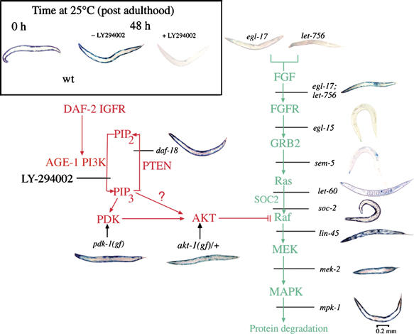
In addition to contractile function, muscle provides a metabolic buffer by degrading protein in times of organismal need. Protein is also degraded during adaptive muscle remodeling upon exercise, but extreme degradation in diverse clinical conditions can compromise function(s) and threaten life. Here, we show how two independent signals interact to control protein degradation. In striated muscles of Caenorhabditis elegans, reduction of insulin-like signaling via DAF-2 insulin/IGF receptor or its intramuscular effector PtdIns-3-kinase (PI3K) causes unexpected activation of MAP kinase (MAPK), consequent activation of pre-existing systems for protein degradation, and progressive impairment of mobility. Degradation is prevented by mutations that increase signal downstream of PI3K or by disruption of autocrine signal from fibroblast growth factor (FGF) via the FGF receptor and its effectors in the Ras-MAPK pathway. Thus, the activity of constitutive protein degradation systems in normal muscle is minimized by a balance between directly interacting signaling pathways, implying that physiological, pathological, or therapeutic alteration of this balance may contribute to muscle remodeling or wasting.


Figures

Figure 1
Figure 1
Intramuscular DAF-2 signaling opposes protein degradation in muscle. Animals were grown to mid-adulthood at 15°C and then shifted to 25°C for an additional 48 h either without or with the protein synthesis inhibitor CHx (400 μg/ml) added 6 h before temperature upshift. (A) Histochemical stain for β-galactosidase activity in muscle (blue). Reporter activity in wild-type animals (top row) remains stable after temperature shift even when treated with CHx. daf-2(m41) animals (second row) show a loss of reporter activity after temperature upshift; this is not prevented by CHx pretreatment. The loss of activity in daf-2(m41) animals is rescued by muscle-specific expression of wild-type DAF-2 (Punc-54daf-2+, third row) or by a gain-of-function mutation pdk-1(mg142) (bottom row). (B) Immunoblot (Zdinak et al, 1997) of 146 kDa myosin-β-galactosidase fusion protein (using monoclonal anti-β-galactosidase antibody) in 30-worm lysates confirms that the loss of staining seen in (A) corresponds to degradation of the reporter protein. (C) Quantitation (NIH Image software) of protein levels in (B) and of β-galactosidase activity by fluorimetric assay (Zdinak et al, 1997) of 10-worm lysates (means±s.d. of three independent determinations) confirms that protein degradation (squares) and loss of activity (circles) proceed at similar rates in daf-2 animals (red) whereas no degradation occurs in wild-type (black) or daf-2; pdk-1 (gf) animals (green).
Figure 2
Figure 2
Intramuscular DAF-2 and AGE-1 signaling opposes loss of movement. Animals were grown to mid-adulthood at 15°C (0 h) and then an additional 24 or 48 h at 25°C. Measurements of animal movement rate (Szewczyk et al, 2002) were made for each strain (10 replicates on each of 10 animals of each genotype) at each time point. (A) daf-2(m41ts) mutant animals without (white) or with (striped) the reporter protein display a time-dependent loss of mobility at a rate comparable to reporter protein loss (Figure 1C). Like reporter protein loss, loss of mobility is rescued in animals expressing wild-type daf-2+ only in muscle (shaded) and prevented by gain-of-function mutation in pdk-1 (black). (B) age-1 animals without (white) or with (striped) the reporter protein display a time-dependent loss of movement comparable to daf-2 animals (A). Like reporter protein loss (Figure 3), mobility loss is rescued in animals expressing wild-type age-1+ only in muscle (black). Differences between rescued or suppressed animals compared with unrescued mutant animals were significant (**) at P<0.001 by two-tailed t-test.
Figure 3
Figure 3
Intramuscular AGE-1 (PtdIns-3-kinase) signaling opposes protein degradation. Animals were grown to mid-adulthood at 15°C (left) or for an additional 48 h at 25°C either without (middle) or with (right) the PtdIns-3-kinase inhibitor LY-294002 (LY, 160 μM). Wild-type animals (top) degrade the reporter protein when treated with LY; this is not prevented by CHx pretreatment. age-1(hx546ts) (second row) mutants degrade the reporter protein upon shift to 25°C even in the absence of LY. Reporter degradation in age-1(mg44) null-mutant animals is prevented by muscle-specific expression of wild-type AGE-1 from a transgene (Punc-54age-1+) (third row), but this rescue can be overcome by treatment with LY. A null mutation of the forkhead transcription factor gene daf-16(mgDf50) does not suppress LY-induced protein degradation (fourth row).
Figure 4
Figure 4
DAF-2 signaling (red pathway) opposes degradation-promoting signal from FGFs (green pathway). All animals were grown to mid-adulthood at 15°C, then treated for 48 h at 25°C with the PtdIns-3-kinase inhibitor LY-294002 (160 μM). This causes LacZ degradation in wild-type animals (Inset). Mutations that increase signal downstream of AGE-1 (pdk-1(mg142), daf-18(e1375), or akt-1(mg144), animals at left) block protein degradation triggered by LY treatment. Mutations that decrease signal at Raf (lin-45(sy96)), MEK (mek-2(ku114)), or MAPK (mpk-1(n2521)) also block protein degradation during LY treatment (lower right), but degradation is not prevented by reduction-of-function mutations affecting FGFR (egl-15(n1783)), GRB-2 (sem-5(n1779 or n1619)), or Ras (let-60(n2021)). Degradation is also blocked in a double mutant (egl-17(n1377); let-756(s2613)) deficient in both FGF homologs, but not in either single mutant. Quantitative data for rates of degradation in FGFR pathway mutants have been published (Szewczyk and Jacobson, 2003; Szewczyk et al, 2002).
Figure 5
Figure 5
MPK-1 MAPK is diphosphorylated (activated) after inhibition of PI3K. Adult animals were treated with 160 μM LY-294002 for the indicated times at 25°C. Immunoblotting of pTpY-MPK-1 after electrophoresis on 12% SDS–polyacrylamide gels was conducted with monoclonal anti-pTpY-ERK antibody (Szewczyk and Jacobson, 2003). Each lane contained a 50-worm lysate. The untreated animals (0 h) show no immunoreactive pTpY-MPK-1.
Figure 6
Figure 6
Increased PtdIns-P3 signal blocks protein degradation triggered by activated FGFR. Animals were grown to mid-adulthood at 15°C or for an additional 48 h at 25°C. (A) Histochemical stain for β-galactosidase activity in muscle (blue). Activation of FGFR at 25°C (clr-1(e1745ts) animals, top row) triggers the loss of reporter activity (Szewczyk and Jacobson, 2003). This can be prevented by a mutation (daf-18(e1375) animals, bottom) that increases the level of PtdIns-P3 (see pathway, Figure 4). (B) Immunoblot of 146 kDa β-galactosidase fusion protein in 30-worm lysates confirms that the loss of staining seen in (A) corresponds to degradation of the reporter protein. (C) Quantitation (NIH Image software) of protein levels in (B) and of β-galactosidase activity by fluorimetric assay (Zdinak et al, 1997) of 10-worm lysates (means±s.d. of three independent determinations) confirms that protein degradation (squares) and loss of activity (circles) proceed at similar rates in clr-1 animals (red) whereas little degradation is observed in clr-1; daf-18 animals (green). To facilitate comparison, the dashed lines are taken from Figure 1C and the same scale is used. (D) Immunoblot of pTpY-MPK-1 (30-worm lysate per lane) confirms that MPK-1 is activated in clr-1 animals, and that daf-18 mutation greatly reduces MPK-1 activation (37% as much pTpY-MPK-1 at 24 h and 43% as much at 48 h). Samples were run on the same gel and detected on the same membrane to permit direct comparison of pTpY-MPK-1 levels.
Figure 7
Figure 7
Induced expression of the lin-45AA transgene provokes protein degradation. The transgene, driven by a heat-shock promoter, is carried on an extrachromosomal array marked with a GFP transgene constitutively expressed in gut cells (see Materials and methods). Only animals that expressed the GFP marker were used. Young adult animals were heat-shocked for 40 min at 37°C where indicated, then returned to 20°C for 48 h. (A) Histochemical stain for β-galactosidase activity in muscle (blue). (B) Immunoblot of 146 kDa myosin-LacZ fusion protein in 30-worm lysates confirms that the loss of staining seen in (A) corresponds to degradation of the reporter protein, with 27% of reporter protein remaining at 48 h. (C) Immunoblot of pTpY-MPK-1 (30-worm lysate per lane) confirms that MPK-1 is activated in heat-shocked lin-45AA animals.

Similar articles

Cited by

References

    1. Andreatta C, Nahreini P, Hovland B, Kumar J, Edwards-Prasad J, Prasad K (2001) Use of short-lived green fluorescent protein for the detection of proteasome inhibition. BioTechniques 30: 656–660 - PubMed
    1. Attaix D, Combaret L, Pouch MN, Taillandier D (2001) Regulation of proteolysis. Curr Opin Clin Nutr Metab Care 4: 45–49 - PubMed
    1. Avery AW, Figueroa C, Vojtek AB (2007) UNC-51-like kinase regulation of fibroblast growth factor receptor substrate 2/3. Cell Signal 19: 177–184 - PubMed
    1. Babar P, Adamson C, Walker GA, Walker DW, Lithgow GJ (1999) P13-kinase inhibition induces dauer formation, thermotolerance and longevity in C. elegans. Neurobiol Aging 20: 513–519 - PubMed
    1. Bence NF, Sampat RM, Kopito RR (2001) Impairment of the ubiquitin-proteasome system by protein aggregation. Science 292: 1552–1555 - PubMed

Publication types

MeSH terms