. 2007 Mar;117(3):803-11.
doi: 10.1172/JCI30142. Epub 2007 Feb 8.

Injury enhances TLR2 function and antimicrobial peptide expression through a vitamin D-dependent mechanism

Affiliations

Injury enhances TLR2 function and antimicrobial peptide expression through a vitamin D-dependent mechanism

Jürgen Schauber et al. J Clin Invest. 2007 Mar.

Abstract

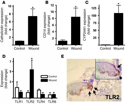
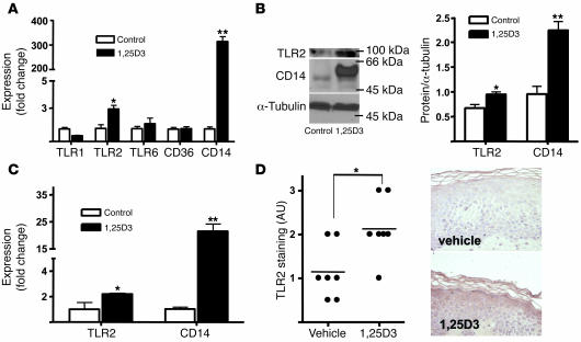
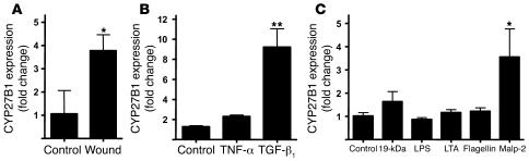
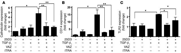
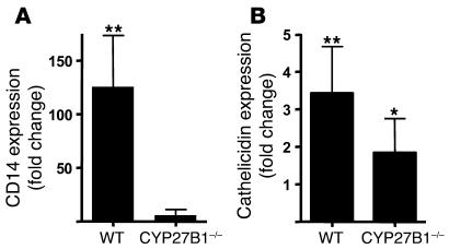
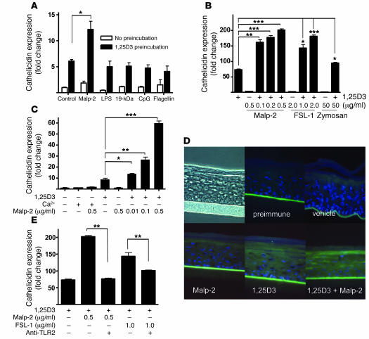
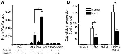
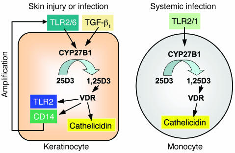
An essential element of the innate immune response to injury is the capacity to recognize microbial invasion and stimulate production of antimicrobial peptides. We investigated how this process is controlled in the epidermis. Keratinocytes surrounding a wound increased expression of the genes coding for the microbial pattern recognition receptors CD14 and TLR2, complementing an increase in cathelicidin antimicrobial peptide expression. These genes were induced by 1,25(OH)2 vitamin D3 (1,25D3; its active form), suggesting a role for vitamin D3 in this process. How 1,25D3 could participate in the injury response was explained by findings that the levels of CYP27B1, which converts 25OH vitamin D3 (25D3) to active 1,25D3, were increased in wounds and induced in keratinocytes in response to TGF-beta1. Blocking the vitamin D receptor, inhibiting CYP27B1, or limiting 25D3 availability prevented TGF-beta1 from inducing cathelicidin, CD14, or TLR2 in human keratinocytes, while CYP27B1-deficient mice failed to increase CD14 expression following wounding. The functional consequence of these observations was confirmed by demonstrating that 1,25D3 enabled keratinocytes to recognize microbial components through TLR2 and respond by cathelicidin production. Thus, we demonstrate what we believe to be a previously unexpected role for vitamin D3 in innate immunity, enabling keratinocytes to recognize and respond to microbes and to protect wounds against infection.


Figures

Figure 1
Figure 1. Injury triggers increased TLR2, CD14, and CYP24A1 in skin.
Human wounds 24 hours after full-thickness sterile skin incision were evaluated for the expression of innate immune recognition and response molecules (n = 5). Transcript abundance was measured by qPCR, normalized to GAPDH expression, and compared with noninjured skin (n = 4). (A) Wounded skin showed an expected increase in expression of cathelicidin, a vitamin D3–responsive antimicrobial gene. Additional vitamin D3–responsive genes, the TLR co-receptor CD14 (B) and the vitamin D3 catabolic enzyme CYP24A1 (C), also increased after injury. (D) Expression of TLR2 mRNA, but not that of TLR1, TLR4, and TLR6, was increased after injury. (E) A corresponding increase in TLR2 protein staining on keratinocytes at the wound edges was seen (original magnification, ×100). The incision site is marked by black arrows. An enlarged sector is displayed in the inset (original magnification, ×400). *P < 0.05, Mann-Whitney test.
Figure 2
Figure 2. The effect of 1,25D3 on the expression of TLRs in cultured keratinocytes.
(A) Expression of TLR2 and CD14 mRNA was increased by 1,25D3 (100 nM) in cultured keratinocyte monolayers after 24 hours. (B) Expression of TLR2 and CD14 protein was increased by 1,25D3 (100 nM) in monolayer keratinocyte extracts evaluated by Western blot and quantified by image density analysis. (C) Keratinocytes grown in differentiated epidermal constructs stimulated with 1,25D3 (100 nM) also showed an increase in CD14 and TLR2 transcript abundance. Data are mean ± SD of a representative experiment performed in triplicate. *P < 0.05, **P < 0.01, Student’s t test. (D) Skin from healthy volunteers (n = 7) was treated with 1,25D3 (1.0 mM applied once). Controls are contralateral skin treated with vehicle. After 4 days, punch biopsies from both sites were obtained, and skin sections were stained for TLR2 expression. Staining intensity — graded according to the intensity of immunoreactivity (0, no expression; 3, strong expression) — increased in patients treated with topical 1,25D3, as determined by an investigator blinded to treatment group. Sections from 1 representative study participant are displayed. *P < 0.05, Mann-Whitney test.
Figure 3
Figure 3. CYP27B1 is increased in response to injury, TGF-β1, or activation of TLR2.
(A) The expression of CYP27B1, which converts inactive 25D3 to active 1,25D3, was evaluated in skin wounds as in Figure 1. Wounded skin (n = 5) increased CYP27B1 mRNA compared with controls (n = 4). *P < 0.05, Mann-Whitney test. (B) Keratinocytes were cultured in the presence of TNF-α (20 ng/ml) or TGF-β1 (1 μg/ml) for 24 hours, after which RNA was isolated and CYP27B1 transcript abundance was analyzed by qPCR. (C) Keratinocytes were cultured with different TLR ligands for 24 hours, after which CYP27B1 expression analyzed as described above. Both TGF-β1 and the TLR2 ligand Malp-2 induced CYP27B1 expression. 19-kDa, 19-kDa lipopeptide; LTA, lipoteichoic acid. Data are mean ± SD of a single experiment performed in triplicate and are representative of 3 independent experiments. *P < 0.05, **P < 0.01, Student’s t test.
Figure 4
Figure 4. TGF-β1 leads to an increase of 1,25D3-responsive genes in wounds by activation of CYP27B1.
Keratinocytes were pretreated with the VDR antagonist ZK159222 (VAZ; 10–7 M) or the CYP27B1 antagonist itraconazole (ITRA; 10–7 M) and then stimulated with TGF-β1 (1 ng/ml) in the presence or absence of 25D3 (10 nM) for 24 hours. Expression of cathelicidin (A), CD14 (B), and TLR2 (C) mRNA was determined as described in Figure 2. Induction of these innate immune effector and response genes by TGF-β1 found in wounds was dependent on the availability of 25D3 and the activity of CYP27B1 or a functional VDR. Data are mean ± SD of a single experiment performed in triplicate and are representative of 3 independent experiments. *P < 0.05, **P < 0.01, Student’s t test.
Figure 5
Figure 5. Activation of CYP27B1 is responsible for increased vitamin D3 signaling in wounds.
Wounds from CYP27B1–/– mice and age-matched wild-type littermates 24 hours after full-thickness sterile skin incision were evaluated for the expression of innate immune recognition and response molecules (n = 5 per group). Transcript abundance in wounded skin was measured by qPCR and normalized to noninjured skin from the same animal. (A) CD14 increased in wild-type animals after injury but not in mice lacking CYP27B1. (B) Cathelicidin was induced in wounds from both wild-type and CYP27B1–/– animals. Although cathelicidin induction was less in CYP27B1–/– mice, the difference was not statistically significant. *P < 0.05, **P < 0.01 versus control uninjured skin, Mann-Whitney test.
Figure 6
Figure 6. TLR2 function in keratinocytes is increased by 1,25D3.
(A) To test the functional relevance of the increased expression of TLR2 and CD14 in keratinocytes exposed to 1,25D3, cells were incubated with a low dose of 1,25D3 (0.1 nM for 24 hours) and then stimulated with TLR ligands for an additional 24 hours. Cathelicidin mRNA abundance was measured as in Figure 2. Only TLR2/6 ligand Malp-2 increased expression above that induced by low-dose 1,25D3 alone. (B) Dose-dependent response of cathelicidin expression following administration of TLR2/6 agonists Malp-2, FSL-1, or zymosan at the indicated concentrations in the presence of 1,25D3 (100 nM). (C) Cathelicidin mRNA expression in keratinocytes incubated with a low dose of 1,25D3 (0.1 nM) or a high calcium concentration (1.7 mM) for 24 hours and then stimulated with Malp-2 for another 24 hours. (D) Organotypic epidermal constructs were stimulated with Malp-2 (0.1 μg/ml) alone or in the presence of 1,25D3 (100 nM), and cathelicidin peptide expression was determined by immunostaining. Constructs were stained with a polyclonal anti–LL-37 antibody (green), and nuclei were detected with DAPI (blue). Original magnification, ×400. (E) Cathelicidin mRNA expression was inhibited by a TLR2-neutralizing antibody applied to keratinocytes stimulated with Malp-2 or FSL-1 in the presence of 1,25D3 (100 nM). Data are mean ± SD of a single experiment performed in triplicate and are representative of 3 independent experiments. *P < 0.05, **P < 0.01, ***P < 0.001, Student’s t test.
Figure 7
Figure 7. TLR2 activation signals cathelicidin induction through the vitamin D3 pathway.
(A) A functional VDRE was required for transcriptional activation of cathelicidin by Malp-2 and 1,25D3. HaCaT keratinocytes containing cathelicidin promoter reporter constructs (pGL3 1,500) were treated with Malp-2 (0.1 μg/ml) in the presence or absence of 1,25D3 (100 nM). Promoter constructs with a deleted VDRE at position –619 bp to –633 bp (pGL3 1,500–VDRE) lost transcriptional activity in all experiments. Values represent the ratio between firefly and Renilla luciferase activities. (B) In addition, treatment of keratinocytes with the VDR antagonist ZK159222 (10–7 M) blocked Malp-2–induced cathelicidin. Data are mean ± SD of a single experiment performed in triplicate and are representative of 3 independent experiments. *P < 0.05, Student’s t test.
Figure 8
Figure 8. Schematic model for 1,25D3-regulated innate immune functions in keratinocytes and monocytes.
Two distinct 1,25D3-dependent pathways in keratinocytes and monocytes are shown. In skin injury, keratinocytes are activated by TGF-β1 or TLR2/6 ligands, which then leads to induction of CYP27B1. As a consequence 25D3 is converted to 1,25D3, which, upon activation of the VDR, induces cathelicidin, TLR2, and CD14. The 1,25D3-induced TLR2 enables the response of keratinocytes to TLR2 activation, resulting in further increased cathelicidin expression. In contrast, circulating monocytes are activated by TLR2/1 agonists. As a consequence, the genes encoding the VDR and CYP27B1 are induced. CYP27B1 converts 25D3 to 1,25D3 and subsequently increases cathelicidin.

Similar articles

Cited by

References

    1. Roth D.E., et al. Association between vitamin D receptor gene polymorphisms and response to treatment of pulmonary tuberculosis. J. Infect. Dis. 2004;190:920–927. - PubMed
    1. Wayse V., Yousafzai A., Mogale K., Filteau S. Association of subclinical vitamin D deficiency with severe acute lower respiratory infection in Indian children under 5 y. Eur. J. Clin. Nutr. 2004;58:563–567. - PubMed
    1. Barnes P.F., Modlin R.L., Bikle D.D., Adams J.S. Transpleural gradient of 1,25-dihydroxyvitamin D in tuberculous pleuritis. J. Clin. Invest. 1989;83:1527–1532. - PMC - PubMed
    1. Liu P.T., et al. Toll-like receptor triggering of a vitamin D-mediated human antimicrobial response. Science. 2006;311:1770–1773. - PubMed
    1. Zasloff M. Fighting infections with vitamin D. Nat. Med. 2006;12:388–390. - PubMed

Publication types

MeSH terms