

Relationship among neuroimaging indices of cerebral health during normal aging
- PMID: 17290369
- PMCID: PMC6870647
- DOI: 10.1002/hbm.20369
Relationship among neuroimaging indices of cerebral health during normal aging
Abstract
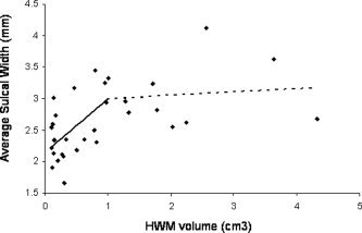
Sensitive measures of brain aging show great promise for gauging factors that affect aging and degenerative processes, such as risk genes or therapy. Here we examined age-related trends for three indices of cerebral health: gyral gray matter (GM) thickness, dilation of sulcal spaces with CSF, and the volume of T2-hyperintense white matter (HWM) lesions. The study involved 31 healthy adults age 57-82 years old. Measurements of average GM thickness, average sulcal span and HWM volume were performed using high-resolution 3D T1- and T2-weighted brain MR images. Age-related trends for the three cerebral health indices were consistent with previously published work though the analysis of their covariance led to a previously unreported relationship. Simultaneous multiple regression found that dilation of cortical sulci were primarily (t = 2.59, P < 0.01) related to the increases in HWM volume and secondarily related (t = -2.51, P < 0.01) to the reductions of the cortical GM thickness. The are-corrected correlation between reduction in GM thickness and increases in HWM volume, was not significant (P = 0.34). These findings are of interest in designing quantitative measures of brain aging for monitoring individual patients and in large-scale clinical trials.
Figures


Similar articles
-
Limited relationships between two-year changes in sulcal morphology and other common neuroimaging indices in the elderly.Neuroimage. 2013 Dec;83:12-7. doi: 10.1016/j.neuroimage.2013.06.058. Epub 2013 Jun 22. Neuroimage. 2013. PMID: 23800792
-
Age-related morphology trends of cortical sulci.Hum Brain Mapp. 2005 Nov;26(3):210-20. doi: 10.1002/hbm.20198. Hum Brain Mapp. 2005. PMID: 16161162 Free PMC article.
-
Loss of cerebral white matter structural integrity tracks the gray matter metabolic decline in normal aging.Neuroimage. 2009 Mar 1;45(1):17-28. doi: 10.1016/j.neuroimage.2008.11.010. Epub 2008 Nov 25. Neuroimage. 2009. PMID: 19095067 Free PMC article.
-
Relationship between white matter fractional anisotropy and other indices of cerebral health in normal aging: tract-based spatial statistics study of aging.Neuroimage. 2007 Apr 1;35(2):478-87. doi: 10.1016/j.neuroimage.2006.12.021. Epub 2006 Dec 23. Neuroimage. 2007. PMID: 17292629
-
Positron emission tomography metabolic data corrected for cortical atrophy using magnetic resonance imaging.Alzheimer Dis Assoc Disord. 1996 Fall;10(3):141-70. doi: 10.1097/00002093-199601030-00005. Alzheimer Dis Assoc Disord. 1996. PMID: 8876777
Cited by
-
Identification of Early-Stage Alzheimer's Disease Using Sulcal Morphology and Other Common Neuroimaging Indices.PLoS One. 2017 Jan 27;12(1):e0170875. doi: 10.1371/journal.pone.0170875. eCollection 2017. PLoS One. 2017. PMID: 28129351 Free PMC article.
-
Blood pressure and cerebral white matter share common genetic factors in Mexican Americans.Hypertension. 2011 Feb;57(2):330-5. doi: 10.1161/HYPERTENSIONAHA.110.162206. Epub 2010 Dec 6. Hypertension. 2011. PMID: 21135356 Free PMC article.
-
Age-related effects on cortical thickness patterns of the Rhesus monkey brain.Neurobiol Aging. 2012 Jan;33(1):200.e23-31. doi: 10.1016/j.neurobiolaging.2010.07.010. Epub 2010 Aug 30. Neurobiol Aging. 2012. PMID: 20801549 Free PMC article.
-
Alterations of the cerebral cortex in sporadic small vessel disease: A systematic review of in vivo MRI data.J Cereb Blood Flow Metab. 2016 Apr;36(4):681-95. doi: 10.1177/0271678X15625352. Epub 2016 Jan 19. J Cereb Blood Flow Metab. 2016. PMID: 26787108 Free PMC article.
-
Abuse of amphetamines and structural abnormalities in the brain.Ann N Y Acad Sci. 2008 Oct;1141:195-220. doi: 10.1196/annals.1441.031. Ann N Y Acad Sci. 2008. PMID: 18991959 Free PMC article. Review.
References
-
- Bastos Leite AJ,Scheltens P,Barkhof F ( 2004): Pathological aging of the brain: An overview. Top Magn Reson Imaging 15: 369–389. - PubMed
-
- Csernansky JG,Wang L,Miller JP,Galvin JE,Morris JC ( 2005): Neuroanatomical predictors of response to donepezil therapy in patients with dementia. Arch Neurol 62: 1718–1722. - PubMed
-
- de Leeuw FE,de Groot JC,Achten E,Oudkerk M,Ramos LM,Heijboer R,Hofman A,Jolles J,van Gijn J,Breteler MM ( 2001): Prevalence of cerebral white matter lesions in elderly people: A population based magnetic resonance imaging study. The Rotterdam Scan Study. J Neurol Neurosurg Psychiatry 70: 9–14. - PMC - PubMed
Publication types
MeSH terms
Grants and funding
LinkOut - more resources
Full Text Sources
Medical
