. 2007 Mar 30;355(1):194-9.
doi: 10.1016/j.bbrc.2007.01.138. Epub 2007 Feb 2.

The mouse dead-end gene isoform alpha is necessary for germ cell and embryonic viability

Affiliations

The mouse dead-end gene isoform alpha is necessary for germ cell and embryonic viability

Chitralekha Bhattacharya et al. Biochem Biophys Res Commun. .

Abstract

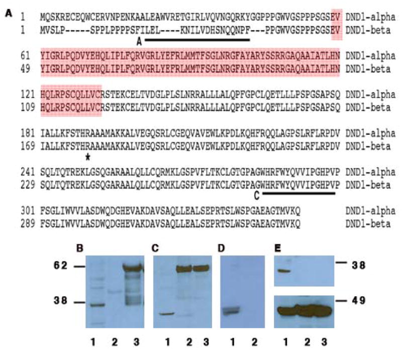
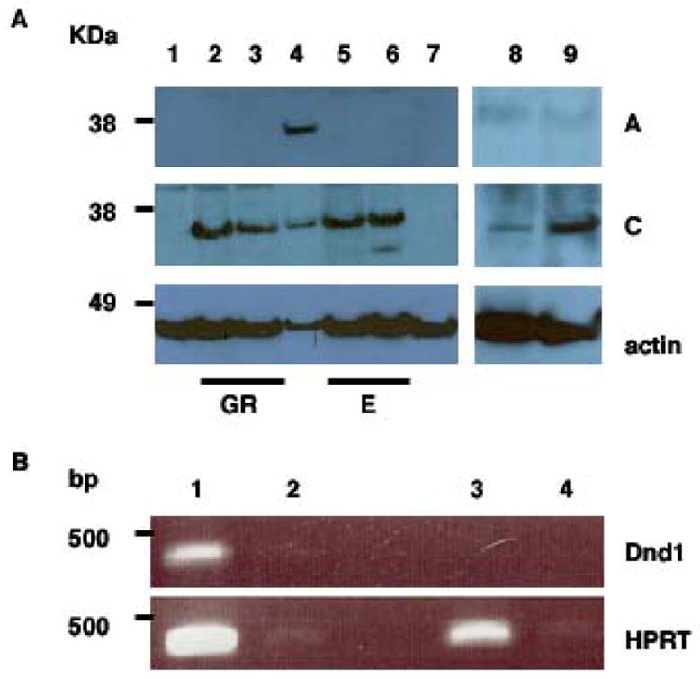
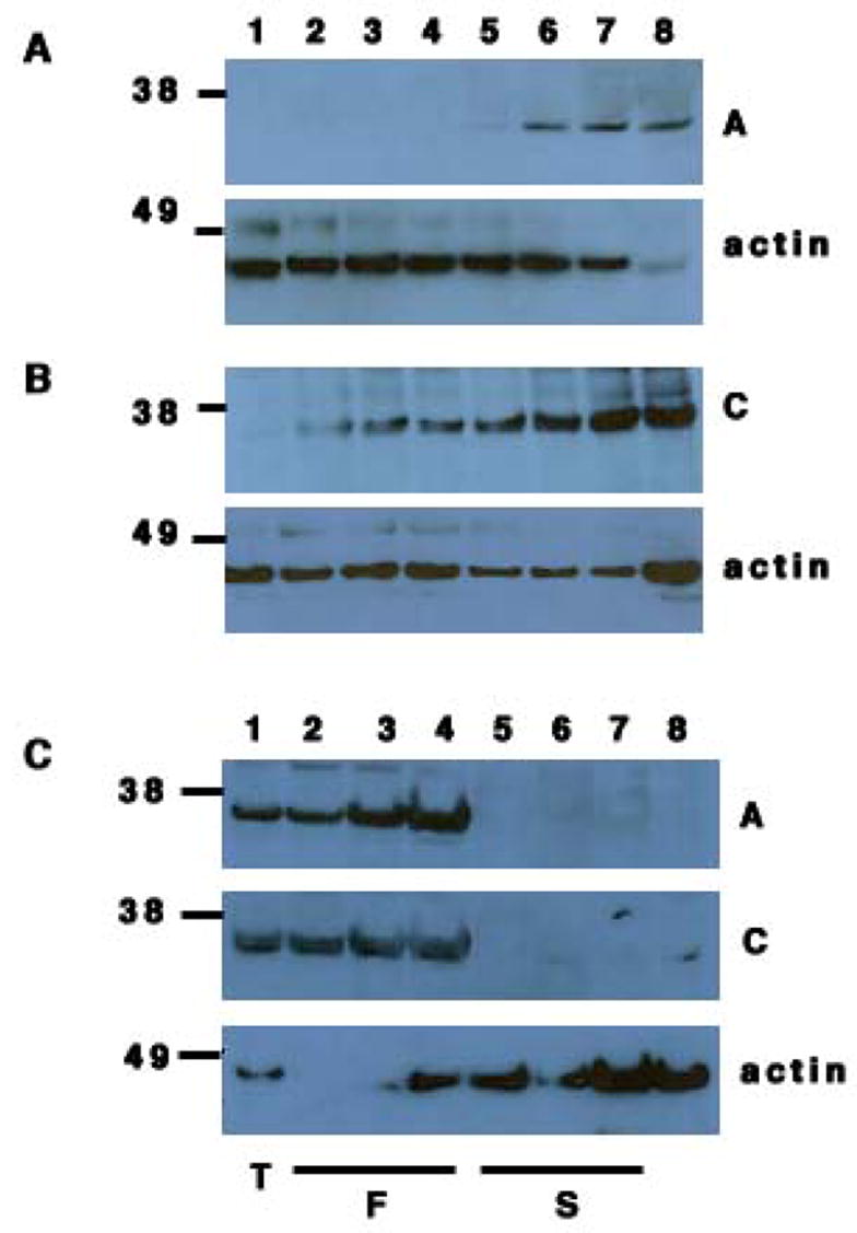
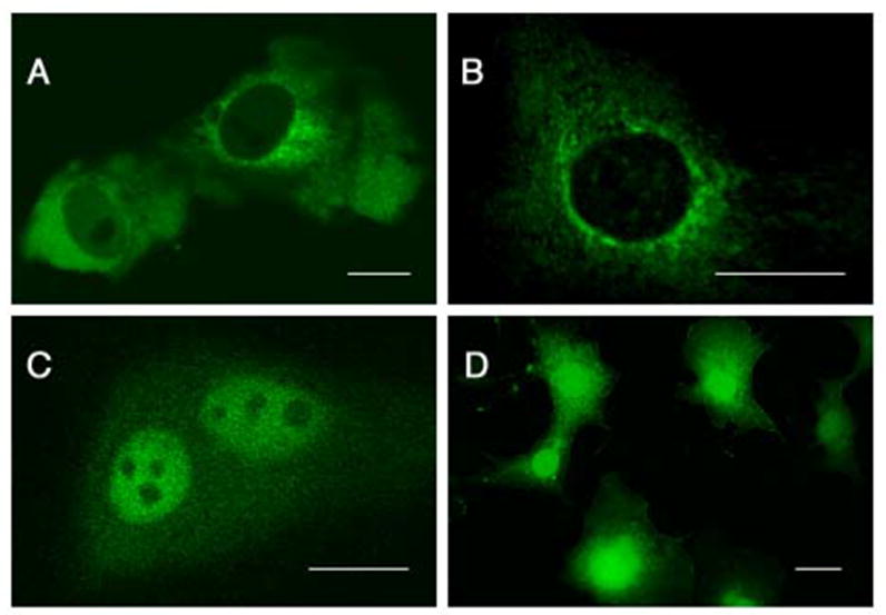
Inactivation of the dead-end (Dnd1) gene in the Ter mouse strain results in depletion of primordial germ cells (PGCs) so that mice become sterile. However, on the 129 mouse strain background, loss of Dnd1 also increases testicular germ cell tumor incidence in parallel to PGC depletion. We report that inactivation of Dnd1 also affects embryonic viability in the 129 strain. Mouse Dnd1 encodes two protein isoforms, DND1-isoform alpha (DND1-alpha) and DND1-isoform beta (DND1-beta). Using isoform-specific antibodies, we determined DND1-alpha is expressed in embryos and embryonic gonads whereas DND1-beta expression is restricted to germ cells of the adult testis. Our data implicate DND1-alpha isoform to be necessary for germ cell viability and therefore its loss in Ter mice results in PGC depletion, germ cell tumor development and partial embryonic lethality in the 129 strain.


Figures

Figure 1
Figure 1. The mouse DND1-α and DND1-β protein isoforms
(A) Sequence comparison of DND1 isoforms (GenBank:AAQ63636 and AAH34897). A and C (underlined) mark the sequences to generate antibody A and C. The red box indicates the RNA recognition motif. The asterisk (*) marks the amino acid (R) that is mutated to a stop codon in Ter mice. (B) Western blotting using antibody A of testes lysate (lane 1); GST-DND1-α (lane 2) and GST-DND1-β (lane 3). (C) Western blotting using antibody C of testes lysate (lane 1); GST-DND1-α (lane 2) and GST-DND1-β (lane 3). (D) Western blotting using both antibody A and C of normal testes (lane 1) and spleen (lane 2). (E) (top panel) Western blotting using antibody C of normal testes (lane 1); germ cell deficient testes from Ter/Ter (lane 2); testicular tumor from Ter/Ter (lane 3). Rehybridization of the blot with anti-βactin antibody (bottom panel).
Figure 2
Figure 2. Expression of DND1-α and DND1-β in embryonic tissues
(A) Lysates from embryonic testes (GR) at E13.5 (lane 2) and E15.5 (lane 3), adult testes (lane 4), E13.5 embryo (lane 5), E15.5 embryo (lane 6), EG (lane 8) and ES cells (lane 9), were electrophoresed before western blotting with antibody A (top panel); antibody C (middle panel) and anti-β actin (bottom panel). Negative control: mouse spleen (lane 1) and germ cell tumor lysates from Ter/Ter mice (lane 7). GR = embryonic testis/genital ridges; E = embryos. (B) (top panel) RT-PCR of total RNA from EG cells (lanes 1 and 2) grown on Mitomycin-C treated MEF cells and Mitomycin-C treated MEF cells alone (lanes 3 and 4). Lanes 2 and 4 are control lanes where no superscript was added during RT. (bottom panel) Control RT-PCR for HPRT.
Figure 3
Figure 3. Expression of DND1-α and DND1-β adult testis
(A) Western botting of lysates from post-natal testes of day 1, 6, 10, 15, 20, 25, 30 and 50 (lanes 1, 2, 3, 4, 5, 6, 7 and 8 respectively) with antibody A (top panel) and anti-β actin (bottom panel). (B) Western blotting of lysates from post-natal testes of day 1, 6, 10, 15, 20, 25, 30 and 50 (lanes 1, 2, 3, 4, 5, 6, 7 and 8 respectively) with antibody C (top panel) and anti-β actin (bottom panel). (C) Western blotting of lysates from adult testes (lane 1), fractionated cells from adult testes: pachytene spermatids (lane 2), round spermatids (lane 3) and elongated spermatids (lane 4); Sertoli cell lines, TM4 (lane 5), 15P-1 (lane 6), MSC1 (lane 7) and germ cell deficient Ter/Ter testis (lane 8) with antibody A (top panel); antibody C (middle panel) and anti-β actin (bottom panel).
Figure 4
Figure 4. Mammalian DND1 localizes to the cytoplasm or nucleus in different cell types
Subcellular localization of mouse GFP-DND1-α (A) and GFP-DND1-β (B) in COS-7 cells and He La cells (C). Control transfection of vector encoding GFP only (D) in COS-7 results in GFP expression in both cytoplasm and nucleus. Line indicates 20 μm.

Similar articles

Cited by

References

    1. Oosterhuis JW, Looijenga LHJ. Testicular germ-cell tumours in a broader perspective. Nature Rev Cancer. 2005;5:210–222. - PubMed
    1. Rescorla FJ. Pediatric germ cell tumors. Semin Surgical Oncology. 1999;16:144–158. - PubMed
    1. Horwich A, Shipley J, Huddart R. Testicular germ-cell cancer. Lancet. 2006;367:754–765. - PubMed
    1. Noguchi T, Noguchi M. A recessive mutation (ter) causing germ cell deficiency and a high incidence of congenital testicular teratomas in 129/Sv-ter mice. J Natl Cancer Inst. 1985;75:385–392. - PubMed
    1. Stevens LC. A new inbred subline of mice (129/terSv) with a high incidence of spontaneous congenital testicular teratomas. J Natl Cancer Inst. 1973;50:235–242. - PubMed

Publication types

MeSH terms