Comparative Study
. 2007 Apr 1;304(1):382-93.
doi: 10.1016/j.ydbio.2006.12.039. Epub 2006 Dec 23.

Transcription reactivation steps stimulated by oocyte maturation in C. elegans

Affiliations
Comparative Study

Transcription reactivation steps stimulated by oocyte maturation in C. elegans

Amy K Walker et al. Dev Biol. .

Abstract

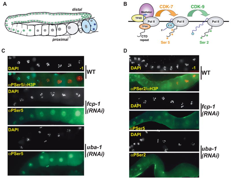
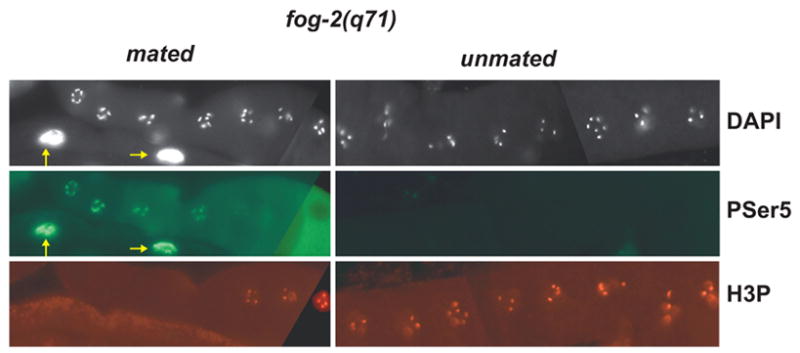
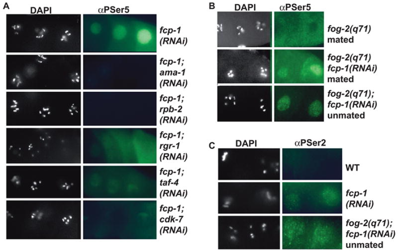
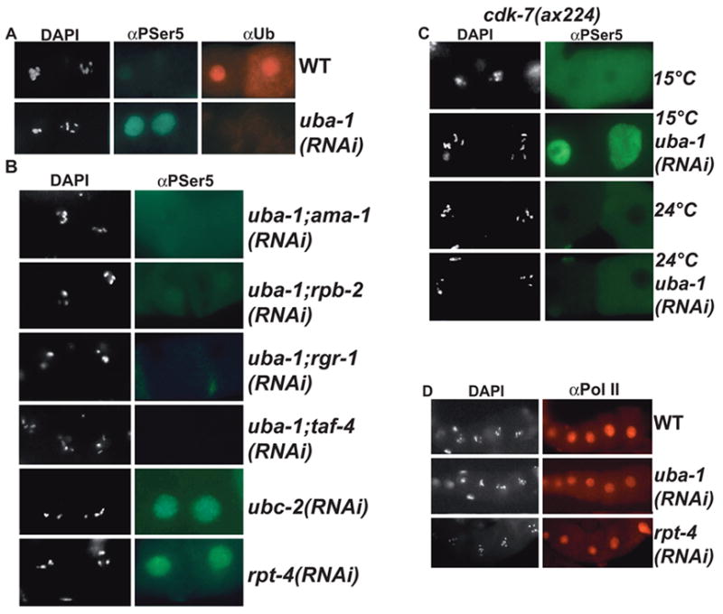
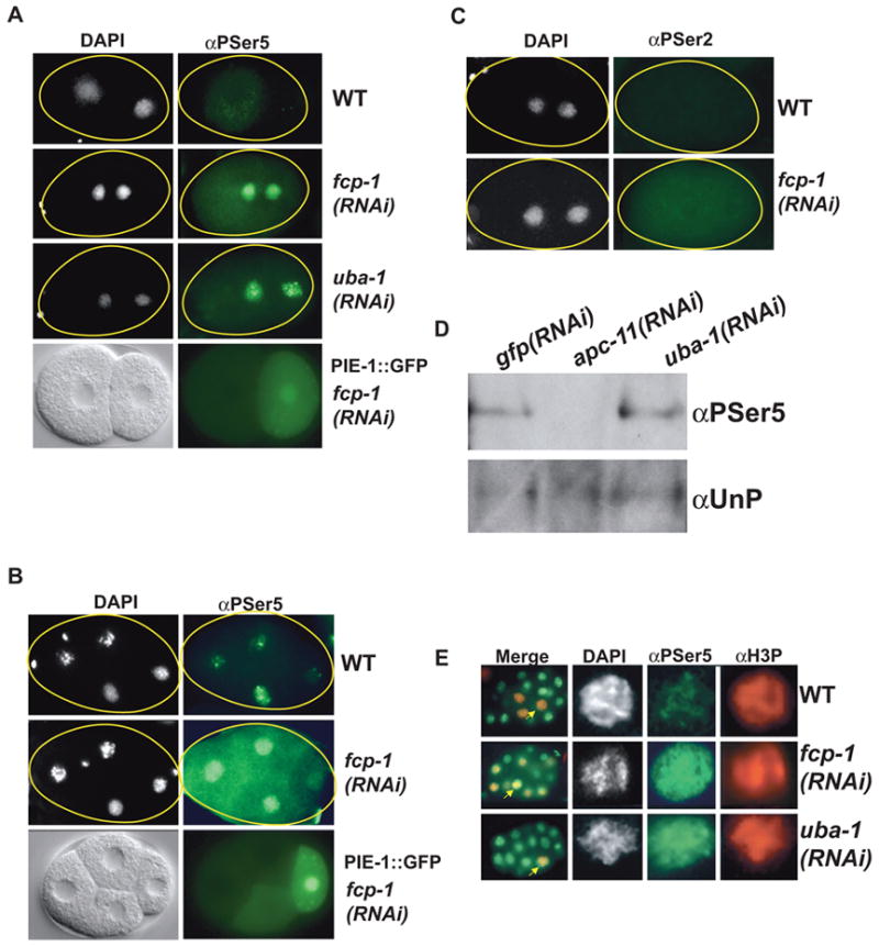
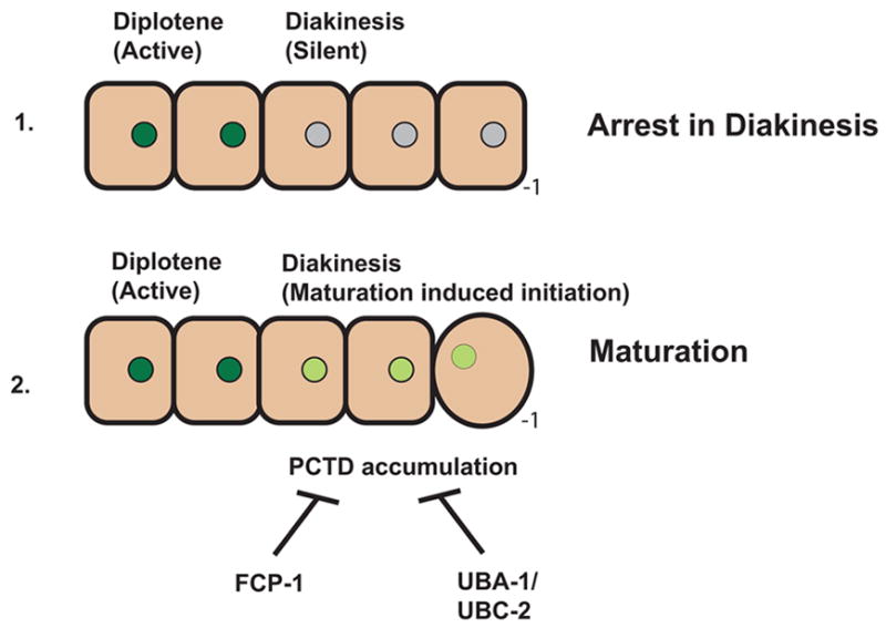
Developing oocytes produce materials that will support early embryonic development then cease transcription before fertilization. Later, a distinct transcription program is established in the embryo. Little is understood about how these global gene regulation transitions are effected. We have investigated in C. elegans how oocyte transcription is influenced by maturation, a process that releases meiotic arrest and prepares for fertilization. By monitoring transcription-associated phosphorylation of the RNA polymerase II (Pol II) C-terminal domain (CTD), we find that oocyte transcription shuts down independently of maturation. Surprisingly, maturation signals then induce CTD phosphorylation that is associated specifically with transcription initiation steps and accumulates to high levels when expression of the CTD phosphatase FCP-1 is inhibited. This CTD phosphorylation is also uncovered when a ubiquitylation pathway is blocked, or when maturation is stimulated precociously. CTD phosphorylation is similarly detected during embryonic mitosis, when transcription is also largely silenced. We conclude that oocyte maturation signals induce abortive transcription events in which FCP-1 may recycle phosphorylated Pol II and that analogous processes may occur during mitosis. Our findings suggest that maturation signals may initiate preparations for embryonic transcription, possibly as part of a broader program that begins the transition from maternal to zygotic gene expression.


Figures

Fig. 1
Fig. 1. Pol II CTD phosphorylation in proximal oocytes
(A) An adult C. elegans hermaphrodite gonad arm, in which oogenesis proceeds distally to proximally (relative to the uterus) in an assembly-line fashion (Greenstein, 2005; Schedl, 1997). A stem cell population at the distal end gives rise to approximately 160 sperm during larval development, then during adulthood produces only oocytes. Transcriptionally active nuclei, which are apparent from the distal mitotic region through the diplotene stage (Kelly et al., 2002; Schisa et al., 2001)(see panels C and D), are marked in green. Proximal oocyte nuclei that have entered diakinesis and are transcriptionally silent are shown in grey. The most proximal oocyte, which is designated as -1, assumes a rounded shape shortly before ovulation into the spermatheca, where fertilization occurs. Embryos (blue) initiate transcription in somatic nuclei (dark blue) at the 4-cell stage. (B) RNA Pol II transcription cycle (Bentley, 2005; Meinhart et al., 2005; Orphanides and Reinberg, 2002). A partial PIC is shown at a promoter. The PIC components that are shown are brought together specifically at promoters. The multi-copy CTD repeat (52 in human, 38 in C. elegans) must be unphosphorylated for Pol II to be recruited into the PIC, which also includes a set of general transcription factors and the Mediator complex. During initiation, the CTD repeat is phosphorylated on Ser 5 by CDK-7, a subunit of the general transcription factor TFIIH. This modification is required for promoter clearance and recruitment of the 5’ capping enzyme (C. E.), after which Ser 2 is phosphorylated by CDK-9. Ser 2 phosphorylation promotes transcription elongation, and is important for recruiting factors involved in mRNA processing. (C) Proximal oocytes of wild type and RNAi gonads (top panels) stained with DAPI (white), and antibodies to PSer5 (αPSer5) (green) and phosphohistone H3 (Ser10) (αH3P) (red). αH3P staining of RNAi gonads is not shown either here or in (D), but was indistinguishable from wild type. The –1 oocyte is pictured farthest to the right here and in subsequent figures, unless otherwise noted. Identical exposure times are shown within the panel sets here and in all other figures. (D) Proximal oocytes of wild type and RNAi gonads stained with DAPI (white), and antibodies to PSer2 (αPSer2) (green) and αH3P (red, shown for wild type only).
Fig. 2
Fig. 2. Oocyte transcription shuts down independently of sperm-dependent maturation signals
Proximal fog-2(q71) oocytes are shown as in Fig. 1. Unmated fog-2(q71) females accumulated many oocytes in diakinesis, all of which were H3P-positive and none of which showed nuclear PSer5 staining. The cytoplasmic αPSer5 staining apparent in the -1 oocyte in the mated sample was seen in some experiments. This staining was not detected by antibodies to two other AMA-1 (Pol II large subunit) epitopes (Fig. 4D; data not shown) and did not disappear when AMA-1 was depleted by RNAi, indicating that it was background. Intestinal nuclei are indicated by arrows.
Fig. 3
Fig. 3. Ser 5 phosphorylation in diakinetic fcp-1(RNAi) oocytes requires transcription initiation factors and maturation signals
(A) Proximal oocytes in the indicated RNAi backgrounds were stained with DAPI and αPSer5. In each combinatorial RNAi experiment, input RNA amounts were carefully maintained by addition of a control dsRNA where appropriate (Materials and Methods). (B) Maturation signals stimulate de novo CTD phosphorylation in diakinetic fcp-1(RNAi) oocytes. Mated fog-2(q71) and mated or unmated fog-2(q71); fcp-1(RNAi) gonads were stained with DAPI and αPSer5. PSer5 staining was present at low levels and did not increase progressively in unmated fcp-1(RNAi);fog-2 gonads, which contain approximately 8–10 oocytes in diakinesis. (C) PSer2 is present at low levels in diakinetic fcp-1(RNAi) oocytes. Wild type, fcp-1(RNAi), or fog-2(q71); fcp-1(RNAi) gonads were stained with DAPI and αPSer2. Similarly low and variable levels of PSer2 staining were observed in diakinetic oocytes in fcp-1(RNAi) and mated or unmated fog-2(q71); fcp-1(RNAi) animals (Supplementary Table 2; data not shown). This PSer2 staining was abolished by ama-1 (Pol II large subunit) RNAi (data not shown).
Fig. 4
Fig. 4. Regulation of transcription-associated PSer5 accumulation by a ubiquitylation pathway
(A) Proximal oocytes of wild type (top row) or uba-1(RNAi) gonads stained with DAPI, and antibodies to PSer5 and mono-/poly-ubiquitinated proteins (αUb). (B) Proximal oocytes stained with DAPI and αPSer5 in the indicated RNAi backgrounds. An additional proteasome component, pbs-7, was also required for PSer5 to appear in diakinetic oocytes after uba-1 RNAi (data not shown). In each combinatorial RNAi experiment, input RNA amounts were carefully maintained by addition of a control dsRNA where appropriate (Materials and Methods). (C) Proximal oocytes from a conditional cdk-7 mutant (Wallenfang and Seydoux, 2002) at the permissive (15°C) or non-permissive temperature (24°C) were stained with DAPI and αPSer5. This cdk-7 mutant was used in these uba-1 experiments because uba-1 and cdk-7 RNAi effects become manifest in embryos at different times after injection. (D) Levels of the Pol II large subunit AMA-1 in oocytes lacking ubiquitylation or proteasome function. Columns show DAPI or an antibody to a non-CTD epitope of Pol II (ARNA3).
Fig. 5
Fig. 5. Induction of CTD Ser 5 phosphorylation by oocyte maturation signals
(A) fog-2(q71) proximal oocytes stained with αPSer5 (green) and αH3P (red) to mark oocytes in diakinesis. A somatic sheath cell nucleus is marked by an asterisk. The top and bottom panels on the right (mated fog-2(q71); uba-1(RNAi)) show αPSer5 or αH3P staining of the same gonad. (B) oma-1(zu405te33); oma-2(te51) gonads were stained with αPSer5 and αH3P to mark oocytes in diakinesis. (C) Constitutive maturation promotes CTD Ser 5 phosphorylation. The top row shows proximal oocytes from unmated vab-1(dx31); fog-2(q71); ceh-18(mg57) females. For wee-1.3(RNAi) gonads (bottom) the –2 and -3 oocytes are shown. (D) Wild type (top) or uba-1(RNAi) (bottom) gonads were stained with an antibody to activated MAP kinase. Staining levels and patterns were comparable between wild type and RNAi gonads, although staining intensities varied among individuals. (E) Oocyte maturation is not accelerated by uba-1 RNAi. Maturation rates in wild type and uba-1(RNAi) hermaphrodites. Error bars show standard deviation among independent experiments.
Fig. 6
Fig. 6. fcp-1 and a ubiquitylation pathway prevent PSer5 accumulation in transcriptionally silent embryonic cells
(A) Pre-ZGA embryos accumulate PSer5 after fcp-1 or uba-1 RNAi. Staining with DAPI (left column) or PSer5 (right) is shown in the top three rows. In multiple RNAi experiments the levels of PSer5 staining observed in one- and two-cell embryos were comparable to those seen in transcriptionally active cells in later stage embryos on the same slide (data not shown). The bottom row shows Nomarski (left) and epifluorescent (right) images of an embryo that expresses a translational fusion of PIE-1 to green fluorescent protein (PIE-1::GFP) (Reese et al., 2000) and has been subjected to fcp-1 RNAi. In panels A-C posterior is to the right. (B) Presence of nucleoplasmic PSer5 staining in the germ cell lineage in fcp-1(RNAi) embryos. The top two rows show wild type or fcp-1(RNAi) embryos stained with DAPI and αPSer5. In the top panel, the two most anterior cells are entering mitosis. The bottom row contains Nomarski (left) or epifluorescent (right) images of a four-cell fcp-1(RNAi) embryo that expresses PIE-1::GFP. The germline precursor P2 is to the right. (C) PSer2 staining is not detected in pre-ZGA fcp-1(RNAi) embryos. Wild type or fcp-1(RNAi) embryos were stained with DAPI and αPSer2. Robust PSer2 staining was apparent in later-stage embryos on the same slides (not shown). (D) High levels of Ser5 phosphorylation in pre-ZGA uba-1(RNAi) embryos. This western blot assayed extracts prepared from the same number of gfp(RNAi) embryos (ranging in age from 1 cell to around 250 cells), apc-11(RNAi) embryos (>80% arrested at less than 4 cell stage) and uba-1(RNAi) embryos (>90% arrested at less than 4 cell stage). αUnP (8WG16) recognizes the unphosphorylated Pol II CTD. (E) fcp-1 and a ubiquitylation pathway prevent PSer5 accumulation during mitosis. The left column shows merged staining with αPSer5 and αH3P, which identifies mitotic cells. Arrows show nuclei expanded in the other panels. PSer5 staining uniformly appeared in mitotic cells in fcp-1(RNAi), uba-1(RNAi), and ubc-2(RNAi) embryos at all stages (data not shown).
Fig. 7
Fig. 7. Induction of transcription steps by oocyte maturation
During C. elegans oogenesis transcription ceases upon entry into diakinesis, in which meiosis arrests in the absence of maturation. Signals that trigger maturation release this arrest and induce incomplete transcription events in which CTD Ser 5 is phosphorylated. The FCP-1 phosphatase and a ubc-2-dependent ubiquitylation pathway are required to prevent this phosphorylated Pol II from accumulating within the nucleoplasm.

Similar articles

Cited by

References

    1. Akoulitchev S, Reinberg D. The molecular mechanism of mitotic inhibition of TFIIH is mediated by phosphorylation of CDK7. Genes Dev. 1998;12:3541–50. - PMC - PubMed
    1. Batchelder C, Dunn MA, Choy B, Suh Y, Cassie C, Shim EY, Shin TH, Mello C, Seydoux G, Blackwell TK. Transcriptional repression by the C. elegans germline protein PIE-1. Genes Dev. 1999;13:202–212. - PMC - PubMed
    1. Baugh LR, Hill AA, Slonim DK, Brown EL, Hunter CP. Composition and dynamics of the Caenorhabditis elegans early embryonic transcriptome. Development. 2003;130:889–900. - PubMed
    1. Bean CJ, Schaner CE, Kelly WG. Meiotic pairing and imprinted X chromatin assembly in Caenorhabditis elegans. Nat Genet. 2004;36:100–5. - PMC - PubMed
    1. Bentley DL. Rules of engagement: co-transcriptional recruitment of pre-mRNA processing factors. Curr Opin Cell Biol. 2005;17:251–6. - PubMed

Publication types

LinkOut - more resources