. 2007 Apr 15;304(2):848-59.
doi: 10.1016/j.ydbio.2007.01.005. Epub 2007 Jan 9.

Utp14b: a unique retrogene within a gene that has acquired multiple promoters and a specific function in spermatogenesis

Affiliations

Utp14b: a unique retrogene within a gene that has acquired multiple promoters and a specific function in spermatogenesis

Ming Zhao et al. Dev Biol. .

Abstract

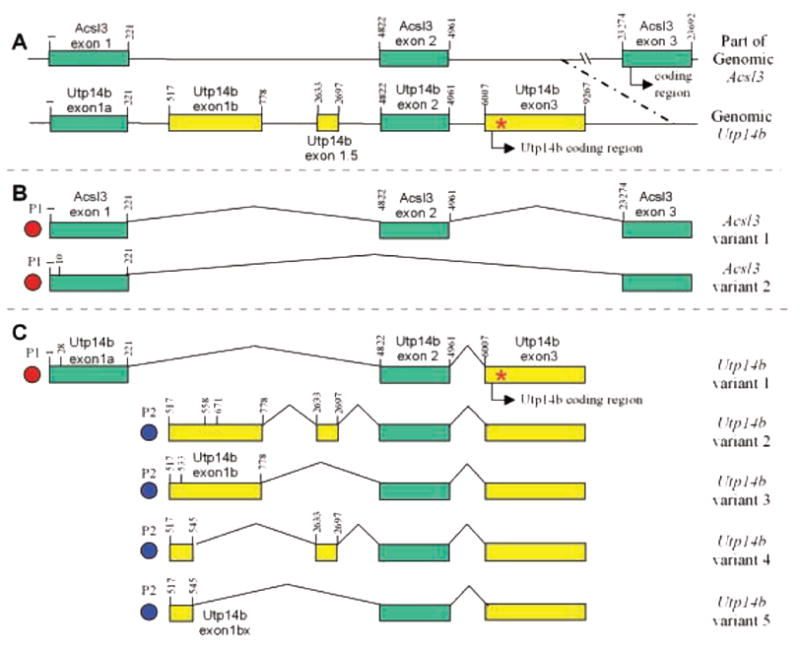
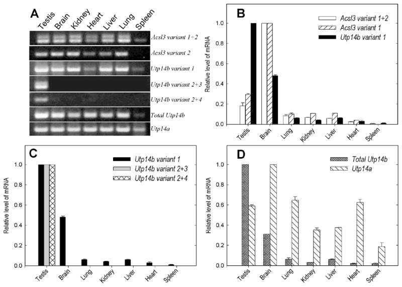
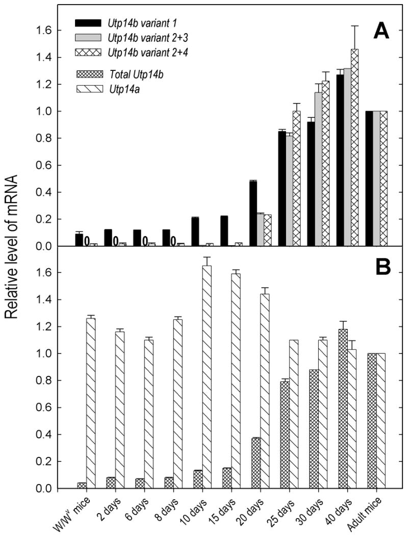
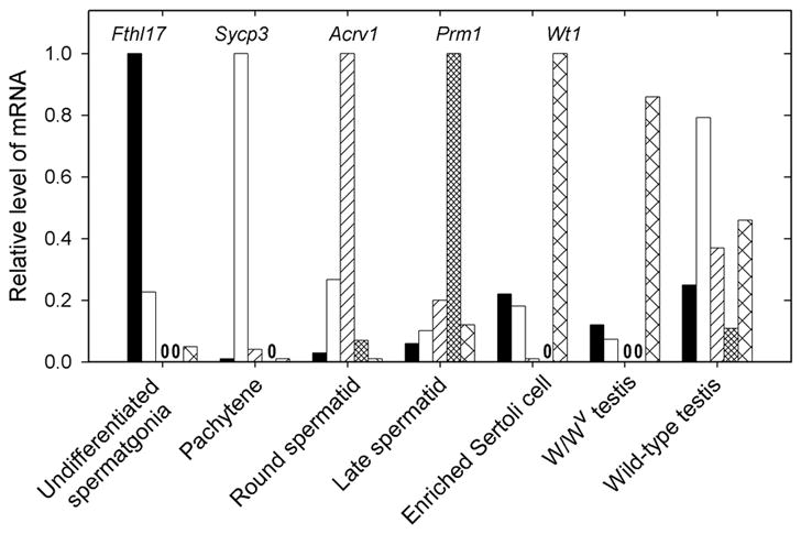
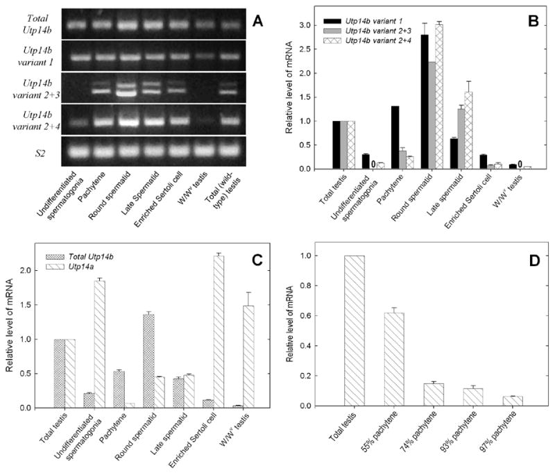
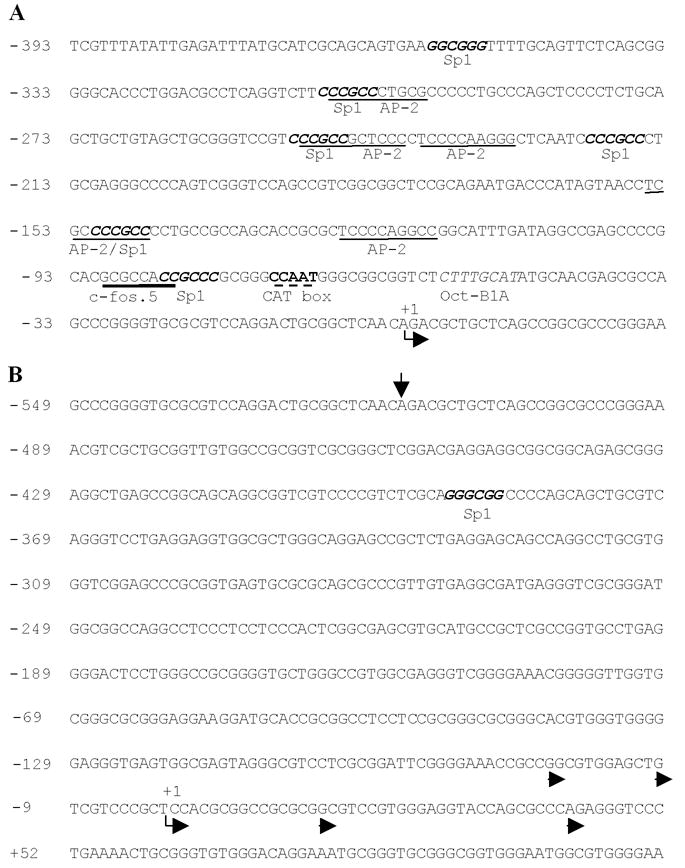
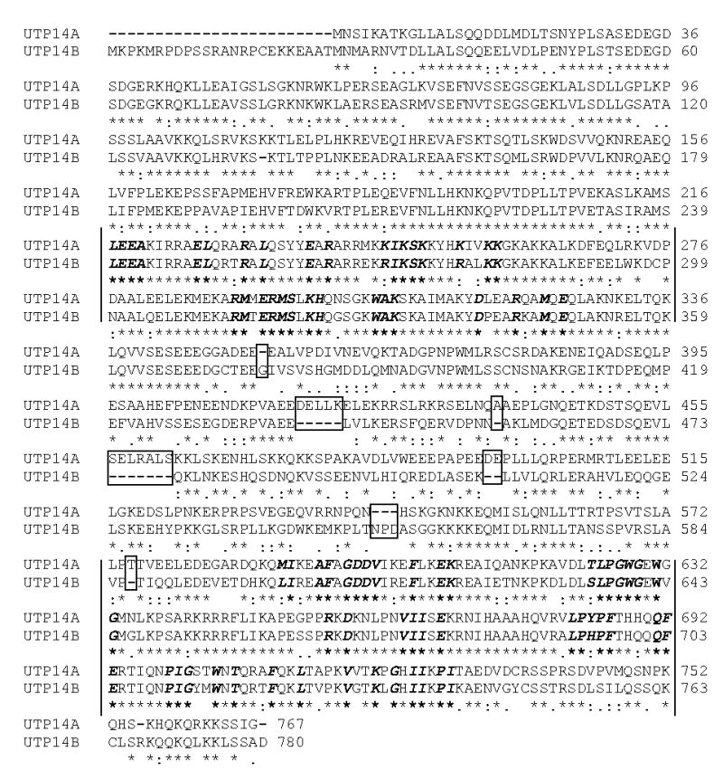
The mouse retrogene Utp14b is essential for male fertility, and a mutation in its sequence results in the sterile juvenile spermatogonial depletion (jsd) phenotype. It is a retrotransposed copy of the Utp14a gene, which is located on the X chromosome, and is inserted within an intron of the autosomal acyl-CoA synthetase long-chain family member 3 (Acsl3) gene. To elucidate the roles of the Utp14 genes in normal spermatogenic cell development as a basis for understanding the defects that result in the jsd phenotype, we analyzed the various mRNAs produced from the Utp14b retrogene and their expression in different cell types. Two classes of transcripts were identified: variant 1, a transcript driven by the host gene promoter, that is predominantly found in germ cells but is ubiquitously expressed at low levels; and variants 2-5, a group of alternatively spliced transcripts containing some unique untranslated exons that are transcribed from a novel promoter that is germ-cell-specific. Utp14b (predominantly variant 1) is expressed at moderately high levels in pachytene spermatocytes, the developmental stage at which the expression of the X-linked Utp14a is suppressed. The levels of both classes of Utp14b transcripts were highest in round spermatids despite the transcription of Utp14a in these cells. We propose that when Utp14b initially inserted into Acsl3, it utilized the Acsl3 promoter to drive expression in pachytene spermatocytes to compensate for inactivation of Utp14a expression. The novel cell-type-specific promoter for Utp14b likely evolved later, as the protein may have acquired a germ cell-specific function in spermatid development.


Figures

Fig. 1
Fig. 1
(A) Genomic maps of Acsl3 and Utp14b (not drawn to scale) showing the exons that can be included in transcripts. (B) Transcripts of Acsl3 observed by 5′-RACE. (C) Transcripts of Utp14b observed by 5′-RACE. The nucleotide numbers of the exon boundaries, relative to the major transcriptional start site of Acsl3 and Utp14b variant 1, are indicated vertically above the exons. Downstream alternative transcription start sites, differing by more than a few base pairs, are indicated as vertical lines with nucleotide number within the first exon of the transcript. P1, P2: putative promoters. *Utp14b mutation site for jsd phenotype.
Fig. 2
Fig. 2
Tissue-specific expression of Acsl3, Utp14b, and Utp14a. (A) Agarose gel electrophoresis analysis of transcript-specific RT-PCR products taken from products of real-time PCR (40 cycles). (B–D) Tissue-specific transcript levels determined by real time-PCR. Expression of (B) Acsl3 (variants 1 and 2) and Utp14b variant 1, (C) Utp14b variant 1, variants 2+3 and variants 2+4, and (D) total Utp14b and Utp14a. Expression levels were first normalized to ribosomal protein Rps2 mRNA levels, and then the values for the tissues with the most abundant expression were set at 1.0.
Fig. 3
Fig. 3
Age-dependent expression of Utp14a and of different Utp14b transcript variants in wild-type mouse testes and expression in adult, germ-cell deficient W/Wv mutants. (A) Comparison of Utp14b variant 1 expression with that of variants 2+3 and 2+4. (B) Comparison of total Utp14b with Utp14a expression. Expression levels were first normalized to ribosomal protein Rps2 mRNA levels, and then the values for the adult wild-type testes were set at 1.0. Zero’s in place of bars, indicate that no transcript-specific RT-PCR product was produced.
Fig. 4
Fig. 4
Analysis of purities of cellular fractions with real-time PCR for markers specific for particular cell types. Fthl17, Sycp3, Acrv1, Prm1 and Wt1 were used as markers of spermatogonia, pachytene spermatocytes, round spermatids, late spermatids, and Sertoli cells, respectively. Expression levels were first normalized to ribosomal protein Rps2 mRNA levels and then the values for each gene in the cell type most enriched in that gene were set at 1.0. Zero’s in place of bars, indicate that no transcript-specific RT-PCR product was produced.
Fig. 5
Fig. 5
Expression of Utp14a and of different Utp14b variants in testes of wild-type and germ-cell deficient W/Wv mice and in purified cell fractions from wild-type mice testis. (A) Agarose gel analysis of real-time PCR products. (B–D) Cell-type specific transcript levels determined by real time-PCR. Comparison of expression of (B) Utp14b variant 1 with variant 2+3 and variant 2+4, and (C) total Utp14b with Utp14a. (D) Effect of further purification of pachytene spermatocytes on levels of Utp14a expression in fractions. Purities of 55%, 74%, and 93% were obtained with cell suspensions prepared by trypsinization (Meistrich, 1977) and elutriation alone; elutriation plus Percoll gradients; and a combination of elutriation, Percoll gradients, and plating on DSA, respectively. The procedure for obtaining 97% purity is described in the Materials and Methods. Normalization was done as in Fig. 3. Zero’s in place of bars, indicate that no transcript-specific RT-PCR product was produced.
Fig. 6
Fig. 6
Identification of potential promoter sequences and the transcriptional start sites of Acsl3 and Utp14b. (A) The core promoter region upstream of the Acsl3/Utp14b variant 1 start site contained a single CCAAT (cat) box (bold letters and dashed underline) and an oct-B1A binding site (italics). Upstream there were six Sp1 binding sites (bold and italics), five AP-2 sites (underline), and a single c-fos.5 site (bold underline), some of which were overlapping. An arrow below the text indicates the transcriptional start site. (B) The region upstream of the Utp14b testis-specific transcript start site does not contain any known basal promoter elements or enhancers. There is a single Sp1 site (bold and italics). An arrow above the text indicts the transcriptional start site of Acsl3/Utp14b variant 1. The major consensus start site (start site at that nucleotide or with 3 bp) is indicated by an arrow below the text and other observed start sites are indicated by arrowheads.
Fig. 7
Fig. 7
Alignment of UTP14A and UTP14B proteins showing identities, similarities, and conserved structural motifs. Two regions of containing a large percentage of highly conserved amino acids are shown between the bars; the ones shown in bold italics are conserved (absolutely or as a conserved substitution) across phyla. There is spatial conservation (i.e. the number of amino acids between those marked as conserved) across phyla between the first and last conserved amino acid within these regions. Amino acid deletions and insertions within the core of the UTP14B peptide are indicated by dashes and boxes around the amino acids. At the amino terminus of UTP14B there are an additional 24 amino acids. An asterisk (*) indicates amino acid identity, two dots (:) indicates conserved substitution, one dot (.) indicates similarity, and no symbol indicates other random substitutions. The two peptides share 66% identity and 76% homology.

Similar articles

Cited by

References

    1. Ayoub N, Richler C, Wahrman J. Xist RNA is associated with the transcriptionally inactive XY body in mammalian male meiosis. Chromosoma. 1997;106:1–10. - PubMed
    1. Ballow D, Meistrich ML, Matzuk M, Rajkovic A. Sohlh1 is essential for spermatogonial differentiation. Dev Biol. 2006;294:161–167. - PubMed
    1. Bedell MA, Mahakali Zama A. Genetic analysis of Kit ligand functions during mouse spermatogenesis. J Androl. 2004;25:188–199. - PubMed
    1. Boer PH, Adra CN, Lau YF, McBurney MW. The testis-specific phosphoglycerate kinase gene pgk-2 is a recruited retroposon. Mol Cell Biol. 1987;7:3107–3112. - PMC - PubMed
    1. Boettger-Tong HL, Johnston DS, Russell LD, Griswold MD, Bishop CE. Juvenile spermatogonial depletion (jsd) mutant seminiferous tubules are capable of supporting transplanted spermatogenesis. Biol Reprod. 2000;63:1185–1191. - PubMed

Publication types

MeSH terms