

Heterotypic RPE-choroidal endothelial cell contact increases choroidal endothelial cell transmigration via PI 3-kinase and Rac1
- PMID: 17292356
- PMCID: PMC2270476
- DOI: 10.1016/j.exer.2006.12.012
Heterotypic RPE-choroidal endothelial cell contact increases choroidal endothelial cell transmigration via PI 3-kinase and Rac1
Abstract
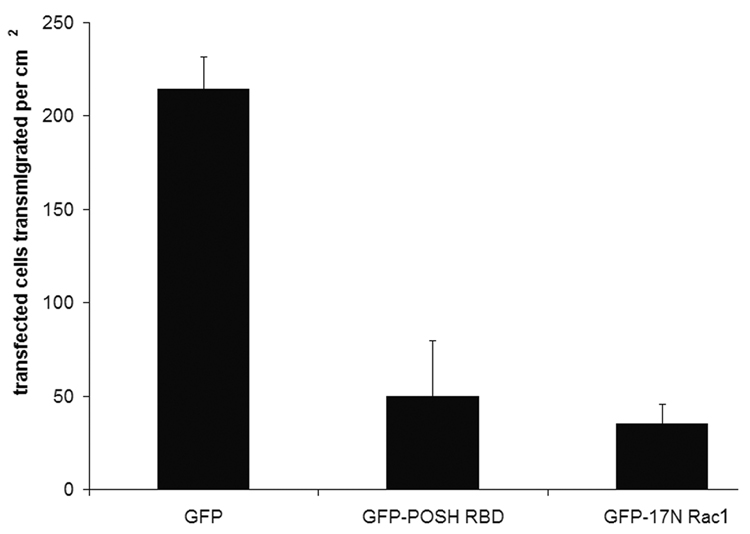
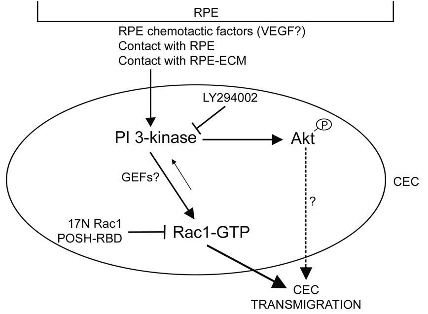
Age-related macular degeneration (AMD) is the major cause of non-preventable blindness. Severe forms of AMD involve breaching of the retinal pigment epithelial (RPE) barrier by underlying choroidal endothelial cells (CECs), followed by migration into, and subsequent neovascularization of the neurosensory retina. However, little is known about the interactions between RPE and CECs and the signaling events leading to CEC transmigration. While soluble chemotactic factors secreted from RPE can contribute to inappropriate CEC transmigration, other unidentified stimuli may play an additional role. Using a coculture model that maintains the natural structural orientation of CECs to the basal aspect of RPE, we show that "contact" with RPE and/or RPE extracellular matrix increases CEC transmigration of the RPE barrier. From a biochemical standpoint, contact between CECs and RPE results in an increase in the activity of the GTPase Rac1 within the CECs; this increase is dependent on upstream activation of PI 3-K and Akt1. To confirm a link between these signaling molecules and increased CEC transmigration, we performed transmigration assays while inhibiting both PI 3-K and Rac1 activity, and observed that both decreased CEC transmigration. We hypothesize that contact between CECs and RPE stimulates a signaling pathway involving PI 3-K, Akt1, and Rac1 that facilitates CEC transmigration across the RPE barrier, an important step in the development of neovascular AMD.
Figures


Similar articles
-
Choroidal endothelial cells transmigrate across the retinal pigment epithelium but do not proliferate in response to soluble vascular endothelial growth factor.Exp Eye Res. 2006 Apr;82(4):608-19. doi: 10.1016/j.exer.2005.08.021. Epub 2005 Nov 2. Exp Eye Res. 2006. PMID: 16259980
-
The role of RPE cell-associated VEGF₁₈₉ in choroidal endothelial cell transmigration across the RPE.Invest Ophthalmol Vis Sci. 2011 Feb 1;52(1):570-8. doi: 10.1167/iovs.10-5595. Print 2011 Jan. Invest Ophthalmol Vis Sci. 2011. PMID: 20811045 Free PMC article.
-
Breaking barriers: insight into the pathogenesis of neovascular age-related macular degeneration.Eye Brain. 2011;3:19-28. doi: 10.2147/EB.S24951. Epub 2011 Sep 27. Eye Brain. 2011. PMID: 27795668 Free PMC article.
-
Regulation of signaling events involved in the pathophysiology of neovascular AMD.Mol Vis. 2016 Feb 27;22:189-202. eCollection 2016. Mol Vis. 2016. PMID: 27013848 Free PMC article. Review.
-
Regulation of Rac1 Activation in Choroidal Endothelial Cells: Insights into Mechanisms in Age-Related Macular Degeneration.Cells. 2021 Sep 14;10(9):2414. doi: 10.3390/cells10092414. Cells. 2021. PMID: 34572063 Free PMC article. Review.
Cited by
-
7-ketocholesterol induces endothelial-mesenchymal transition and promotes fibrosis: implications in neovascular age-related macular degeneration and treatment.Angiogenesis. 2021 Aug;24(3):583-595. doi: 10.1007/s10456-021-09770-0. Epub 2021 Feb 28. Angiogenesis. 2021. PMID: 33646466 Free PMC article.
-
Thy-1 Regulates VEGF-Mediated Choroidal Endothelial Cell Activation and Migration: Implications in Neovascular Age-Related Macular Degeneration.Invest Ophthalmol Vis Sci. 2016 Oct 1;57(13):5525-5534. doi: 10.1167/iovs.16-19691. Invest Ophthalmol Vis Sci. 2016. PMID: 27768790 Free PMC article.
-
IQGAP1 causes choroidal neovascularization by sustaining VEGFR2-mediated Rac1 activation.Angiogenesis. 2020 Nov;23(4):685-698. doi: 10.1007/s10456-020-09740-y. Epub 2020 Aug 11. Angiogenesis. 2020. PMID: 32783108 Free PMC article.
-
Activation of Rap1 inhibits NADPH oxidase-dependent ROS generation in retinal pigment epithelium and reduces choroidal neovascularization.FASEB J. 2014 Jan;28(1):265-74. doi: 10.1096/fj.13-240028. Epub 2013 Sep 16. FASEB J. 2014. PMID: 24043260 Free PMC article.
-
Influence of Dll4 via HIF-1α-VEGF signaling on the angiogenesis of choroidal neovascularization under hypoxic conditions.PLoS One. 2011 Apr 19;6(4):e18481. doi: 10.1371/journal.pone.0018481. PLoS One. 2011. PMID: 21526177 Free PMC article.
References
-
- Arefieva TI, Kuktina NB, Antonova OA, Krasnikova TL. MCP-1 stimulated chemotaxis of monocytic and endothelial cells is dependent on activation of different signaling cascades. Cytokine. 2005:439–446. - PubMed
-
- Burridge K, Wennerberg K. Rho and Rac take center stage. Cell. 2004;116:167–179. - PubMed
-
- Cheng HL, Steinway ML, Russell JW, Feldman EL. GTPases and phosphatidylinositol 3-kinase are critical for insulin-like growth factor-1-mediated Schwamm cell motility. J Biol Chem. 2000;275:27197–27204. - PubMed
Publication types
MeSH terms
Substances
Grants and funding
LinkOut - more resources
Full Text Sources
Medical
Research Materials
Miscellaneous
