

Early vulnerability to ischemia/reperfusion injury in motor terminals innervating fast muscles of SOD1-G93A mice
- PMID: 17292357
- PMCID: PMC2097955
- DOI: 10.1016/j.expneurol.2006.12.021
Early vulnerability to ischemia/reperfusion injury in motor terminals innervating fast muscles of SOD1-G93A mice
Abstract
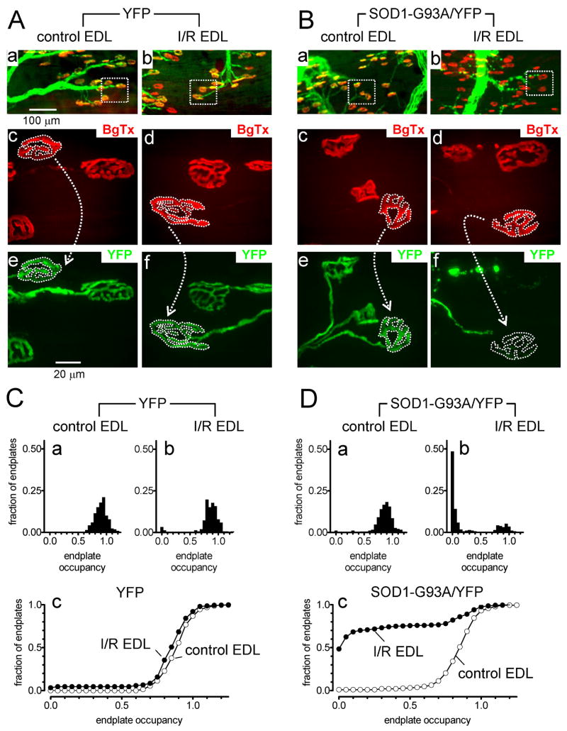
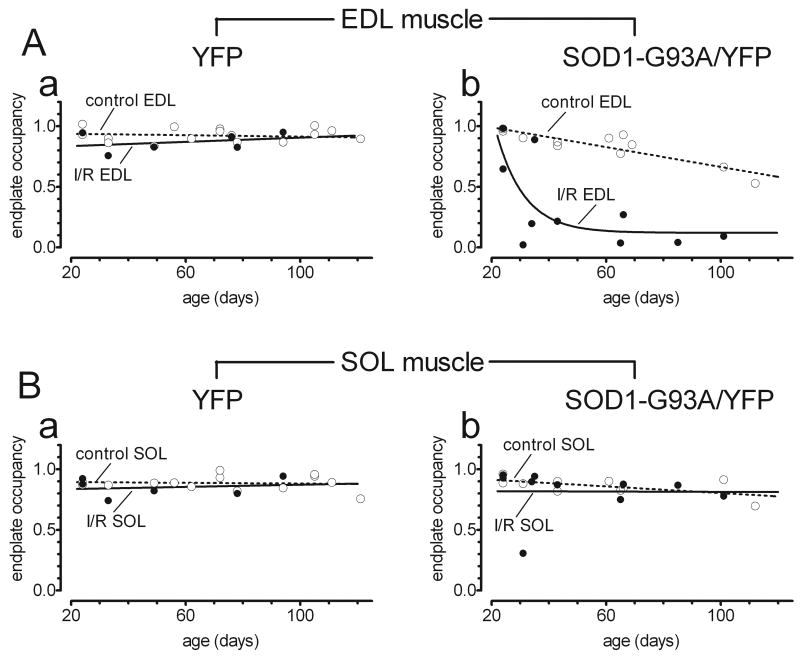
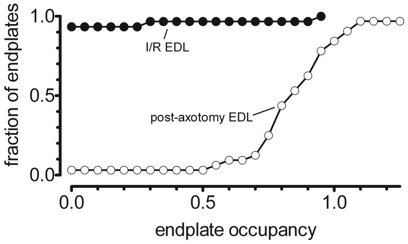
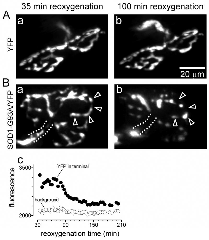
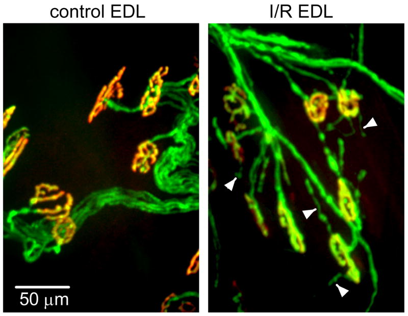
In mouse models of familial amyotrophic lateral sclerosis (fALS), motor neurons are especially vulnerable to oxidative stresses in vitro. To determine whether this increased vulnerability also extends to motor nerve terminals in vivo, we assayed the effect of tourniquet-induced ischemia/reperfusion (I/R) injury on motor terminals innervating fast and slow hindlimb muscles in male G93A-SOD1 mice and their wild-type littermates. These mice also expressed yellow fluorescent protein (YFP) in motor neurons. We report that in SOD1-G93A/YFP mice the motor terminals innervating two predominantly fast muscles, extensor digitorum longus (EDL) and plantaris, were more vulnerable to I/R injury than motor terminals innervating the predominantly slow soleus muscle. The mean duration of EDL ischemia required to produce a 50% reduction in endplate innervation in SOD1-G93A/YFP mice was 26 min, compared to 45 min in YFP-only mice. The post-I/R destruction of EDL terminals in SOD1-G93A mice was rapid (<2 h) and was not duplicated by cutting the sciatic nerve at the tourniquet site. The increased sensitivity to I/R injury was evident in EDL muscles of SOD1-G93A/YFP mice as young as 31 days, well before the onset of motor neuron death at approximately 90 days. This early vulnerability to I/R injury may correlate with the finding (confirmed here) that in fALS mice motor nerve terminals innervating fast hindlimb muscles degenerate before those innervating slow muscles, at ages that precede motor neuron death. Early vulnerability of fast motor terminals to I/R injury thus may signal, and possibly contribute to, early events involved in motor neuron death.
Figures

Similar articles
-
Functional over-load saves motor units in the SOD1-G93A transgenic mouse model of amyotrophic lateral sclerosis.Neurobiol Dis. 2010 Feb;37(2):412-22. doi: 10.1016/j.nbd.2009.10.021. Epub 2009 Oct 29. Neurobiol Dis. 2010. PMID: 19879358
-
Muscle fiber-type specific terminal Schwann cell pathology leads to sprouting deficits following partial denervation in SOD1G93A mice.Neurobiol Dis. 2020 Nov;145:105052. doi: 10.1016/j.nbd.2020.105052. Epub 2020 Aug 20. Neurobiol Dis. 2020. PMID: 32827689
-
The effect of peripheral nerve injury on disease progression in the SOD1(G93A) mouse model of amyotrophic lateral sclerosis.Neuroscience. 2005;130(4):897-910. doi: 10.1016/j.neuroscience.2004.09.069. Neuroscience. 2005. PMID: 15652988
-
Mitochondria in motor nerve terminals: function in health and in mutant superoxide dismutase 1 mouse models of familial ALS.J Bioenerg Biomembr. 2011 Dec;43(6):581-6. doi: 10.1007/s10863-011-9392-1. J Bioenerg Biomembr. 2011. PMID: 22089637 Free PMC article. Review.
-
Mechanisms of Muscle Denervation in Aging: Insights from a Mouse Model of Amyotrophic Lateral Sclerosis.Aging Dis. 2015 Oct 1;6(5):380-9. doi: 10.14336/AD.2015.0506. eCollection 2015 Sep. Aging Dis. 2015. PMID: 26425392 Free PMC article. Review.
Cited by
-
A role for motoneuron subtype-selective ER stress in disease manifestations of FALS mice.Nat Neurosci. 2009 May;12(5):627-36. doi: 10.1038/nn.2297. Epub 2009 Mar 29. Nat Neurosci. 2009. PMID: 19330001
-
The Role of Skeletal Muscle in Amyotrophic Lateral Sclerosis.Brain Pathol. 2016 Mar;26(2):227-36. doi: 10.1111/bpa.12350. Brain Pathol. 2016. PMID: 26780251 Free PMC article. Review.
-
Morphological Regeneration and Functional Recovery of Neuromuscular Junctions after Tourniquet-Induced Injuries in Mouse Hindlimb.Front Physiol. 2017 Apr 6;8:207. doi: 10.3389/fphys.2017.00207. eCollection 2017. Front Physiol. 2017. PMID: 28428759 Free PMC article.
-
Activity-dependent degeneration of axotomized neuromuscular synapses in Wld S mice.Neuroscience. 2015 Apr 2;290:300-20. doi: 10.1016/j.neuroscience.2015.01.018. Epub 2015 Jan 21. Neuroscience. 2015. PMID: 25617654 Free PMC article.
-
Calcium dependence of damage to mouse motor nerve terminals following oxygen/glucose deprivation.Exp Neurol. 2012 Mar;234(1):95-104. doi: 10.1016/j.expneurol.2011.12.020. Epub 2011 Dec 27. Exp Neurol. 2012. PMID: 22206924 Free PMC article.
References
-
- Annex BH, Torgan CE, Lin P, Taylor DA, Thompson MA, Peters KG, Kraus WE. Induction and maintenance of increased VEGF protein by chronic motor nerve stimulation in skeletal muscle. Am J Physiol Heart Circ Physiol. 1998;274:H860–H867. - PubMed
-
- Atkin JD, Scott RL, West JM, Lopes E, Quah AK, Cheema SS. Properties of slow- and fast-twitch muscle fibres in a mouse model of amyotrophic lateral sclerosis. Neuromuscul Disord. 2005;15:377–388. - PubMed
-
- Azzouz M, Leclerc N, Gurney M, Warter JM, Poindron P, Borg J. Progressive motor neuron impairment in an animal model of familial amyotrophic lateral sclerosis. Muscle Nerve. 1997;20:45–51. - PubMed
-
- Boillée S, Yamanaka K, Lobsiger CS, Copeland NG, Jenkins NA, Kassiotis G, Kollias G, Cleveland DW. Onset and progression in inherited ALS determined by motor neurons and microglia. Science. 2006;312:1389–1392. - PubMed
Publication types
MeSH terms
Substances
Grants and funding
LinkOut - more resources
Full Text Sources
Molecular Biology Databases
Miscellaneous
