. 2007 May;73(3):173-81.
doi: 10.1016/j.mvr.2006.12.004. Epub 2007 Jan 3.

Signaling pathways involved in OxPAPC-induced pulmonary endothelial barrier protection

Affiliations

Signaling pathways involved in OxPAPC-induced pulmonary endothelial barrier protection

Anna A Birukova et al. Microvasc Res. 2007 May.

Abstract

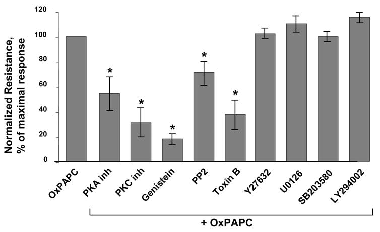
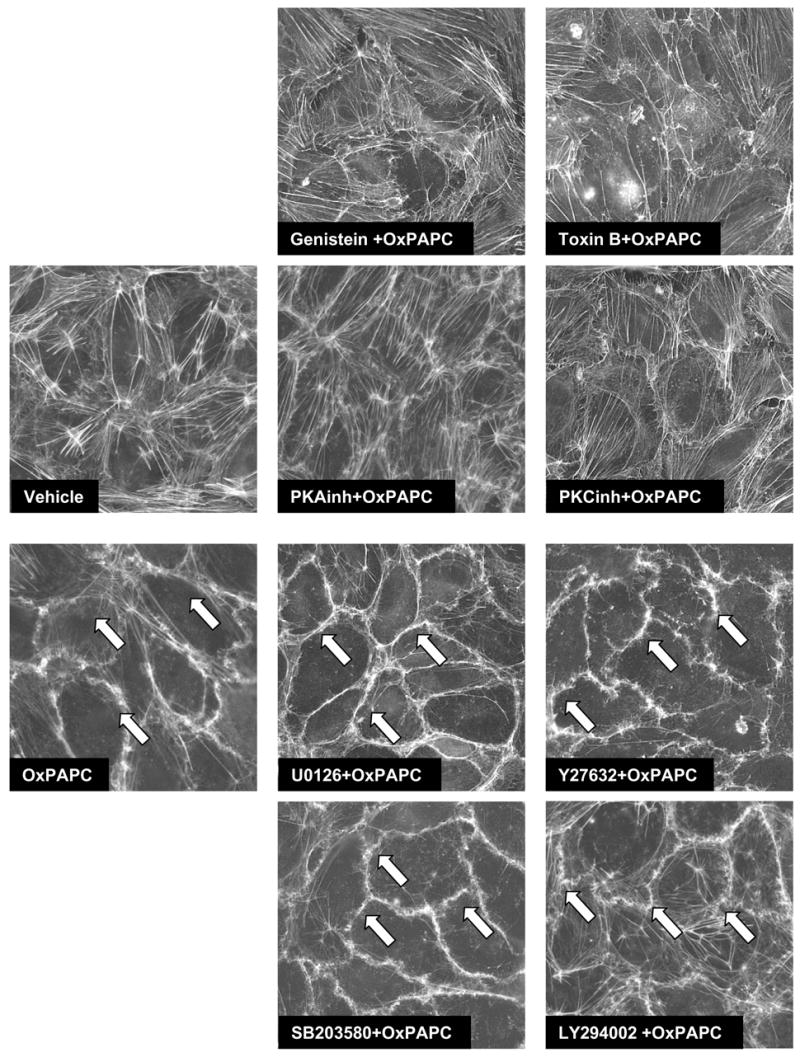
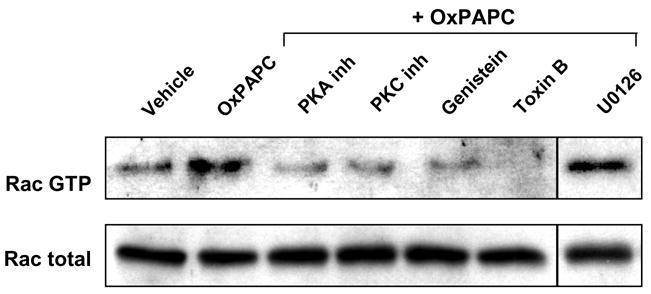
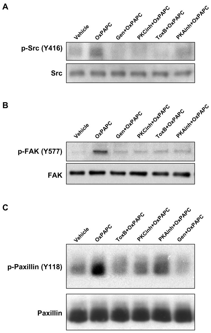
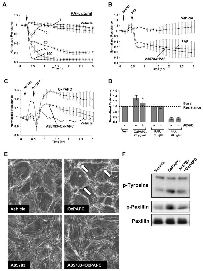
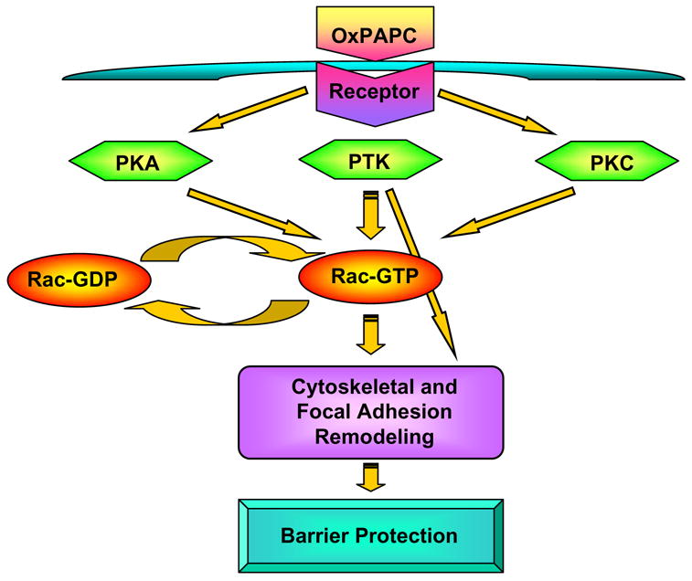
Increased tissue or serum levels of oxidized phospholipids have been detected in a variety of chronic and acute pathological conditions such as hyperlipidemia, atherosclerosis, heart attack, cell apoptosis, acute inflammation and injury. We have recently described signaling cascades activated by oxidized 1-palmitoyl-2-arachidonoyl-sn-glycero-3-phosphocholine (OxPAPC)in the human pulmonary artery endothelial cells (EC) and reported potent barrier-protective effects of OxPAPC, which were mediated by small GTPases Rac and Cdc42. In this study we have further characterized signal transduction pathways involved in the OxPAPC-mediated endothelial barrier protection. Inhibitors of small GTPases, protein kinase A (PKA), protein kinase C (PKC), Src family kinases and general inhibitors of tyrosine kinases attenuated OxPAPC-induced barrier-protective response and EC cytoskeletal remodeling. In contrast, small GTPase Rho, Rho kinase, Erk-1,2 MAP kinase and p38 MAP kinase and PI3-kinase were not involved in the barrier-protective effects of OxPAPC. Inhibitors of PKA, PKC, tyrosine kinases and small GTPase inhibitor toxin B suppressed OxPAPC-induced Rac activation and decreased phosphorylation of focal adhesion kinase (FAK) and paxillin. Barrier-protective effects of OxPAPC were not reproduced by platelet activating factor (PAF), which at high concentrations induced barrier dysfunction, but were partially attenuated by PAF receptor antagonist A85783. These results demonstrate for the first time upstream signaling cascades involved in the OxPAPC-induced Rac activation, cytoskeletal remodeling and barrier regulation and suggest PAF receptor-independent mechanisms of OxPAPC-mediated endothelial barrier protection.


Figures

Figure 1
Figure 1. Inhibitory analysis of signaling pathways involved in OxPAPC-mediated increase in EC resistance
Transendothelial resistance was monitored across confluent endothelial monolayers pretreated with vehicle, PKA inhibitor (20 μM), PKC inhibitor (20 μM), genistein (100 μM), PP2 (2 μM), toxin B (20 ng/ml), Y27632 (5 μM), U0125 (5 μM), SB 203580 (20 μM), or LY294002 (25 μM) for 30 min followed by addition of OxPAPC (20 μg/ml). Bar graphs represent measurements of TER after 30 min of OxPAPC stimulation. Shown are results of three to seven independent experiments represented as mean ± SE. *p<0.01 vs OxPAPC alone.
Figure 2
Figure 2. Analysis of signaling pathways involved in OxPAPC-induced cytoskeletal remodeling
EC grown on glass coverslips were preincubated with genistein (100 μM), small GTPase inhibitor toxin B (20 ng/ml), PKA inhibitor (20 μM), PKC inhibitor (20 μM), Rho kinase inhibitor Y27632 (5 μM), MEK/Erk1,2 inhibitor U0125 (5 μM), p38 MAP kinase inhibitor SB203580 (20 μM), or PI3-kinase inhibitor LY294002 (25 μM) for 30 min followed by addition of OxPAPC (20 μg/ml) for 30 min. Cytoskeletal remodeling was assessed by immunofluorescent staining for F-actin with Texas Red phalloidin. Results are representative of three independent experiments.
Figure 3
Figure 3. Effect of PKA and PKC inhibition on OxPAPC-induced Rac activation
HPAEC were preincubated with PKA inhibitor (20 μM), PKC inhibitor (20 μM), toxin B (20 ng/ml), genistein (100 μM) or U0125 (5 μM) for 30 min followed by addition of OxPAPC (20 μg/ml) for 5 min. Rac activation was analyzed as described in Methods section. Results are representative of three independent experiments.
Figure 4
Figure 4. Effect of PKA and PKC on OxPAPC-induced protein tyrosine phosphorylation
EC were pretreated with genistein (100 μM), PKA inhibitor (20 μM), PKC inhibitor (20 μM), or toxin B (20 ng/ml) for 30 min followed by stimulation with OxPAPC (20 μg/ml) for 30 min. Phosphorylation of specific proteins was detected by western blot with specific antibodies to phospho-Src (Tyr416) (panel A), phospho-FAK (Tyr576/577) (panel B) and phospho-paxillin (Tyr118) (panel C). Equal protein loadings were confirmed by reprobing the membranes with Src, FAK or paxillin antibodies, respectively. Shown are representative results of three independent experiments.
Figure 5
Figure 5. Analysis of potential involvement of PAF receptor in OxPAPC-induced signaling and endothelial barrier protection
Panels A – D: TER measurements in EC monolayers. Cells grown on gold microelectrodes were stimulated with PAF (1 μg/ml, 5 μg/ml, 20 μg/ml, 50 μg/ml, 100 μg/ml) (panel A); or were preincubated with PAF receptor antagonist A85783 (5 μM, 30 min) followed by stimulation with 20 μg/ml of PAF (panel B) or OxPAPC (panel C). Effects of PAF receptor antagonist on PAF- and OxPAPC-mediated permeability changes are summarized in panel D. Shown are pooled data of three independent experiments represented as mean + SD. *p<0.01 vs OxPAPC alone. Panel E: EC were preincubated with A85783 (5 μM, 30 min) followed by stimulation with OxPAPC (20 μg/ml, 30 min) and immunofluorescent analysis of F-actin remodeling by staining with Texas Red phalloidin. Panel F: EC preincubated with vehicle or A85783 (5 μM, 30 min) were treated with OxPAPC, and total protein tyrosine phosphorylation and site-specific paxillin phosphorylation was detected by immunoblotting with phospho-tyrosine or phospho-paxillin (Tyr118) antibodies. Equal protein loading was confirmed by reprobing the membranes with paxillin antibody.
Figure 6
Figure 6. Proposed mechanism of OxPAPC-mediated activation of Rac and regulation of cytoskeletal rearrangement and barrier function
OxPAPC treatment stimulates PKA, PKC and protein tyrosine kinases. PKA, PKC and tyrosine kinases promote Rac activation via stimulation of Rac specific GEF(s). Activated Rac interacts with downstream cytoskeletal and cell adhesion effectors and promotes cytoskeletal remodeling and EC barrier enhancement. In addition, activation of Rac stimulates Src kinase and leads to phosphorylation of FAK and paxillin, which may further promote assembly of new focal adhesion protein complexes and attachment of newly formed actin filaments to the peripherally redistributed focal adhesions. Mechanisms of tyrosine kinase activation upstream of Rac may be also involved in OxPAPC-induced tyrosine phosphorylation of focal adhesion proteins These events result in formation of peripheral actin rim, focal adhesion remodeling and increased EC monolayer barrier properties.

References

    1. Birukov KG. Oxidized lipids: the two faces of vascular inflammation. Curr Atheroscler Rep. 2006;8(3):223–31. - PubMed
    1. Birukov KG, Birukova AA, et al. Shear stress-mediated cytoskeletal remodeling and cortactin translocation in pulmonary endothelial cells. Am J Respir Cell Mol Biol. 2002;26(4):453–64. - PubMed
    1. Birukov KG, V, Bochkov N, et al. Epoxycyclopentenone-containing oxidized phospholipids restore endothelial barrier function via Cdc42 and Rac. Circ Res. 2004;95(9):892–901. - PubMed
    1. Birukov KG, Leitinger N, et al. Signal transduction pathways activated in human pulmonary endothelial cells by OxPAPC, a bioactive component of oxidized lipoproteins. Microvasc Res. 2004;67(1):18–28. - PubMed
    1. Birukova AA, Adyshev D, et al. ALK5 and Smad4 are involved in TGF-beta1-induced pulmonary endothelial permeability. FEBS Lett. 2005;579(18):4031–7. - PubMed

Publication types

MeSH terms

LinkOut - more resources