. 2007 Jul;21(5):581-91.
doi: 10.1016/j.bbi.2006.12.003. Epub 2007 Feb 9.

CXCR4 chemokine receptor signaling mediates pain hypersensitivity in association with antiretroviral toxic neuropathy

Affiliations

CXCR4 chemokine receptor signaling mediates pain hypersensitivity in association with antiretroviral toxic neuropathy

Sonia K Bhangoo et al. Brain Behav Immun. 2007 Jul.

Abstract

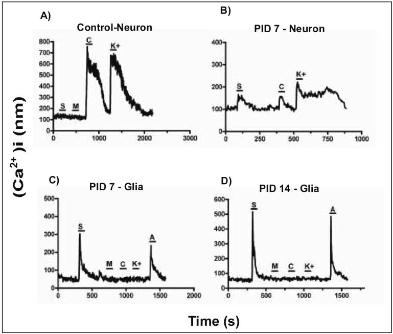
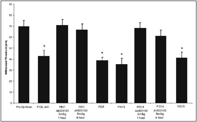
Nucleoside reverse transcriptase inhibitors (NRTIs) are known to produce painful neuropathies and to enhance states of pain hypersensitivity produced by HIV-1 infection. It has also been observed that in some neuropathic pain models, chemokines and their receptors are upregulated, perhaps contributing to the pain state. In order to understand if chemokines are involved in NRTI-mediated sensory neuropathies, we treated rats with the anti-retroviral drug, 2',3'-dideoxycytidine (ddC), which is known to produce an extended period of hyperalgesia and allodynia. Using in situ hybridization, we observed that under normal conditions, CXCR4 chemokine receptors were widely expressed by satellite glia in the dorsal root ganglia (DRG) and Schwann cells in the sciatic nerve. A limited number of DRG neurons also expressed CXCR4 receptors. The chemokine SDF-1/CXCL12 was similarly expressed in glial cells in the DRG and peripheral nerve. Following a single administration of ddC, expression levels of CXCR4 mRNA in glia and neurons and SDF-1 mRNA in glia increased considerably. The functional nature of increased CXCR4 mRNA expression was confirmed by measuring SDF-1 induced [Ca2+]i increases in acutely isolated DRG neurons and glia. In contrast, the expression of the chemokine receptors CCR2 and CCR5 did not change following ddC treatment. Pain hypersensitivity produced by ddC could be inhibited by treatment with the CXCR4 antagonist, AMD3100. Hence, we postulate that NRTIs produce pain hypersensitivity through the upregulation of CXCR4 signaling in the DRG. Increased numbers of CXCR4 receptors would also explain the synergism observed between NRTI treatment and the proalgesic effects of HIV-1 infection.


Figures

Figure 1
Figure 1. The effects of ddC on paw withdrawal thresholds in response to mechanical stimulation
Baseline responses to tactile stimulation with von Frey filaments were determined before NRTI injection and for 3-42 days following NRTI treatment. Each bar represents the combined bilateral changes in mean threshold force (±SE) eliciting a withdrawal to mechanical indentation of the hindpaw. The threshold force was significantly reduced starting 3 days after the intraperitoneal injection of ddC, and lasted through 42 days (*, p<0.001, n=12).
Figure 2
Figure 2. ddC induced neuropathic pain is accompanied by nerve pathology
The sciatic nerve was removed and analyzed for resulting pathology three days after a single i.p injection of ddC. A & B) Low and high power photomicrographs of the sciatic nerve from a vehicle-treated rat. C & D) Low and high power photomicrographs of a PID3 sciatic nerve after a single i.p. injection of ddC. Note the increased myelin sheath thickness of the largest diameter nerve fibers in the sciatic nerve. The mean diameter of normal and pathological axons did not differ, suggesting that the increase in myelin area diminishes the area of the axon cytoplasm. Also observed is a degeneration of the Remak bundles associated with unmyelinated axons as well (see arrows). Scale bar for A and C is 20μm; B and D is 5μm.
Figure 3
Figure 3. CXCR4 receptor mRNA expression in the DRG following ddC administration
We used in situ hybridization to visualize changes in CXCR4 receptor mRNA levels. A) Lumbar DRG removed from saline-treated rat exhibited a basal level of CXCR4 mRNA receptor expression. This was mainly observed in non-neuronal cells (densely black small cells), however, a few neurons were also positive. B) At one day post-injection, there was an upregulation of the CXCR4 receptor mRNA in the DRG. C and D) At 7 and 14 days post-injection, there was an even greater level of CXCR4 mRNA expression in the DRG. This appeared to be mainly in non-neuronal cells; however, there is some neuronal CXCR4 expression as well. (Arrows: neurons negative for CXCR4 mRNA transcripts, arrowheads: CXCR4 mRNA transcript positive neurons). Scale bar is 250 μm for panels A-C. Panel D is 75 μm (n=4 for each time point).
Figure 4
Figure 4. SDF1 mRNA expression in the DRG following ddC injection
In situ hybridization was used to assess the expression pattern of SDF1 mRNA. A) Low power and (B) high power photomicrograph of basal expression of SDF1 mRNA was observed in the lumbar DRG from saline injected rats, mainly in non-neuronal cells (small, densely stained cells). After a single injection of ddC, the level of SDF1 mRNA expression increased at post-injection day (PID) -7 (C, low power magnification; D, high power magnification) and PID14 days (E, low power magnification; F, high power magnification). Most of the expression appeared to be in non-neuronal cells, however some neurons were also positively labeled. (Arrows, glia positive for SDF1 mRNA; arrowheads, neurons positive for SDF1 mRNA). Scale bar A, C and E is 100 μm; B, D and F is 50 μm (n = 4 for each time point).
Figure 5
Figure 5. SDF1 increases [Ca2+]i levels in DRG cells following ddC administration
Examples of [Ca2+]i responses of cells acutely isolated from rat DRG at 7 and 14 days post-ddC injection. A) Under normal conditions, neuronal cells (capsaicin and/or K+ sensitive) were generally unresponsive to SDF1 (100nM) application. At post-ddC injection day 7 (PID7), the number of cells responding to SDF1 application increased (see table 1). Some of those responding cells were classified as neurons (B), while most responding to SDF1 application were classified as glial cells (capsaicin,K+ insensitive,ATP sensitive) (C and D) The percentage of cells responding to SDF1 at PID 14 also increased (see table 1 and D). For all experiments, MCP1 (100nM) was also applied. The percentage of cells responding to MCP-1 did not change (see table 1) [MCP-1 (M), SDF-1(S), Capsaicin (C), high K (K) and ATP (A)].
Figure 6
Figure 6. The effect of the CXCR4 antagonist, AMD3100, on ddC-induced neuropathic pain
After the induction of neuropathic pain using ddC, AMD3100 was administered and bilateral pain behavior was assessed using the Von-Frey filament test. Behavior was tested at 1, 4 and 24 hours after the administration of AMD3100 (5mg/kg) on post-injection day (PID) -7 and PID14. Following a single injection of ddC, the bilateral paw withdrawal threshold required to elicit a response was significantly reduced at post-injection PID 6 and PID13 compared to vehicle-treated rats (n=6;*p<0.001). Following administration of AMD3100 on PID7 and again at PID14, bilateral paw withdrawal thresholds increased to pre-ddC levels and attenuation of nociceptive behavior lasted for at least 4 hours. Twenty-four hours after AMD3100 treatment, paw withdrawal threshold levels did not differ from PID6 or PID13.

References

    1. Abbadie C, Lindia JA, Cumiskey AM, Peterson LB, Mudgett JS, Bayne EK, DeMartino JA, MacIntyre DE, Forrest MJ. Impaired neuropathic pain responses in mice lacking the chemokine receptor CCR2. Proc Natl Acad Sci U S A. 2003;100:7947–7952. - PMC - PubMed
    1. Adlkofer K, Martini R, Aguzzi A, Zielasek J, Toyka KV, Suter U. Hypermyelination and demyelinating peripheral neuropathy in Pmp22-deficient mice. Nat Genet. 1995;11:274–280. - PubMed
    1. Belmadani A, Tran PB, Ren D, Miller RJ. Chemokines regulate the migration of neural progenitors to sites of neuroinflammation. J Neurosci. 2006;26:3182–3191. - PMC - PubMed
    1. Berger JR, Levy RM. The neurologic complications of human immunodeficiency virus infection. Med Clin North Am. 1993;77:1–23. - PubMed
    1. Bezzi P, Domercq M, Brambilla L, Galli R, Schols D, De Clercq E, Vescovi A, Bagetta G, Kollias G, Meldolesi J, Volterra A. CXCR4-activated astrocyte glutamate release via TNFalpha: amplification by microglia triggers neurotoxicity. Nat Neurosci. 2001;4:702–710. - PubMed

Publication types

MeSH terms