

Structural basis for the actin-binding function of missing-in-metastasis
- PMID: 17292833
- PMCID: PMC1853380
- DOI: 10.1016/j.str.2006.12.005
Structural basis for the actin-binding function of missing-in-metastasis
Abstract
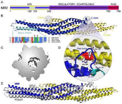
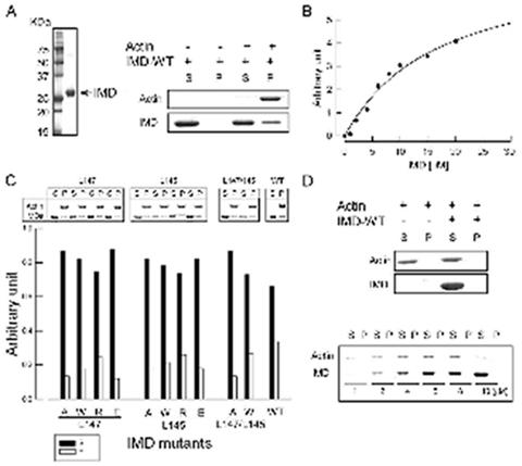
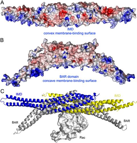
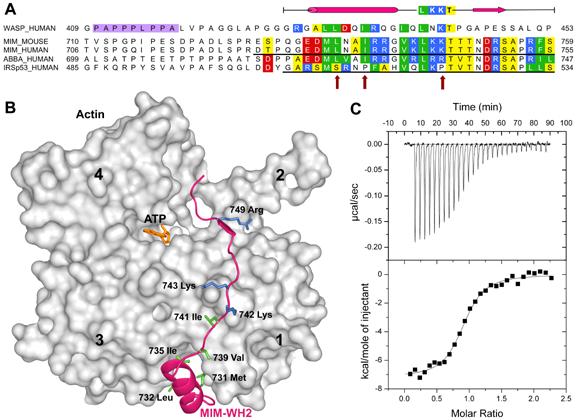
The adaptor protein missing-in-metastasis (MIM) contains independent F- and G-actin binding domains, consisting, respectively, of an N-terminal 250 aa IRSp53/MIM homology domain (IMD) and a C-terminal WASP-homology domain 2 (WH2). We determined the crystal structures of MIM's IMD and that of its WH2 bound to actin. The IMD forms a dimer, with each subunit folded as an antiparallel three-helix bundle. This fold is related to that of the BAR domain. Like the BAR domain, the IMD has been implicated in membrane binding. Yet, comparison of the structures reveals that the membrane binding surfaces of the two domains have opposite curvatures, which may determine the type of curvature of the interacting membrane. The WH2 of MIM is longer than the prototypical WH2, interacting with all four subdomains of actin. We characterize a similar WH2 at the C terminus of IRSp53 and propose that in these two proteins WH2 performs a scaffolding function.
Figures
Similar articles
-
The structural basis of actin interaction with multiple WH2/beta-thymosin motif-containing proteins.Structure. 2006 Mar;14(3):469-76. doi: 10.1016/j.str.2005.12.011. Structure. 2006. PMID: 16531231
-
A novel actin bundling/filopodium-forming domain conserved in insulin receptor tyrosine kinase substrate p53 and missing in metastasis protein.J Biol Chem. 2004 Apr 9;279(15):14929-36. doi: 10.1074/jbc.M309408200. Epub 2004 Jan 29. J Biol Chem. 2004. PMID: 14752106
-
Actin-bound structures of Wiskott-Aldrich syndrome protein (WASP)-homology domain 2 and the implications for filament assembly.Proc Natl Acad Sci U S A. 2005 Nov 15;102(46):16644-9. doi: 10.1073/pnas.0507021102. Epub 2005 Nov 7. Proc Natl Acad Sci U S A. 2005. PMID: 16275905 Free PMC article.
-
WH2 domain: a small, versatile adapter for actin monomers.FEBS Lett. 2002 Feb 20;513(1):92-7. doi: 10.1016/s0014-5793(01)03242-2. FEBS Lett. 2002. PMID: 11911886 Review.
-
Control of actin assembly by the WH2 domains and their multifunctional tandem repeats in Spire and Cordon-Bleu.Int Rev Cell Mol Biol. 2011;290:55-85. doi: 10.1016/B978-0-12-386037-8.00005-3. Int Rev Cell Mol Biol. 2011. PMID: 21875562 Review.
Cited by
-
Structured and intrinsically disordered domains within Amphiphysin1 work together to sense and drive membrane curvature.Soft Matter. 2019 Nov 21;15(43):8706-8717. doi: 10.1039/c9sm01495k. Epub 2019 Oct 17. Soft Matter. 2019. PMID: 31621751 Free PMC article.
-
The ArfGAP ASAP1 Controls Actin Stress Fiber Organization via Its N-BAR Domain.iScience. 2019 Dec 20;22:166-180. doi: 10.1016/j.isci.2019.11.015. Epub 2019 Nov 14. iScience. 2019. PMID: 31785555 Free PMC article.
-
I-BAR protein antagonism of endocytosis mediates directional sensing during guided cell migration.J Cell Biol. 2010 Apr 19;189(2):353-67. doi: 10.1083/jcb.200910136. Epub 2010 Apr 12. J Cell Biol. 2010. PMID: 20385776 Free PMC article.
-
Molecular basis for the dual function of Eps8 on actin dynamics: bundling and capping.PLoS Biol. 2010 Jun 1;8(6):e1000387. doi: 10.1371/journal.pbio.1000387. PLoS Biol. 2010. PMID: 20532239 Free PMC article.
-
The BAR domain of the Arf GTPase-activating protein ASAP1 directly binds actin filaments.J Biol Chem. 2020 Aug 7;295(32):11303-11315. doi: 10.1074/jbc.RA119.009903. Epub 2020 May 22. J Biol Chem. 2020. PMID: 32444496 Free PMC article.
References
Publication types
MeSH terms
Substances
Grants and funding
LinkOut - more resources
Full Text Sources
Other Literature Sources
Molecular Biology Databases
