

Structural rigidity of a large cavity-containing protein revealed by high-pressure crystallography
- PMID: 17292912
- PMCID: PMC1853337
- DOI: 10.1016/j.jmb.2006.12.021
Structural rigidity of a large cavity-containing protein revealed by high-pressure crystallography
Abstract
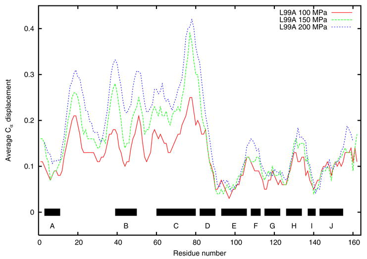
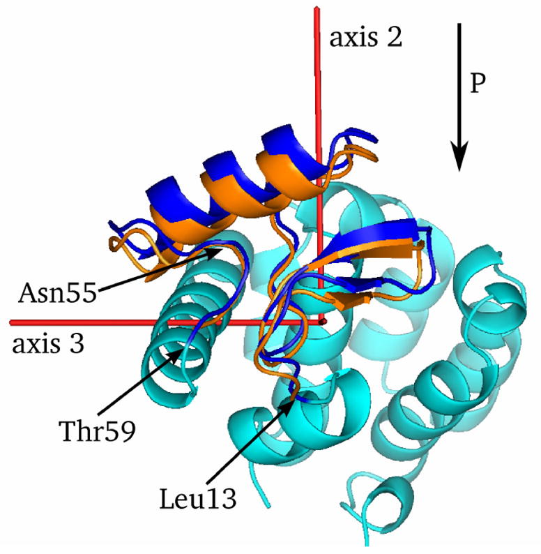
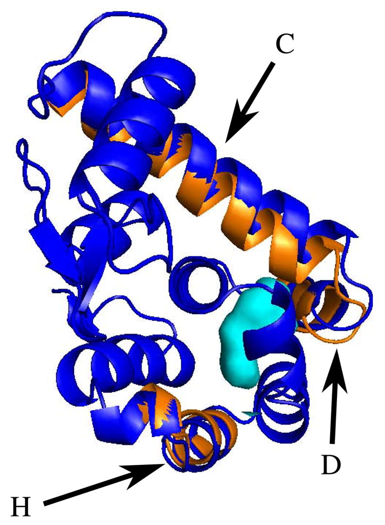
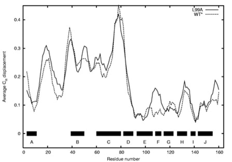
Steric constraints, charged interactions and many other forces important to protein structure and function can be explored by mutagenic experiments. Research of this kind has led to a wealth of knowledge about what stabilizes proteins in their folded states. To gain a more complete picture requires that we perturb these structures in a continuous manner, something mutagenesis cannot achieve. With high pressure crystallographic methods it is now possible to explore the detailed properties of proteins while continuously varying thermodynamic parameters. Here, we detail the structural response of the cavity-containing mutant L99A of T4 lysozyme, as well as its pseudo wild-type (WT*) counterpart, to hydrostatic pressure. Surprisingly, the cavity has almost no effect on the pressure response: virtually the same changes are observed in WT* as in L99A under pressure. The cavity is most rigid, while other regions deform substantially. This implies that while some residues may increase the thermodynamic stability of a protein, they may also be structurally irrelevant. As recently shown, the cavity fills with water at pressures above 100 MPa while retaining its overall size. The resultant picture of the protein is one in which conformationally fluctuating side groups provide a liquid-like environment, but which also contribute to the rigidity of the peptide backbone.
Figures

Similar articles
-
Structural and thermodynamic characterization of T4 lysozyme mutants and the contribution of internal cavities to pressure denaturation.Biochemistry. 2008 Oct 21;47(42):11097-109. doi: 10.1021/bi801287m. Epub 2008 Sep 25. Biochemistry. 2008. PMID: 18816066 Free PMC article.
-
Structural analysis of a non-contiguous second-site revertant in T4 lysozyme shows that increasing the rigidity of a protein can enhance its stability.J Mol Biol. 1999 Oct 8;292(5):1111-20. doi: 10.1006/jmbi.1999.3102. J Mol Biol. 1999. PMID: 10512706
-
Role of cavities and hydration in the pressure unfolding of T4 lysozyme.Proc Natl Acad Sci U S A. 2014 Sep 23;111(38):13846-51. doi: 10.1073/pnas.1410655111. Epub 2014 Sep 8. Proc Natl Acad Sci U S A. 2014. PMID: 25201963 Free PMC article.
-
Lessons from the lysozyme of phage T4.Protein Sci. 2010 Apr;19(4):631-41. doi: 10.1002/pro.344. Protein Sci. 2010. PMID: 20095051 Free PMC article. Review.
-
Structural and genetic analysis of the folding and function of T4 lysozyme.FASEB J. 1996 Jan;10(1):35-41. doi: 10.1096/fasebj.10.1.8566545. FASEB J. 1996. PMID: 8566545 Review.
Cited by
-
Pushed to extremes: distinct effects of high temperature versus pressure on the structure of STEP.Commun Biol. 2024 Jan 12;7(1):59. doi: 10.1038/s42003-023-05609-0. Commun Biol. 2024. PMID: 38216663 Free PMC article.
-
Differential Responses in the Core, Active Site and Peripheral Regions of Cytochrome c Peroxidase to Extreme Pressure and Temperature.J Mol Biol. 2024 Nov 15;436(22):168799. doi: 10.1016/j.jmb.2024.168799. Epub 2024 Sep 26. J Mol Biol. 2024. PMID: 39332669
-
Universality and Structural Implications of the Boson Peak in Proteins.Biophys J. 2019 Jul 23;117(2):229-238. doi: 10.1016/j.bpj.2019.06.007. Epub 2019 Jun 14. Biophys J. 2019. PMID: 31255295 Free PMC article.
-
Spontaneous drying of non-polar deep-cavity cavitand pockets in aqueous solution.Nat Chem. 2020 Jul;12(7):589-594. doi: 10.1038/s41557-020-0458-8. Epub 2020 May 18. Nat Chem. 2020. PMID: 32424255 Free PMC article.
-
Coupling of pressure-induced structural shifts to spectral changes in a yellow fluorescent protein.Biophys J. 2009 Sep 16;97(6):1719-27. doi: 10.1016/j.bpj.2009.06.039. Biophys J. 2009. PMID: 19751677 Free PMC article.
References
-
- Eriksson AE, Baase WA, Matthews BW. Similar hydrophobic replacements of Leu99 and Phe153 within the core of T4 lysozyme have different structural and thermodynamic consequences. J Mol Biol. 1993;229:747–769. - PubMed
-
- Eriksson AE, Baase WA, Zhang XJ, Blaber M, Baldwin EP, Matthews BW. Response of a protein structure to cavity-creating mutations and its relation to the hydrophobic effect. Science. 1992;255:178–183. - PubMed
-
- Baldwin E, Baase WA, Zhang X, Feher V, Matthews BW. Generation of ligand binding sites in T4 lysozyme by deficiency-creating substitutions. J Mol Biol. 1998;277:467–85. - PubMed
Publication types
MeSH terms
Substances
Associated data
- Actions
- Actions
- Actions
- Actions
- Actions
- Actions
- Actions
- Actions
- Actions
- Actions
- Actions
- Actions
- Actions
Grants and funding
LinkOut - more resources
Full Text Sources
Research Materials
